Figures & data
Figure 1. Preoperative planning software (Materialise NV) and postoperative CT measurements for frontal femoral component angle (FFCA), sagittal femoral component angle (SFCA), axial femoral component angle (AFCA), frontal tibial component angle (FTCA), and sagittal tibial component angle (STCA).
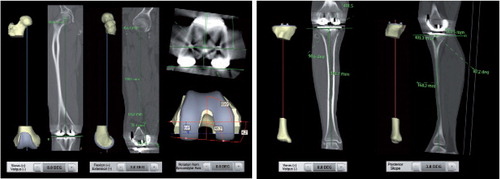
Table 1. The target angle from the preoperatively planned alignment from planning software and postoperative CT measurements depicted as mean and SD from the 2 observers
Table 2. Intraclass correlation (ICC) between observer 1 and observer 2 in postoperative CT measurements
Figure 2. Box plot showing the distributions of FFCA, SFCA, AFCA, FTCA, and STCA. The boxes show the median and interquartile range (IQR). The whiskers represent the range where the lowest and highest values are not more than 1.5 × IQR above the median. Circles represent outliers, i.e. values more than 1.5 × IQR from the median.
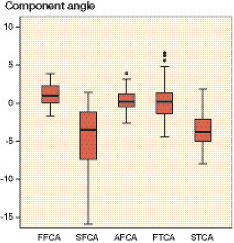
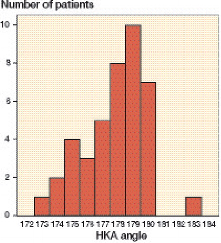
Figure 3. Distribution of the postoperative HKA angle. Angles greater and less than 180 degrees represent valgus and varus, respectively.