Figures & data
Figure 1. Plain AP (A) and lateral view (B) showing a normal alpha angle. A 12 × 14 mm calcified irregular-shaped image was seen at the superolateral acetabular rim. Excluding the “os acetabuli”, the center-edge angle was 15º (25º in the contralateral hip). The Tönnis angle was 24º.
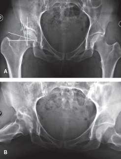
Figure 2. Sagittal plane CT scan image optimized for bone density, showing the “os acetabuli” (arrow).
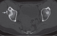
Figure 3. View of the peripheral compartment from the anteromedial portal with a 30º scope. The close relation between the detached labrum (L) and the “os acetabuli” (O) can be seen. F: femoral head.
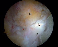
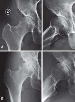
Figure 4. A. At 4-month follow-up, complete resection of the “os acetabuli” and a slight narrowing of the joint space. B. At 10-month follow-up anterosuperior subluxation and clear degenerative joint disease with sclerotic joint line and subchondral cyst formation.