Figures & data
Figure 1. The unsterile O-arm closed around the patient for an intraoperative 3D scan. The patient is draped with 2 sterile drapings, which are removed after scanning so that sterility is not jeopardized.
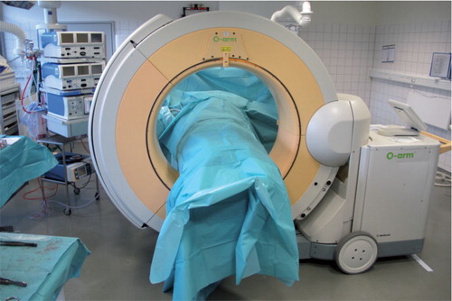
Table 1. Distribution of fractures according to Judet-Letournel, and the operative result achieved for the group operated on with intraoperative 3D control (3D) and the group operated on with intraoperative fluoroscopic control alone (Control)
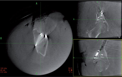
Figure 2. The intraoperative 2D reconstructed views of a posterior column lag screw visualized in a 2-column fracture. The axial reconstruction is shown to the left, coronal view is shown to the upper right, and sagittal view is shown to the lower right. The surgeon can orientate the planes parallel to the screw, enabling a precise evaluation of the screw path and confirming the extra-articular position in the cytoloid fossa.
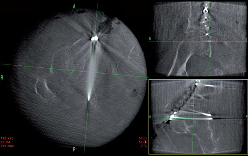
Figure 3. Intraoperative 2D reconstructions of a reduced and stabilized posterior wall fracture. Axial view to the left, coronal view to the upper right, and sagittal view to the lower right. This clearly shows good reduction and extra-articular screw placement.
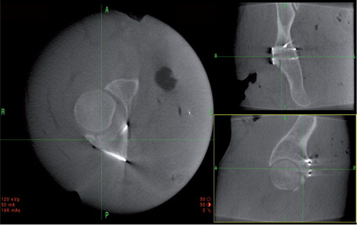
Figure 4. Intraoperative 2D reconstructed views of the unreduced fragment in the supra-acetabular region of a 2-column fracture. Axial view to the left, coronal view to the upper right, and sagittal view to the lower right. The fracture can be reduced with a ball spike through a stab incision laterally and the screw paths for the screws that indirectly fixate the fracture can be planned. The result achieved is shown in .
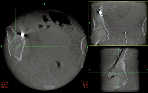
Figure 5. 2D reconstructed images of the 2-column fracture seen in after reduction and indirect screw fixation.