
Figures & data
Figure 1. Flow chart of study.
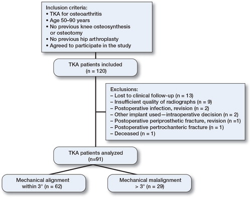
Figure 2. Standing anterior-posterior radiograph with a varus malalignment. Mechanical axis as hip-knee-ankle (HKA) and component alignment angles are represented. The HKA angle is the angle formed between the mechanical femoral and tibial axes. The femoral component (FC) angle is the angle medially between the distal surfaces of the femoral component and the femoral mechanical axis. The tibial component (TC) angle is the medial angle between the tibial component plateau and the tibial mechanical axis.

Table 1. Postoperative radiological data (mean (SD); °) for patients with hip-knee-ankle (HKA) angle and component alignment within ± 3° or > 3° (varus or valgus) from a straight mechanical axis
Figure 3. Comparisons of mean (with 95% CI; whiskers) ROM (°), KSS (points), muscle torques (Nm), and hamstring-quadriceps ratios (%) (n = 91) preoperatively and postoperatively.
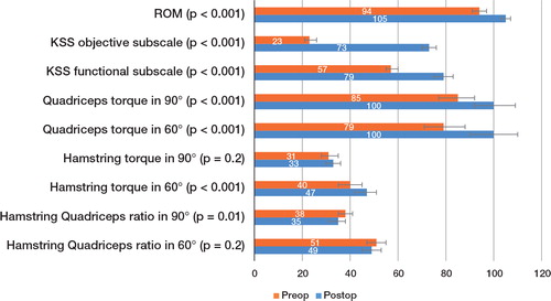
Table 4. Postoperative ROM, KSS objective, functional and pain assessment subscale (ss) results (mean (SD), mean difference with 95% confidence intervals (CI), or rates) between patients with mechanical and component alignment within ± 3° and > 3°