Figures & data
Table 1. Basic clinical and genetic traits of endometriosis and uterine leiomyoma.
Figure 1. Epigenetic landscape and gene network of endometriosis (EM) and uterine leiomyoma (UL). (Top center) ‘Epigenetic landscape’ of mesenchymal stem cell (mSC) development resulting in EM or UL. ‘Epigenetic landscape’ shows branching channels (paths) - feasible ways taken by differentiating mSC. After the onset of pathological process provoked by some alien intrinsic and extrinsic factors mSC undergo blast transformation and proceed further development with many variations resulting in different clinical manifestations of the diseases. (Remaining columns) Main functional groups of the gene-networks associated with the origin and development of EM and UL. Meaningful associated genes are in bolds. The rest are candidate genes identified in functional mapping studies [Rahmioglu et al. Citation2014; Baranov et al. Citation2015].
![Figure 1. Epigenetic landscape and gene network of endometriosis (EM) and uterine leiomyoma (UL). (Top center) ‘Epigenetic landscape’ of mesenchymal stem cell (mSC) development resulting in EM or UL. ‘Epigenetic landscape’ shows branching channels (paths) - feasible ways taken by differentiating mSC. After the onset of pathological process provoked by some alien intrinsic and extrinsic factors mSC undergo blast transformation and proceed further development with many variations resulting in different clinical manifestations of the diseases. (Remaining columns) Main functional groups of the gene-networks associated with the origin and development of EM and UL. Meaningful associated genes are in bolds. The rest are candidate genes identified in functional mapping studies [Rahmioglu et al. Citation2014; Baranov et al. Citation2015].](/cms/asset/9b227e5b-08b6-4c34-9434-39523e77058f/iaan_a_1123325_f0001_oc.jpg)
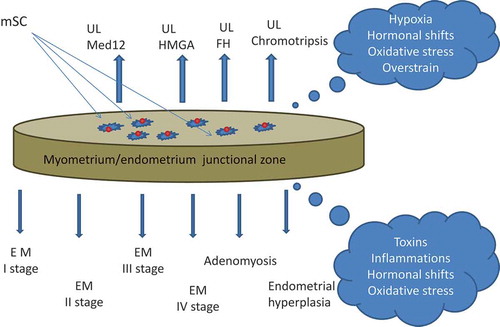
Figure 2. Junctional zone (JZ) of the uterus as ‘the same coin’. Myometrial/endometrial JZ of the uterus at the boundary of endometrium/myometrium layers as a principal source of mesenchymal stem cells (mSC), generating under unfavorable conditions endometriosis (EM) lesions or uterine leiomyoma (UL) fibroids. The mSC from the upper (myometrial) side of the ‘coin’ are epigenetically programmed to generate UL, and the mSC from the bottom (endometrial) side are prone to give EM lesions.