Figures & data
Figure 1. Bunnell's intrinsic tightness test involves passively holding the patient's MCP joint extended, and then passively flexing the PIP joint. There is intrinsic tightness if the PIP joint is difficult to flex.
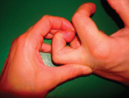
Figure 2. The extensor apparatus of a finger. The red triangle shows the area that should be resected in the distal ulnar intrinsic release procedure. The triangular piece consists of oblique fibres and the ulnar lateral band.
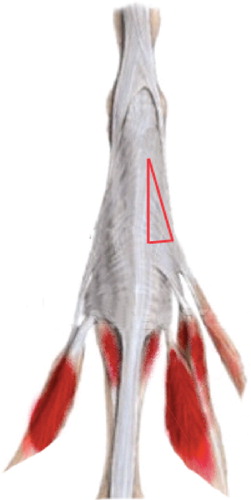
Figure 3. Distal ulnar intrinsic release. An oblique dorsal incision on the proximal phalanx exposes the extensor hood apparatus on the ulnar side and a triangular piece is resected. The triangle is roughly 5 mm at the base, 12–15 mm at the side, and the hypotenuse is 15–16 mm.
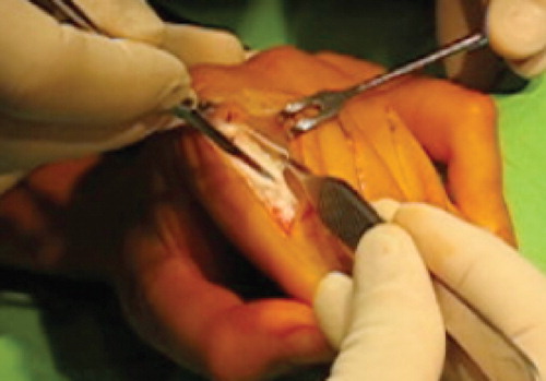
Figure 4. Mean (SD) results over time at follow-up at 1, 3, and 6 months. The biggest improvement of range of motion was during the first month after the operation.
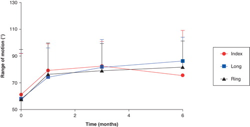
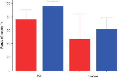
Figure 5. Mean (SD) passive range of motion of the PIP joints preoperatively compared with 6 months postoperatively in the severe group. There were significant increases in range of motion postoperatively in the long and ring finger. Red = before, and blue = after, operation.
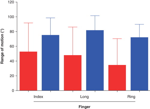
Figure 6. Mean (SD) passive range of motion of the PIP joints preoperatively compared with 6 months postoperatively in the mild group. There was a significant increase in the range of motion for the long finger. Red = before, and blue = after, operation.
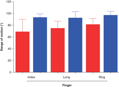
Figure 7. The patients in the mild group had a better range of motion at 1 month follow-up than the severe group, who had more operations. The severe group kept improving their range of motion during the following months. Red = before, and blue = after, operation.