Figures & data
Figure 1. Viability and proliferation of human CB MSCs in RGD-alginate. (A) The effect of RGD-alginate on viability of encapsulated cells. The ratio of viable cells in RGD-alginate to the viable cells in alginate was calculated using the MTT assay. (B) Comparison of MSC proliferation in RGD-alginate and alginate (no significant difference). The ratio of viable cells at different days per viable cells at Day 1 was calculated using the MTT assay. Data are means ± SD, n = 3, Student's t-test. *Significant difference from a value of 1.0, P < 0.05.
Figure 2. FIX secretion by CB MSCs in RGD-alginate. FIX secretion was measured using the ELISA and reported as the mass of FIX (ng) secreted from 1 ml of microcapsules per 24 hr. Data are means ± SD, n ≥ 3. P < 0.01, Student's t-test. No significant difference.
Figure 3. Effect of RGD or fibrinogen modification on viability and FIX secretion of encapsulated cells. (A) Effect of RGD modification versus fibrinogen modification on viability of the encapsulated MSC. (B) Effect of RGD modification versus fibrinogen modification on FIX secretion from the encapsulated MSCs. *Significant difference from a value of 1.0, P < 0.05.
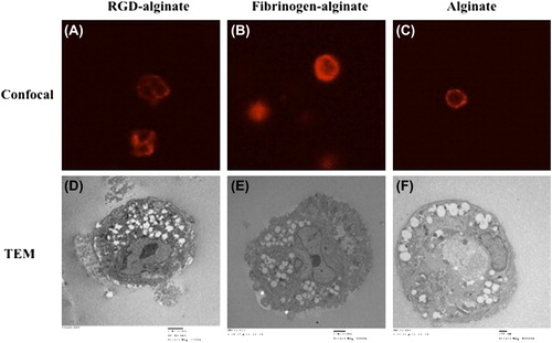
Figure 4. (A–C) F-actin staining of encapsulated cells. (D–F) TEM images of encapsulated cells.