Figures & data
Figure 1. H-NMR spectrum of bacterial PHBV produced by using propionic acid.
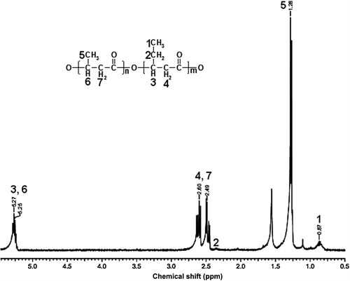
Figure 2. SEM photographs were exhibiting the alignment of the PHBV nanofiber scaffolds at different magnifications.
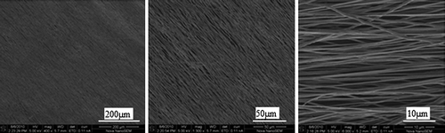
Figure 3. Surgical implantation of autograft (A) and aligned PHBV graft (B) for nerve regeneration in rat sciatic nerve under a microscope (10×).
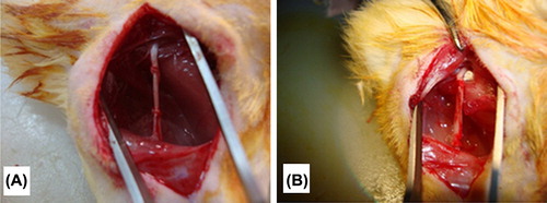
Figure 4. 8th (n = 18) and 16th (n = 12) weeks after surgery, electrophysiological assessments of autograph and PHBV graft group rats. *Statistical significance (p < 0.05).
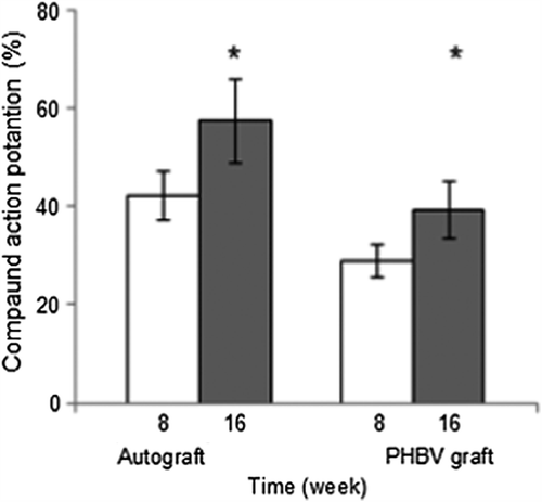
Figure 5. After 4th, 8th (n = 18), and 16th (n = 12) weeks of operations, autograft and PHBV graft group SFI values. *Statistical significance (p < 0.05).
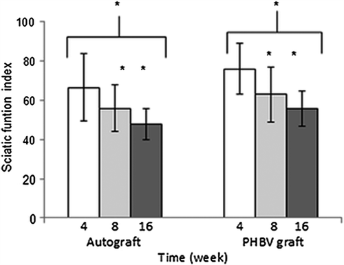
Figure 6. Footprints of PHBV graft-applied legs of the rats, after 4th (A, B); 8th (C, D); 16th (E, F) weeks and not-operated footprint (G).
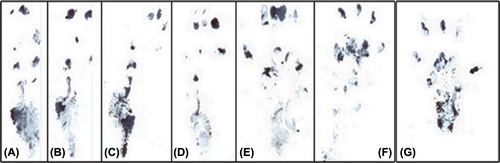
Figure 7. The regenerating numerous axons (arrow) form regular fasicles in 8th (A) and 16th (C) weeks of autograft group. The PHBV graft is surrounded by an inflammatory capsule (c) consisting of phagocytic and fibroblastic cells at its inner side at 8th week (B). In PHBV graft group at 16th week (D) the tubes lumen is filled with fibrovascular connective tissue with small blood vessels (asterisk) and regenerating axon clusters with higher numbers at the 16th week (D) compared to the 8th week (B).
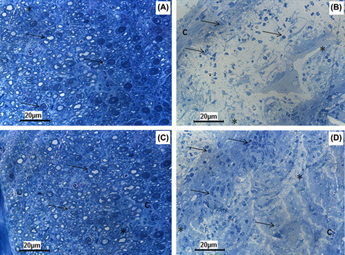
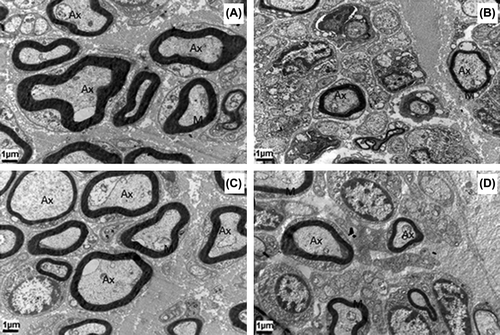
Figure 8. Transmission electron micrographs have the same magnification (12000×). All the groups show the regenerating clusters of myelinating or myelinated axons. Note that the number of axons and the thickness of their myelin sheath is higher in autograft group at the 8th (A) and 16th weeks (C) compared to PHBV graft group at the 8th (B) and 16th weeks (D). Ax: Axon; M: Myelin. Uranyl acetate and lead citrate.