3,858
Views
68
CrossRef citations to date
0
Altmetric
Article

Biosynthesis of silver nanoparticles using Saccharomyces cerevisiae

, , , &
Pages 235-239 | Received 01 Jun 2014, Accepted 19 Jun 2014, Published online: 07 Aug 2014

Figures & data

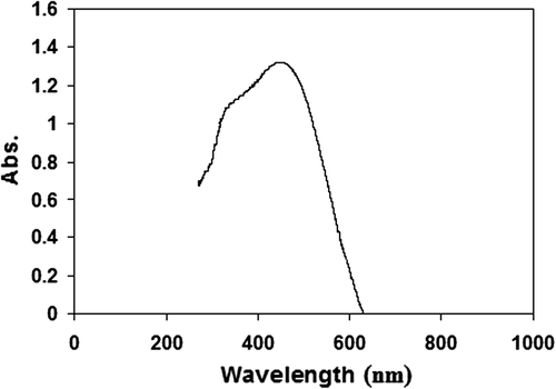
Figure 1. UV-Vis absorption spectrum of the colloidal silver produced. The spectrum was obtained 72 h after the start of AgNO3 (1 mM) reduction using S. cerevisiae.
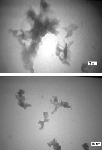
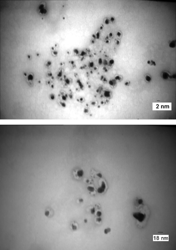
Figure 2. TEM micrographs of the NPs produced 72 h after the start of AgNO3 (1 mM) reduction using dried commercially available S. cerevisiae- Magnification is 25,000.
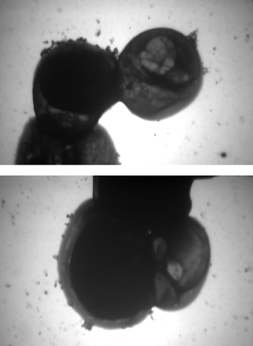
Figure 3. TEM micrographs of the NPs produced 24 h after the start of AgNO3 (1 mM) reduction using freshly cultured S. cerevisiae. Magnification is 4000, and the micrograph is focused on the NPs outside of the yeast, and the ones attached to the membrane.
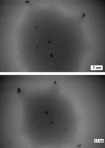
Figure 4. TEM micrographs of the NPs produced 24 h after the start of AgNO3 (1 mM) reduction using freshly cultured S. cerevisiae. Magnification is 25,000, and the micrograph is focused on the location of the NPs inside the yeast.
Figure 5. TEM micrographs of the NPs produced 5 h after the start of AgNO3 (1 mM) reduction using freshly cultured S. cerevisiae. Magnification is 63,000, and the micrograph is focused on the corona or film around the NPs.
Figure 6. TEM micrographs of the NPs produced 24 h after the start of AgNO3 (1 mM) reduction using freshly cultured S. cerevisiae. The micrograph is focused on the location of the NPs in the membrane, and the steps in the secretion of the NPs out of the yeast.
Figure 7. Particle size analysis of NPs produced after 48 h of biotransformation.
Figure 8. TEM micrographs of the NPs produced 24 h after the start of AgNO3 (1 mM) reduction using freshly cultured S. cerevisiae. The micrograph is focused on disintegration of the yeast by NPs.

Reprints and Corporate Permissions

Please note: Selecting permissions does not provide access to the full text of the article, please see our help page How do I view content?

To request a reprint or corporate permissions for this article, please click on the relevant link below:

Academic Permissions

Please note: Selecting permissions does not provide access to the full text of the article, please see our help page How do I view content?

Obtain permissions instantly via Rightslink by clicking on the button below:

If you are unable to obtain permissions via Rightslink, please complete and submit this Permissions form. For more information, please visit our Permissions help page.