7,913
Views
72
CrossRef citations to date
0
Altmetric
Review

Advances in preparation and characterization of chitosan nanoparticles for therapeutics

, , , &
Pages 305-314 | Received 21 May 2014, Accepted 28 May 2014, Published online: 19 Aug 2014

Figures & data

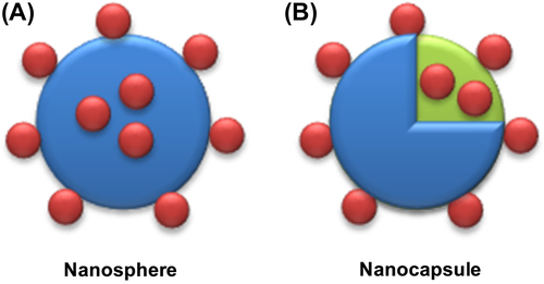
Figure 1. Different types of nanoparticles (A) Nanospheres where drug is either entrapped in the polymer matrix or adsorbed onto surface or both. (B) Nanocapsules where drug is either entrapped inside the hollow capsule or adsorbed onto surface or both.
Figure 2. Schematic representation of preparation of chitosan from chitin by deacetylation.

Table I. Methods for preparation of chitosan nanoparticles.
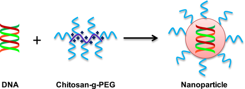
Figure 3. Polyelectrolyte complex method: the nanoparticles formation takes place due to electrostatic interaction between anion (DNA) and cation (chitosan), followed by charge neutralization.
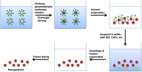
Figure 4. Microemulsion method: in the reverse micelles, the reaction takes place in the aqueous core of the reverse micellar droplets. Here the aqueous solutions of monomer, cross-linking agent and other hydrophilic compounds remain in the aqueous core (host nanoreactor) of the reverse micelles. Polymerization reaction, which leads to the formation of the nanoparticles, takes place within these aqueous cores by a primary growth process.

Table II. Techniques for characterization of chitosan nanoparticles.
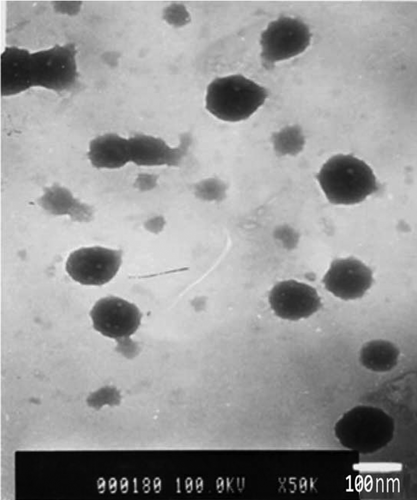
Figure 5. Transmission electron microscopy image of chitosan nanoparticles crosslinked with glutaraldehyde. The average particle size is 90 nm. (Adapted from CitationManchanda and Nimesh 2010).
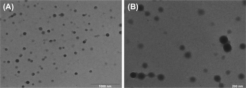
Figure 6. Transmission electron image (TEM) of chitosan/DNA complexes. The average size of complexes is 200 nm. The image A is at lower magnification with the bar equal to 1000 nm and image B is at higher magnification with the bar equal to 200 nm. (Adapted from CitationNimesh et al. 2012).

Reprints and Corporate Permissions

Please note: Selecting permissions does not provide access to the full text of the article, please see our help page How do I view content?

To request a reprint or corporate permissions for this article, please click on the relevant link below:

Academic Permissions

Please note: Selecting permissions does not provide access to the full text of the article, please see our help page How do I view content?

Obtain permissions instantly via Rightslink by clicking on the button below:

If you are unable to obtain permissions via Rightslink, please complete and submit this Permissions form. For more information, please visit our Permissions help page.