818
Views
17
CrossRef citations to date
0
Altmetric
ORIGINAL ARTICLE

In vitro and in vivo evaluation of porous TiNi-based alloy as a scaffold for cell tissue engineering

, , , , , & show all
Pages 704-709 | Received 03 Jun 2014, Accepted 28 Oct 2014, Published online: 23 Jan 2015

Figures & data

Figure 1. SEM images showing interconnected pores (a) and rough pore walls with micro-porous surface (b) of porous TiNi-based SMA scaffold.
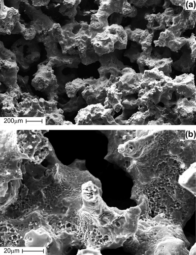
Figure 1. SEM images showing interconnected pores (a) and rough pore walls with micro-porous surface (b) of porous TiNi-based SMA scaffold.

Figure 2. The weight increase percentage calculated with the dry weight of cultured scaffold and the initial weight of the scaffold.
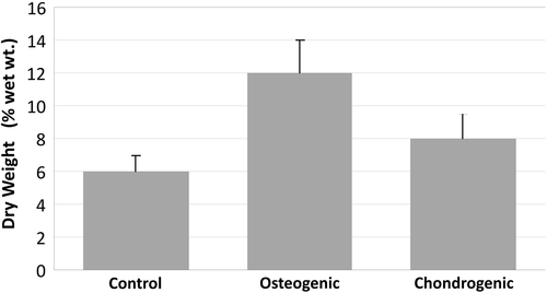
Figure 2. The weight increase percentage calculated with the dry weight of cultured scaffold and the initial weight of the scaffold.

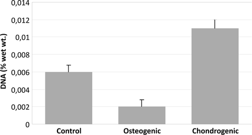
Figure 3. Amount of DNA content in cultured scaffolds.

Figure 3. Amount of DNA content in cultured scaffolds.

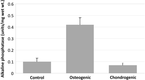
Figure 4. Alkaline phosphatase activity in cultured scaffolds.

Figure 4. Alkaline phosphatase activity in cultured scaffolds.

Figure 5. SEM images showing pancreas islet cells seeded on porous TiNi-based SMA scaffold before implantation: cells fully spread along the network of interconnected pores (a), cells fixed on rough pore walls with micro-porous surface (b).
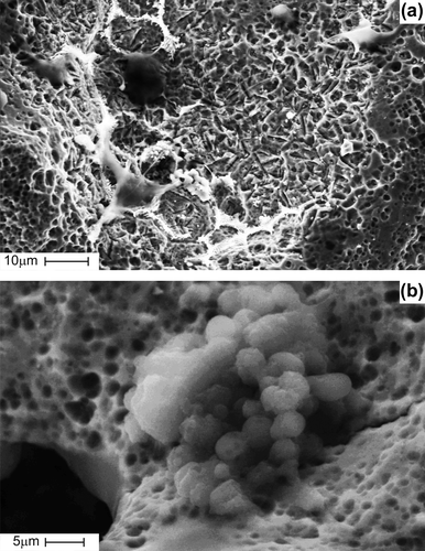
Figure 5. SEM images showing pancreas islet cells seeded on porous TiNi-based SMA scaffold before implantation: cells fully spread along the network of interconnected pores (a), cells fixed on rough pore walls with micro-porous surface (b).

Figure 6. SEM images showing time-periodic pancreas islet cell ingrowth seeded on porous TiNi-based SMA scaffold: at day 7 post-implantation, cells in the process of further proliferation with synthesizing extracellular matrix and forming the spatial pseudopodium (a); at day 7 post-implantation, cells spreading across the pores and relatively small pores fully filled with cells and extracellular matrix (b); at day 14 post-implantation gradual cellular ingrowth from the periphery towards the center (c); at day 28 post-implantation, scaffold entirely filled with cells and extracellular matrix (d).
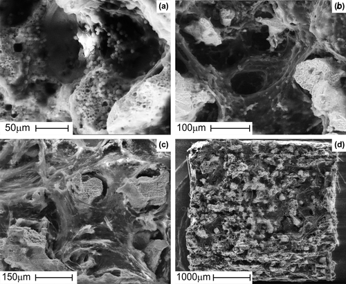
Figure 6. SEM images showing time-periodic pancreas islet cell ingrowth seeded on porous TiNi-based SMA scaffold: at day 7 post-implantation, cells in the process of further proliferation with synthesizing extracellular matrix and forming the spatial pseudopodium (a); at day 7 post-implantation, cells spreading across the pores and relatively small pores fully filled with cells and extracellular matrix (b); at day 14 post-implantation gradual cellular ingrowth from the periphery towards the center (c); at day 28 post-implantation, scaffold entirely filled with cells and extracellular matrix (d).
Reprints and Corporate Permissions

Please note: Selecting permissions does not provide access to the full text of the article, please see our help page How do I view content?

To request a reprint or corporate permissions for this article, please click on the relevant link below:

Academic Permissions

Please note: Selecting permissions does not provide access to the full text of the article, please see our help page How do I view content?

Obtain permissions instantly via Rightslink by clicking on the button below:

If you are unable to obtain permissions via Rightslink, please complete and submit this Permissions form. For more information, please visit our Permissions help page.