2,168
Views
15
CrossRef citations to date
0
Altmetric
ORIGINAL ARTICLE

Electrospun polycaprolactone matrices with tensile properties suitable for soft tissue engineering

, , , , &
Pages 878-884 | Received 08 Nov 2014, Accepted 11 Dec 2014, Published online: 24 Jan 2015

Figures & data

Table I. Tensile property of PCL with different solvents.

Table II. Porosity measurement of binary images with 3 different thresholds.

Table III. Tensile property of the nanofibrous matrices collected for 4 h.

Figure 1. (A), (B), and (C). SEM of electrospun membranes of polycaprolactone, with the concentrations of 10%, 12%, and 15% (Magnification – 5000×).
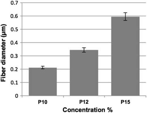
Figure 2. Depicts the graphical representation of the measurement of fiber diameter of electrospun matrices with different concentrations of 10% wt, 12% wt, and 15% wt, using ImageJ software. The fiber diameter increases with an increase in concentration.
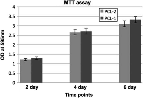
Figure 3. Biocompatibility of the polycaprolactone membrane, evaluated by the MTT assay using L6 rat skeletal myoblast cell growth tested at 3 different time points (2, 4, and 6, days in culture).
Figure 4. Cryosectioned polycaprolactone electrospun matrices seeded with rat skeletal myoblasts were stained for muscle-specific actin with FITC (A), DAPI for nuclear staining (B), and merged (C).
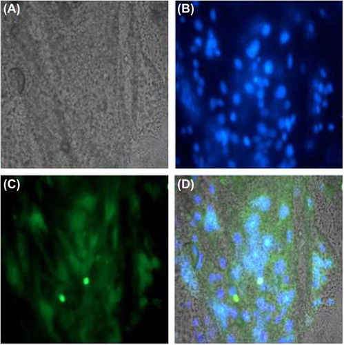
Figure 5. Cryosectioned matrices seeded with rat skeletal myoblasts were stained with (A) Phase contrast, (B) nuclear staining with DAPI, (C) desmin with FITC, and (D) Merged image of all.
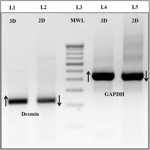
Figure 6. Agarose electrophoresis image of RT-PCR analysis of desmin and GAPDH. (Lanes 1 and 2) – Desmin 3D and 2D. (Lanes 4 and 5) – GAPDH 3D and 2D.

Reprints and Corporate Permissions

Please note: Selecting permissions does not provide access to the full text of the article, please see our help page How do I view content?

To request a reprint or corporate permissions for this article, please click on the relevant link below:

Academic Permissions

Please note: Selecting permissions does not provide access to the full text of the article, please see our help page How do I view content?

Obtain permissions instantly via Rightslink by clicking on the button below:

If you are unable to obtain permissions via Rightslink, please complete and submit this Permissions form. For more information, please visit our Permissions help page.