581
Views
2
CrossRef citations to date
0
Altmetric
ORIGINAL ARTICLE

Clinical test on circulating tumor cells in peripheral blood of lung cancer patients, based on novel immunomagnetic beads

, , , , &
Pages 892-897 | Received 10 Nov 2014, Accepted 06 Dec 2014, Published online: 14 Feb 2015

Figures & data

Figure 1. CTC enrichment and separation equipment. (A) The beaker containing 50mLPBS. (B) Peristaltic pump. (C) Horizontal table and self-made enrichment and separation container. (D) Vacuum pump. E: The beaker for receiving PBS pumped from D.
Figure 2. Recognition and indentation of CTCs. (A) and (B) original cell A549. (C) A549 that reacted with immunomagnetic antibodies. (D) cell A549 and residual white cell after immunomagnetic enrichment and separation.
Figure 3. Comparison of white cells on the slide with different parameters. (A) 20 rpm/min speed with 5° angle of incline. (B) 40 rpm/min speed with 5° angle of incline. (C) 20 rpm/min speed with 15° angle of incline. (D) 20 rpm/min speed with 15° angle of incline .

Table I. Number of CTCs under different parameters.

Figure 4. Regression curve of A549.

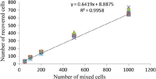
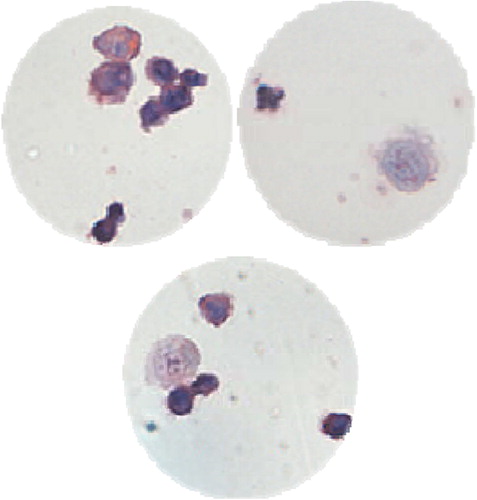
Table II. Evaluation and test results of cell recoveries from samples of peripheral blood from healthy volunteers mixed with A549.

Figure 5. CTCs captured in the peripheral blood of lung cancer patients through the novel immunomagnetic separation method, and white cells around the CTCs, in the slide observed under the 40 × light microscope.

Table III. Results of CTCs detected according to the clinical features of lung cancer patients.

Reprints and Corporate Permissions

Please note: Selecting permissions does not provide access to the full text of the article, please see our help page How do I view content?

To request a reprint or corporate permissions for this article, please click on the relevant link below:

Academic Permissions

Please note: Selecting permissions does not provide access to the full text of the article, please see our help page How do I view content?

Obtain permissions instantly via Rightslink by clicking on the button below:

If you are unable to obtain permissions via Rightslink, please complete and submit this Permissions form. For more information, please visit our Permissions help page.