701
Views
1
CrossRef citations to date
0
Altmetric
ORIGINAL ARTICLE

Sequence-specific label-free nucleic acid biosensor for the detection of the hepatitis C virus genotype 1a using a disposable pencil graphite electrode

, &
Pages 912-917 | Received 20 Nov 2014, Accepted 11 Dec 2014, Published online: 24 Jan 2015

Figures & data

Table I. Oligonucleotide sequences employed (mismatches are indicated by bold and underlined characters).
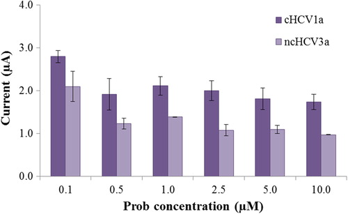
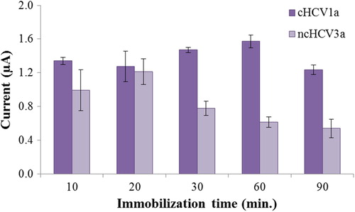
Figure 1. Schematic diagram of experimental procedures.
Figure 2. Effect of probe concentration on the guanine oxidation signal: (a) after hybridization of complementary target oligonucleotide (cHCV1a), and (b) after hybridization of noncomplementary oligonucleotide (ncHCV3a). Error bars represent the standard deviation of three independent measurements.
Figure 3. Effect of the immobilization time on the guanine oxidation signal of the biosensor after hybridization with its complementary target oligonucleotide (c-HCV1a) and noncomplementary oligonucleotide (nc-HCV3a). Probe and target concentrations were 2.5 μM. Error bars represent the standard deviation of three independent measurements.
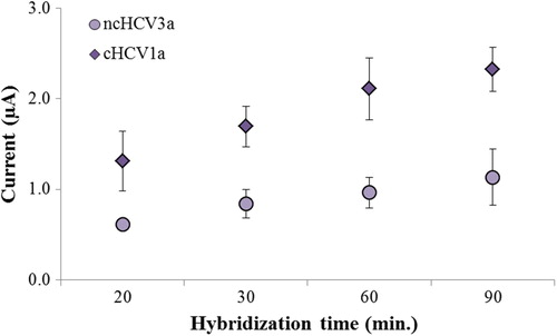
Figure 4. Effect of the hybridization time on the guanine oxidation signal of the biosensor after hybridization with its complementary target oligonucleotide (c-HCV1a) and noncomplementary oligonucleotide (nc-HCV3a). Probe and target concentrations were 2.5 μM. Immobilization time was 60 min. Error bars represent the standard deviation of three independent measurements.
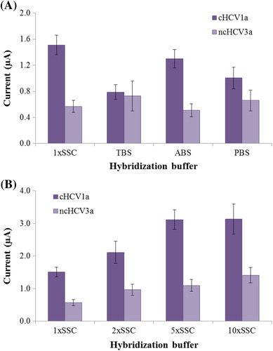
Figure 5. (a) Effect of buffer solution on the hybridization reaction. (b) Effect of ionic strength on hybridization.
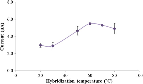
Figure 6. Guanine oxidation signals versus hybridization temperature. Error bars represent the standard deviation of three independent measurements at each target DNA.
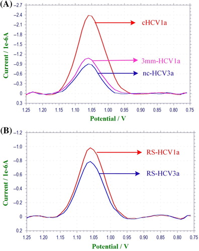
Figure 7. Square wave voltammograms of guanine oxidation on the probe-immobilized PGE. (a) Complementary HCV1a (cHCV1a), noncomplementary HCV3a (ncHCV3a) and 3-base mismatch of HCV1a (3mm-HCV1a) oligonucleotide discrimination (b) Complementary HCV1a (RS-HCV1a) and noncomplementary HCV3a (RS-HCV3a) synthetic single-stranded PCR product analog discrimination.
Figure 8. Plot of the peak current vs. the concentration of HCV1a from 0.05 μM to 5 μM. Inset: related calibration graph at a concentration range of 0.05–0.75 μM.

Reprints and Corporate Permissions

Please note: Selecting permissions does not provide access to the full text of the article, please see our help page How do I view content?

To request a reprint or corporate permissions for this article, please click on the relevant link below:

Academic Permissions

Please note: Selecting permissions does not provide access to the full text of the article, please see our help page How do I view content?

Obtain permissions instantly via Rightslink by clicking on the button below:

If you are unable to obtain permissions via Rightslink, please complete and submit this Permissions form. For more information, please visit our Permissions help page.