Figures & data
Figure 1. (a) Preoperative axial T1-weighted (b) and T2-weighted MR images showing heterogeneous hyperintense well-defined mass (arrows), which has similar intensity with subcutaneous fat tissue, adjacent to extensor digitorum tendon.
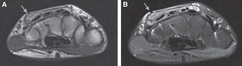
Figure 2. Intraoperative view of the angiofibrolipoma.
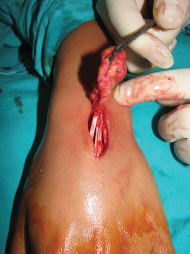
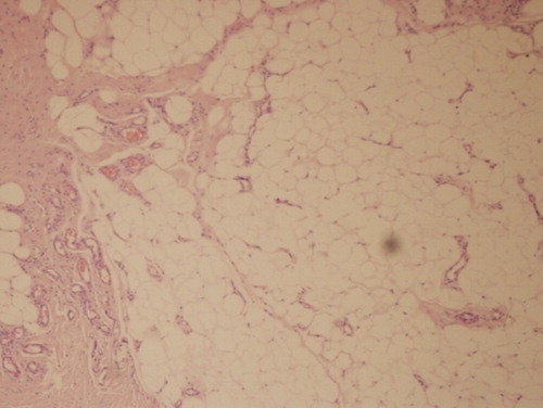
Figure 3. Histological view of the angiofibrolipoma.