Figures & data
Figure 1. Photograph taken by the patient showing a pedicled reverse forearm flap that totally failed to cover the stumps.
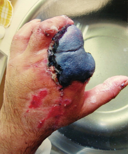
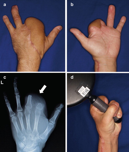
Figure 2. (a, b) Photographs showing the condition at presentation. The stumps of the fingers were almost dry with cicatricial tissue. The patient reported severe pain expanding over the region of the upper arm. (c) Preoperative radiograph showing malposition of the proximal phalanx of the index and middle fingers (arrow) caused by severe scar contracture around the two stumps.
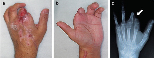
Figure 3. (a) A free anterolateral thigh flap (7 × 23 cm) was designed and harvested with its cutaneous nerve (arrows). (b) Schematic diagram of the digital nerves. (c) Intraoperative photo showing end-to-end (black arrow) and end-to-side (red arrow) neurorrhaphy between the digital nerves and cutaneous nerve of the flap. (d) Schematic diagram of the procedure. After releasing the contracture between the two stumps, the digital nerves between them are coapted to the cutaneous nerve of the flap using end-to-end (black arrow) and end-to-side (red arrow) neurorrhaphy. The other end of the cutaneous nerve of the flap is buried deeply in the soft tissue (green arrow). The other digital nerves are refreshed for about 3 cm and buried deeply in the muscle (blue arrows).
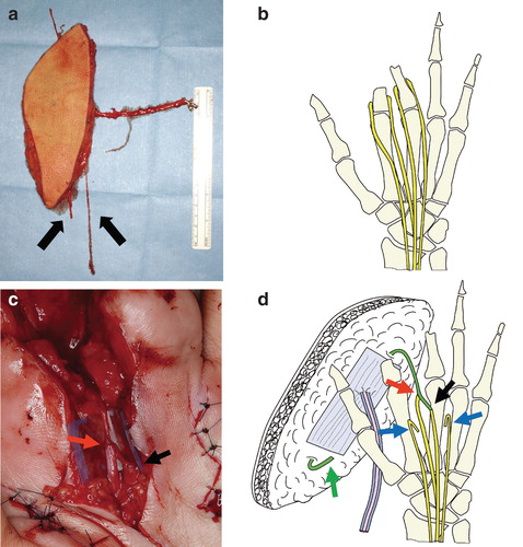
Figure 4. (a, b) Photographs and radiograph at 18 months postoperatively. The skin paddle of the flap was set to divide and cover the two stumps and dorsum of the hand. Almost all pain had disappeared at 18 months postoperatively. (c) Postoperative radiograph shows good alignment of the two stumps (arrow). (d) Range of motion, including that of the other fingers, substantially recovered. The patient can now grasp things easily without pain and enjoys motorbike riding.