Figures & data
Figure 1. (a) Preoperative magnetic resonance T2 images of patient 1. (b, c) Illustrations showing patient 1 just after tumor resection. (b) The mandible osteotomy approach was selected. The resection resulted in a large oropharyngeal defect, and the internal carotid artery was exposed (black arrow). (c) Illustration showing the grossly resected tumor.
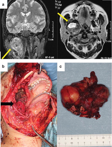
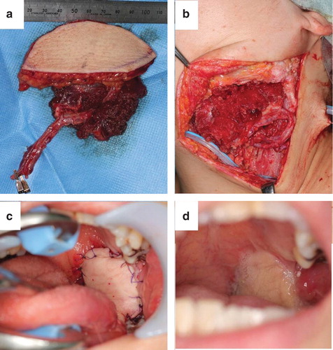
Figure 2. Illustrations showing the reconstruction of patient 1: (a) the rectus abdominis musculocutaneous flap. (b) The flap was transferred into the oroparapharyngeal space. The lingual artery and external jugular vein were used as the recipient vessels. (c, d) Illustrations showing the oropharynx immediately after surgery (c) and 3 weeks after surgery (d).
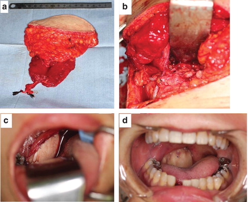
Figure 3. (a) Preoperative magnetic resonance T2 images of patient 2. (b, c) Illustrations showing patient 2 just after tumor resection. (b) Resection was achieved by using the cervical approach. The internal carotid artery was exposed (black arrow). (c) The oropharyngeal defect resulting from the resection is shown.
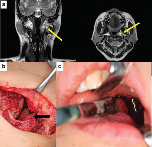
Figure 4. Illustrations showing the reconstruction on patient 2: (a) the anterolateral thigh vastus lateralis musculocutaneous flap. (b) The flap was transferred into the oroparapharyngeal space. The suprathyroid artery and common facial vein were used as the recipient vessels. (c, d) Illustrations showing the oropharynx immediately after surgery (c) and 4 months after surgery (d).