Figures & data
Figure 1. (a) Cutaneous dome shaped nodules peripherally located on the full thickness skin graft, three weeks after excision of squamous cell carcinoma (b) 7 days later, rapid growth and ulceration of the lesions expanding beyond the graft site.
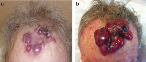
Figure 2. (a) Immature plasma cells, with large eccentric nuclei and scattered mitosis (H&E ×40). (b) Intense staining of CD138 (×10). (c) Positive staining for Ki67 (high proliferation index >80%) (×10).
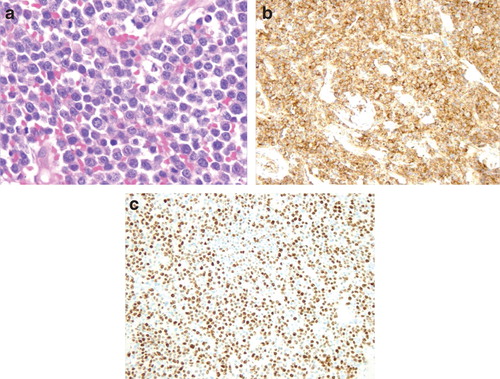
Figure 3. Computed tomography showing destruction of the proximal humerus by a large osteolytic lesion. There is soft tissue plasmacytoma located on the anterior chest wall and partial destruction of the sternum.
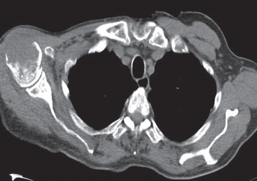
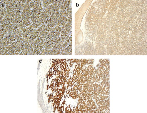
Figure 4. (a) Tumour cells expressing surface IgA. (×40) (b & c) Kappa and lambda light chains, (c) plasma cells showing restriction of immunoglobulin lambda-chain expression (×10).