Figures & data
Figure 1. (a) Preoperative plain film showing an expansile osteolytic lesion in the left second metacarpal bone. (b) Immediate postoperative plain film revealing a bone defect with a hyperdense mass. (c) Three-year postoperative plain film showing consolidation and remodeling of the bone lesion without fracture.
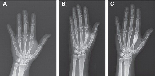
Figure 2. (a) Illustration of left second metacarpal bone showing metastatic adenocarcinoma composed of infiltrating nests of pleomorphic polygonal cells with focal glandular formation and intracytoplasmic vacuoles. (b) Tumor cells of the metastatic lesion (metacarpal bone) are immunoreactive for thyroid transcription factor-1 with nuclear staining. (c) Tumor cells of the metastatic lesion (metacarpal bone) are immunoreactive for cytokeratin 7 with cytoplasmic staining. (d) Histologically, the left lower lung shows features of adenocarcinoma similar to the metastatic lesion.
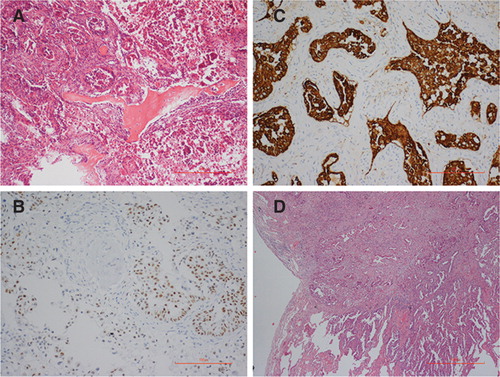
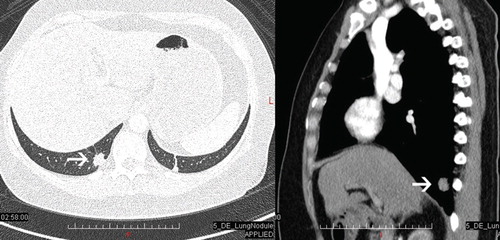
Figure 3. Chest computed tomography showing a 1.5-cm pulmonary nodule with spiculated margin in the right lower lobe (arrow).