Figures & data
Figure 1. Dose-response curves of melanoma cells treated with parthenolide (PN). Cells from disaggregated melanospheres (A), or their adherent counterparts (B), both growing in SCM, were exposed to PN for 2 d. The number of viable cells was assessed with an acid phosphatase activity (APA) assay. All measurements were normalized to cell viability of DMSO-treated controls. (p < 0.05 vs. DMSO-treated control; n = 3).
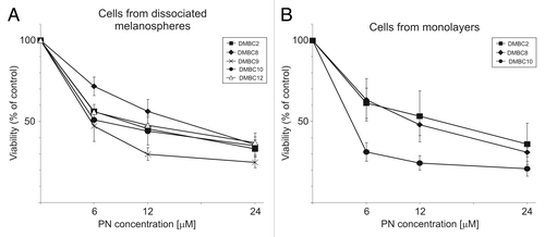
Figure 2. Distinct effects of parthenolide (PN) and dacarbazine (DTIC) on melanoma cell survival after long and short treatment. (A) Microphotographs of DMBC2 populations treated with either 6 μM PN or increasing concentration of dacarbazine (DTIC) for three days followed by few day drug-free intervals were taken after 3 weeks. Scale bars represent 100 μm. (B) Relative number of viable cells was assessed after two day exposure to PN or DTIC at indicated concentrations. The results are the mean of three independent experiments ± SD.
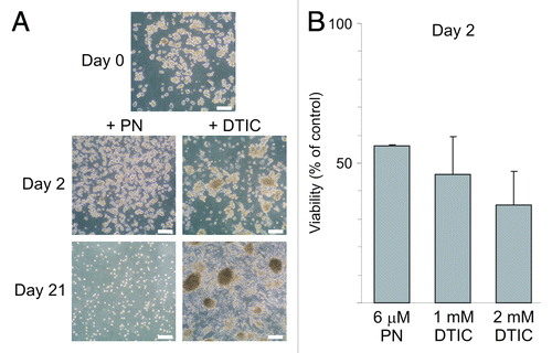
Figure 3. Parthenolide (PN) more efficiently reduces the frequency of cells with a high level of ABCB5 transporter than the frequencies of other subpopulations. (A) The frequency of cells carrying the indicated antigen assessed by flow cytometry showed the high heterogeneity within melanospheres and the variability among populations derived from different nodular melanoma specimens. (B) Treatment with PN decreased the frequency of cells with a high ABCB5 level more efficiently than other subpopulations. (C) Examples of dot plots showing the influence of PN on the frequency of ABCB5-positive cells in DMBC2 and DMBC10 populations. In comparison, changes in the percentage of cells expressing CD49f (DMBC2) and CD90 (DMBC10) were included. (D) Treatment with PN decreased the frequency of ABCB5-positive cells also among adherent counterparts grown in SCM as monolayer culture.
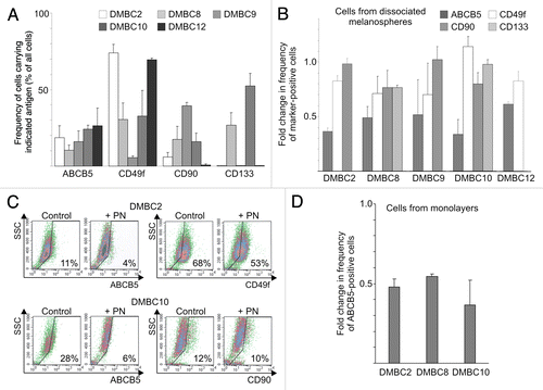
Table 1. Relative change in the frequency (RCF) of marker expressing cells in response to parthenolide (PN) treatment
Figure 4. Parthenolide (PN) reduced viability of sorted ABCB5+ melanoma cells. (A) Fold differences in the drug-reduced viability between unsorted and sorted ABCB5+ populations of DMBC12 were assessed either after PI staining (left panel) or were calculated after viable cells were numbered (right panel). Data are means ± SD of four independent experiments conducted in triplicates using the unsorted population of DMBC12 and the ABCB5-positive subsets obtained in two independent flow-cytometry-based cell-sorting. (p > 0.05). DTIC, dacarbazine; cisPt, cisplatin. (B) PN induced apoptosis in melanoma cells. Typical contour plots are included. Numbers in rectangles indicate the percentages of all Annexin V-positive cells, PI-negative (early apoptosis) and PI-positive (late apoptosis). After sorting of the ABCB5+ subset, no significant changes (p > 0.05) in Annexin V positivity were evident relative to the unsorted populations (right panel). (C) Viability measured four-days after the drug was removed indicate that the ABCB5+ subset was capable of recovering within four days after a short (4 h) treatment with PN less efficiently than the unsorted population. Numbers indicate the percentages of viable, 7-AAD-negative melanoma cells in one of two experiments.
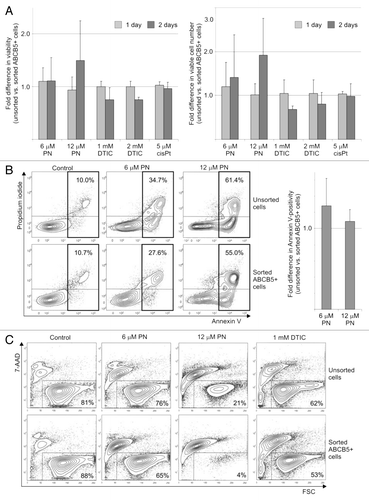
Figure 5. Parthenolide (PN) affects self-renewing capacity of melanoma populations.(A) Images of representative colonies formed by DMBC12 population in soft agar were captured after three weeks. (B) PN-treated melanoma populations lost colony-forming capacity in contrary to dacarbazine (DTIC)-treated populations showing about 2-fold relative increase in the number of colonies formed in soft agar. The results are mean of two independent experiments.
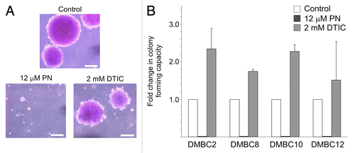
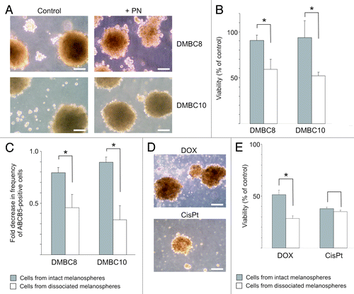
Figure 6. Parthenolide (PN) has a limited capacity to affect cells inside of melanospheres. (A) Microphotographs showing melanosphere integrity following treatment with 12 μM PN. Scale bars represent 100 μm. (B). Comparison of PN-induced changes in viability between populations of cells treated as intact melanospheres and as single melanoma cells from dissociated spheres. Melanoma cells were exposed to drug for one day, and after additional two days in drug-free medium, viability was measured by flow cytometry as percentage of 7-AAD-negative cells in PN- vs. DMSO-treated cells (control) (* p < 0.05; n = 3). (C). Comparison of PN-induced decrease in the ABCB5-positive cell frequency between DMBC10 populations treated as intact melanospheres and those treated as single-cell culture (*p < 0.05; n = 3). (D). Microphotographs showing DMBC10 melanospheres treated with 1 μM doxorubicin (DOX) and 5 μM cisplatin (cisPt) for one day followed by a recovery period. Scale bars represent 100 μm. (E). Comparison of DOX- or cisPt-induced changes in viability between DMBC10 populations of cells treated as intact melanospheres and as single melanoma cells from dissociated spheres (* p < 0.05; n = 3).