Figures & data
Figure 1. Effect of MK2 inhibition and depletion on gemcitabine-induced H2AX phosphorylation in pancreatic cancer cell lines. BxPC-3 (A), MIA PaCa-2 (B), Panc-1 (C), and PaTu 8902 cells (D) were treated with 100 nM gemcitabine and MK2 inhibitor or DMSO for 24 h. H2AX phosphorylation was analyzed by immunoblot. (E) Panc-1 cells were depleted of MK2 by siRNA-mediated knockdown. Forty-eight hours later, the cells were treated with 300 nM gemcitabine for 22 h or left untreated. The cells were fixed and stained for immunofluorescence analysis, and γH2AX fluorescence intensity was quantified. Mean ± SD from 3 technical replicates. (**P = 0.009).
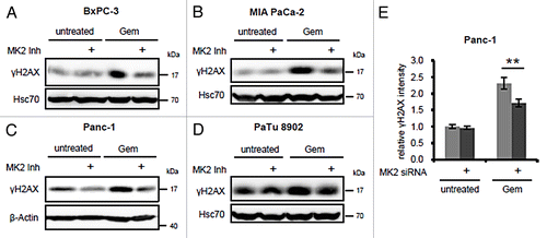
Figure 2. Proliferation of pancreatic cancer cell lines upon treatment with gemcitabine and/or MK2 inhibitor. BxPC-3 (A), MIA PaCa-2 (B), Panc-1 (C), and PaTu 8902 (D) cells were treated with 100 nM gemcitabine and MK2 inhibitor or DMSO for 24 h on day 1. Then the drugs were washed out, and cell confluence was quantified by light microscopy and digital image analysis until day 18.
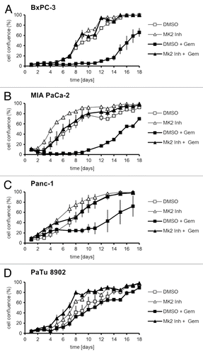
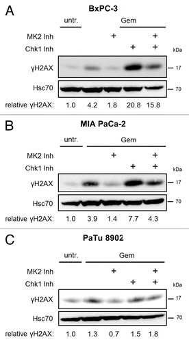
Figure 3. Gemcitabine-induced H2AX phosphorylation in dependence of MK2 and Chk1 inhibition in pancreatic cancer cell lines. BxPC-3 (A), MIA PaCa-2 (B), and PaTu 8902 (C) cells were treated with 100 nM gemcitabine and MK2 inhibitor, Chk1 inhibitor or both for 24 h. Then, H2AX phosphorylation was analyzed by immunoblot. “Relative γH2AX” indicates relative γH2AX intensities normalized to Hsc70 intensities. See Table S1 for raw data.