Figures & data
Figure 1. A C. elegans infection model reveals attenuation of virulence in C. albicans-E. faecalis coinfections. (A-D) Fluorescence imaging shows the two species mixing in the worm and the absence of hyphal growth in the coinfection. Worms were infected with yCherry labeled-C. albicans alone (A and B) or coinfected with GFP-labeled E. faecalis (C and D) and imaged using DIC (A and C) or fluorescence (B and D). Panels (E-H) Ultrastructural analysis of nematode physiology. Worms were infected with C. albicans alone (E and F) or with both species (G and H) before processing for transmission electron microscopy. The disruption of the normal gut physiology in the C. albicans-infected worms relative to the coinfection is apparent. The scale bar in panel F is 1 µm and applied to (E-H).
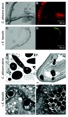
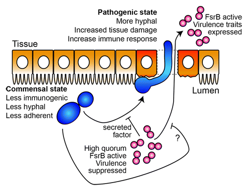
Figure 2. A speculative model for Candida-Enterococcus interactions in the gut. In the worm gut, C. albicans transitions from a commensal mode characterized by yeast-form growth to an invasive, hyphal and pathogenic state. This transition is inhibited by the presence of E. faecalis through molecule(s) secreted in an FsrB-dependent manner, thus maintaining the commensal association with the host. Likewise, mono-infection with E. faecalis imposes significant gut pathology, which is reduced in the presence of C. albicans through an unknown mechanism. In the mammalian gut, the work from Hufnagel and colleagues suggests that these two species also promote a commensal association with the host which we speculate can be disrupted by changes to microbe or host factors leading to infection. C. albicans cells are shown in blue, E. faecalis in red, and gut epithelial cells in orange.