Figures & data
Table 1. Immunocytochemical staining for Insulin, Glucagon, SRIF and IAPP
Figure 1. Control islets. β cells were the most abundant major islet cells (about 60% of total islet cells) with plump and polygonal cytoplasm of variable staining intensity, from moderate to strong staining, followed by α-cells (arrow, about 30%) with strongly stained, round smaller cytoplasm. δ cells accounted for about 15% of islet cells, containing plump or small cytoplasm. β cells and IAPP-positive cells were mostly located in the middle of islets and so were δ-cells whereas strongly immunostained α-cells (arrow) were located at the outer margin of islets and islet lobules. There were globular to sickle-shaped strongly immunostained cytoplasms for insulin and IAPP, which appeared to be dying β-cells (*). L, Large islet; M, Medium-sized islet; Original magnification X 400; (A) Insulin; (B) IAPP; (C) Glucagon; (D) SRIF immunostained.
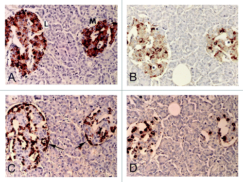
Figure 2. Disbetic islets, Case 1. β cells and α-cells (arrow) were about equal in number in large islet (Left), and α-cells were slightly more in medium-sized islet (Right). β cells were moderately to strongly immunopositive with plump cytoplasm whereas α-cells (arrows) and δ-cells were slightly smaller in the cytoplasm and were strongly immunostained. IAPP-positive cells were about ¼ that of β-cells in large islet, but medium-sized islet had only a few positive cells. (A) Insulin; (B) IAPP; (C) Glucagon; (D) SRIF immunostained; Original magnification X 470.
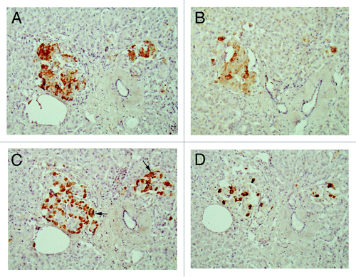
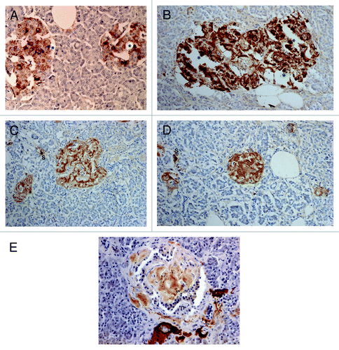
Figure 3. Extra-large diabetic islet, Case 15. This extra-large islet showed less β-cells (A) than α-cells (C). β cells were strongly and granularly immunostained with irregular, fuzzy cell membrane whereas α-cells contained dense positive compact cytoplasm. δ cells consisted of a few large cytoplasm and mostly compact cytoplasm (D). IAPP staining was almost completely negative with only weak residual granular positive staining (B). Stromal amyloid deposits occupied about 20% of the islet area, which was negatively stained for IAPP using a 1: 800 antibody solution (B). (A) Insulin; (B) IAPP; (C) Glucagon; (D) SRIF immunostained; Original magnification X 470.
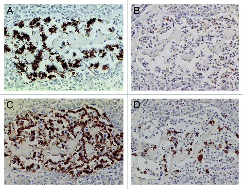
Figure 4. Diabetic islets Case 10, Islets occupied by amyloid deposits in 95% (A, B and C) and Islets occupied by amyloid deposits in > 99% (D, E and F). Islets occupied by amyloid deposits in 95%, A,B and C: Both large islet (Left) and medium-sized islet (Right) consisted of more than 95% amyloid deposits, within which β-cells with partly plump cytoplasm and α cells with dense small cytoplasm were located. IAPP-positive cells were weakly stained in the large islet but were moderately stained in the medium-sized islet. δ cells showed mostly small cytoplasm mixed with a few large cytoplasms. Islets occupied by amyloid deposits > 99%, D, E and F: Both large (Left) and medium-sized islet (Right) contained more than 99% amyloid deposits. Residual β-cells and δ-were minor cells and α-cells were major cells (D). IAPP immunostaing was performed using a 1: 400 diluted antibody solution, revealing moderately positive staining in amyloid deposits (E). In islets containing viable islet cells, residual islets cells with plump cytoplasm and amyloid deposits were stronger stained for IAPP than in . (A and D) Insulin; (B, E and F) IAPP by 1: 400 diluted solution; (C) Glucagon immunostained; Original magnification (A-C) X 320; (D-F) X 420.
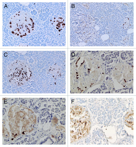
Figure 5. Control (A) and Diabetic islets, Cases 10 (B-D) and Case 14 (E) IAPP immunostaining was performed using a 1: 200 diluted antibody solution. Control islets were strongly immunostained for IAPP in the majority of islet cell cytoplasm (A). Diabetic islets of plump cytoplasm (*) were densely immunostained for IAPP in the cytoplasm, continuous to the moderately immunostained amyloid deposits (B). Diabetic islets occupying 95% amyloid deposits were immunostained moderately to strongly positive whereas viable islet cells surrounded by amyloid deposits were negative for IAPP. Two single cell islets were strongly positive for IAPP (s) (C, D). The end-stage small islets occupied by > 99% amyloid deposits revealed only a few IAPP negative residual islet cells. One strongly IAPP positive, single cell islet was localized (s) (D). Amyloid p immunostaining for the end-stage amyloid deposited islet was moderately positive in lamellar amyloid deposits and peri-islet blood vessel walls were strongly immunostained for amyloid p. Stromal amyloid deposits were moderately positive for amyloid p (E). (A) Control islet; (B-D) Case 10; (E) Case 14. (A-D) IAPP by 1: 200 diluted solution; Case 10; (E) amyloid p; Case 14 immunostained Original magnification X 350.