Figures & data
Figure 1 Immunofluorescence staining with 8H8 mAb of dengue infected cells with Den-2 A15 strain. C6/36-HT cells were infected with Den-2 A15. At 6 (a), 8 (b), 12 (c), 48 (d) and 72 (e) h p.i, the infected cells were reacted 8H8 mAb followed with an FITC-conjugated goat anti-mouse antibody and then examined under a fluorescence microscope. Cytoplasmatic and nuclear staining of infected cells are indicated as C and N, respectively. C6/36-HT cells infected with Den-2A15 reacted with anti-Den-2 polyclonal antibody at 72 h p.i was used as a positive control (f) and non-infected C6/36-HT cells were used as negative control (g).
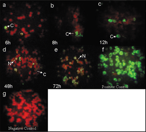
Figure 2 Immunofluorescence staining with 8H8 mab of dengue infected cells. C6/36-HT cells were infected with samples 57 (a–c) and 59 (d–f) from dengue acute patients. At 6 (a), 12 (b), 72 (c) h pi, for sample 57 and 12 (d), 24 (e), 72 (f) h pi for sample 59, the infected cells were reacted 8H8 mab followed with an FITC-conjugated goat anti-mouse antibody and then examined under a fluorescence microscope. Cytoplasmatic and nuclear staining of infected cells is indicated as C and N, respectively. Samples from healthy individuals as negative control (g).
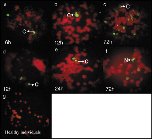
Figure 3 Staining of Dengue capsid protein in suckling mice brain at 24 (a), 48 (b) and 72 h (c) pi Uninfected suckling mouse brain as negative control (d).
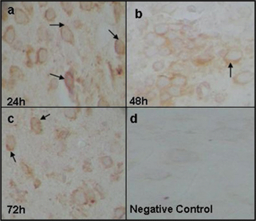
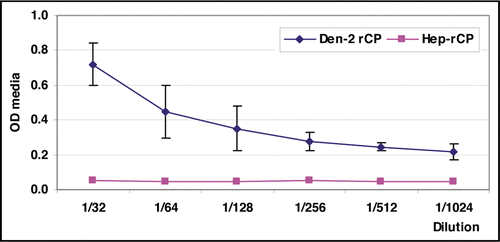
Figure 4 Reactivity of 8H8 mab in the antigen-capture enzyme-linked immunosorbent assay (ELISA) format. The ability to capture the Dengue-2 recombinant protein (Den-2-rCP) of the monoclonal antibody was evaluated at various concentrations of Den-2-rCP.