4,343
Views
92
CrossRef citations to date
0
Altmetric
Review

Monoclonal antibodies in neuro-oncology

Getting past the blood-brain barrier

Pages 153-160 | Received 10 Dec 2010, Accepted 19 Nov 2010, Published online: 01 Mar 2011

Figures & data

Figure 1 Two patterns of tumor growth in the brain. Tumor often grows around blood vessels (left), but some tumors can also infiltrate the brain parenchyma (right).
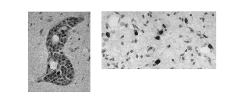
Figure 1 Two patterns of tumor growth in the brain. Tumor often grows around blood vessels (left), but some tumors can also infiltrate the brain parenchyma (right).

Figure 2 Distribution of tumor antigens. A tumor cell displays a characteristic combination of components, many of which are also expressed by normal cells. Even though they may not be unique to the tumor, shared antigens can serve as practical tumor targets.
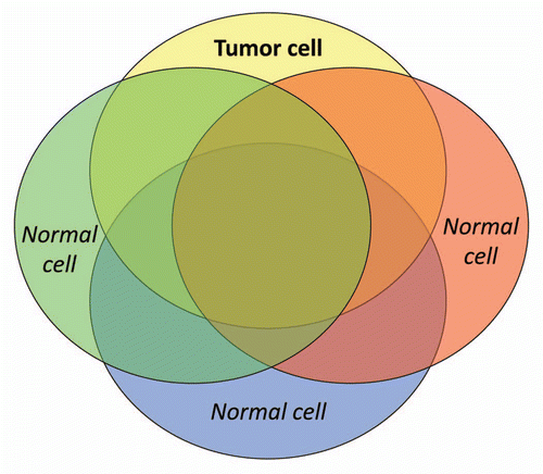
Figure 2 Distribution of tumor antigens. A tumor cell displays a characteristic combination of components, many of which are also expressed by normal cells. Even though they may not be unique to the tumor, shared antigens can serve as practical tumor targets.

Figure 3 A varied role for the BBB. Possible relationships among tumor (black circles), gadolinium (Gd, black dots), antibody (AB, Y shapes), blood vessels (grey) and the blood-brain barrier (BBB), under different conditions of tumor growth are depicted.
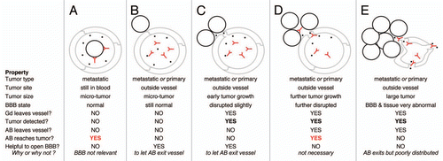
Figure 3 A varied role for the BBB. Possible relationships among tumor (black circles), gadolinium (Gd, black dots), antibody (AB, Y shapes), blood vessels (grey) and the blood-brain barrier (BBB), under different conditions of tumor growth are depicted.

Table 1 Tumor/antibody combinations emphasized in the text