Figures & data
Figure 1 Induction of bioluminescence in V. harveyi reporter strain by CFM from B. anthracis cells. V. harveyi strain BB170 only upregulates the expression of the lux operon [measured as relative light units (RLU)], when AI-2 or AI-2-like molecules are present in its milieu. Cell free medium (CFM) obtained from AI-2-synthesizing bacteria can induce expression of the bioluminescence-generating luxCDABE operon in BB170. (A) In the experiments shown, CFM from 5-h cultures of B. anthracis strains 34F2 and 34F2ΔluxS and sterile CFM alone were used as positive and negative controls. The baseline is the value when uninoculated (sterile) CFM alone at 5 h were used. Each bar represents the mean (±SD) of triplicate experiments. Compared to the negative and positive controls, 34F2ΔluxS:comp showed restored AI-2 activity compared to wild-type 34F2. (B) In the experiment shown, CFM from 5-h cultures of wild-type 34F2 and 34F2ΔluxS:comp were serially diluted 1:1, 1:10 and 1:100. Each bar represents the mean (±SD) of triplicate experiments.
![Figure 1 Induction of bioluminescence in V. harveyi reporter strain by CFM from B. anthracis cells. V. harveyi strain BB170 only upregulates the expression of the lux operon [measured as relative light units (RLU)], when AI-2 or AI-2-like molecules are present in its milieu. Cell free medium (CFM) obtained from AI-2-synthesizing bacteria can induce expression of the bioluminescence-generating luxCDABE operon in BB170. (A) In the experiments shown, CFM from 5-h cultures of B. anthracis strains 34F2 and 34F2ΔluxS and sterile CFM alone were used as positive and negative controls. The baseline is the value when uninoculated (sterile) CFM alone at 5 h were used. Each bar represents the mean (±SD) of triplicate experiments. Compared to the negative and positive controls, 34F2ΔluxS:comp showed restored AI-2 activity compared to wild-type 34F2. (B) In the experiment shown, CFM from 5-h cultures of wild-type 34F2 and 34F2ΔluxS:comp were serially diluted 1:1, 1:10 and 1:100. Each bar represents the mean (±SD) of triplicate experiments.](/cms/asset/2b8385a4-b250-45f1-a1de-5093c400453c/kvir_a_10910752_f0001.gif)
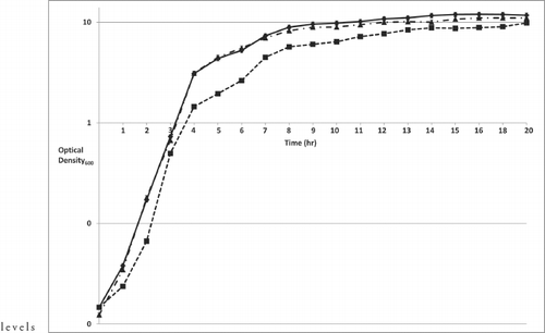
Figure 2 Growth rate analysis of B. anthracis 34F2ΔluxS:comp. B. anthracis strains 34F2, 34F2ΔluxS, and 34F2ΔluxS:comp were grown overnight and diluted in sterile BHI media to an optical density (OD600) of ≈0.01. Cell growth was monitored, CFM removed and passed through a 0.2 µm filter for use in the V. harveyi bioassay. The graph represents the mean (±SD) of triplicate experiments performed on the same day. Solid line with filled diamonds represent strain 34F2, dotted line with filled squares represent strain 34F2ΔluxS and short dash line with filled triangles represents strain 34F2ΔluxS:comp.

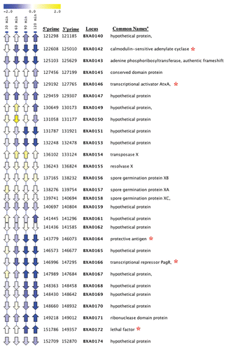
Figure 3 Analysis of pXO1 pathogenicity island gene expression in B. anthracis strain 34F2ΔluxS compared to strain 34F2 using Linear Expression Map (LEM) viewer. Time points are indicated at the top of each column. Arrows indicate the direction of gene orientation. The color scale indicates the log2 changes in expression, according to the scale shown above. Locus ID based on B. anthracis Florida strain A2012. aGenes bolded relate to production or regulation of toxins. bRed asterisk indicates virulence genes.

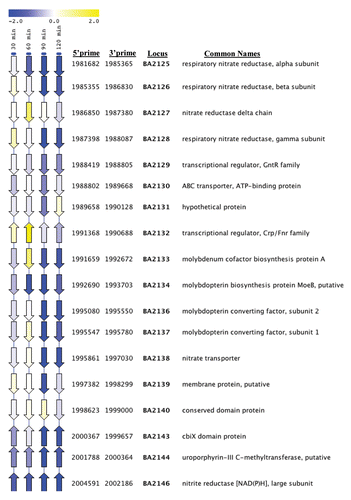
Figure 4 Analysis of respiratory nitrate genes in strain 34F2ΔluxS compared to strain 34F2 and strain 34F2 exposed to 20 µg/ml of fur-2 using LEM. Time points (left-most 4 columns) and sample (exposure to fur-2 for one hour; right-most column) are indicated at the top of each column. Arrows indicate the direction of gene orientation. The color scale indicates the log2 changes in expression, according to the scale shown above. Locus ID based on B. anthracis strain Ames.

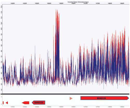
Figure 5 Analysis of small non-coding RNA of strain 34F2ΔluxS compared to strain 34F2 using an Affymetrix tiled array. A select region (between BXA0122 and BXA0124) based on pXO1 from B. anthracis strain A2012, demonstrating transcriptional activity in intergenic regions. Red indicates the transcriptional activity of the parental strain and blue represents strain 34F2ΔluxS.

Table 1 Oligonucleotide primers used in this study
Table 2 B. anthracis chromosomal genes upregulated in the 34F2ΔluxS strain as determined by microarray analysis, sorted by locus
Table 3 B. anthracis chromosomal genes downregulated in the 34F2ΔluxS strain as determined by microarray analysis, sorted by locus
Table 4 Select genes downregulated on pXO1 in B. anthracis 34F2ΔluxS as determined by microarray analysis, sorted by locus
Table 5 Select virulence genes downregulated on pXO1 in 34F2ΔluxS compared to 34F2luxS:comp
Table 6 Select B. anthracis genes on pXO1 downregulated by fur-2