
Abstract
The effect of punicalagin on metabolic risks, oxidative stress, inflammation, cardiac apoptosis and histopathological alterations in experimentally induced diabetes was addressed. Diabetes was induced in male rats by a single injection of streptozotocin (STZ; 40 mg/kg, i.p.), and then punicalagin (1 mg/kg) was i.p. administered every other day for 15 days. The diabetic rats treated with punicalagin exhibited ameliorated hyperglycemia and HbA1c; improved insulin levels, HOMA-IR levels and lipid profiles; and normalized levels of IL-1b, IL-6 and TNF-α. Punicalagin also reduced the increase in the MDA and H2O2 levels; normalized the levels of GSH, SOD and CAT in the heart; and improved serum markers of heart function including the levels of troponin T level and CK-MB and LDH activities. Histopathological examinations of heart sections match these results, confirming the beneficial effect of punicalagin. It also modulated cardiomyocyte apoptosis via enhanced Bcl-2 expression; blocked the increases in P53, Bax and caspases-3, 8 and 9; and ameliorated DNA damage in the heart. The current results suggest that punicalagin protected the heart against apoptosis, necrosis, inflammation and DNA damage by improving the redox state, suppressing caspases and P53 and increasing Bcl-2. In conclusion, punicalagin possesses strong therapeutic potential in treating and regulating diabetes and attenuating its associated complications in the heart.
1 Introduction
The prevalence of the diabetic heart has markedly increased in the past few years and is now considered the main cause of sickness and death among the diabetic and obese population. Diabetic cardiomyopathy is associated with increased oxidative stress that stimulates cellular injury and contributes to the development and progression of complications associated with diabetes [Citation1]. Apoptosis is a key process in diabetic cardiac diseases derived from poor glycemic control. A number of studies have documented an increased occurrence of apoptosis in the cardiomyocytes of diabetic patients and experimentally induced diabetic animals [Citation2,Citation3]. It is mediated, at least in part, by the activation of the mitochondrial pathway, which is often triggered by reactive oxygen species (ROS).
There is evidence from experimental and clinical settings on the benefits of antioxidant therapies in the prevention and management of diabetic complications [Citation4–Citation[5]Citation6]. Pomegranate (Punica granatum L. Punicaceae) juice has been shown to contain the highest antioxidant capacity compared to other polyphenol-rich beverages [Citation7,Citation8]. Clinical studies showed positive effects of pomegranate juice consumption on diabetic patients' blood diabetic and oxidative stress parameters [Citation9]. Furthermore, it is documented that pomegranate juice supplementation showed a protective effect against isoperiotinaol-induced cardiac necrosis in rats [Citation10]. The efficient antioxidant potential of the pomegranate is attributed to its major antioxidant polyphenol, punicalagin [Citation11]. A primary mechanism by which pomegranate fractions exert their beneficial effects on the disease condition is by reducing oxidative stress and lipid peroxidation. This reduction may occur by directly neutralizing the generated ROS, enhancing the activities of specific antioxidant enzymes, prompting metal chelation activity, and inhibiting or activating certain transcriptional factors, such as nuclear factor kappaB and peroxisome proliferator-activated receptor gamma [Citation12].
Punicalagin is a bioactive ellagitannin, a type of phenolic compound isolated from pomegranate and exhibits high antioxidant and free radical scavenging activities [Citation13], with several health benefits [Citation14]. It demonstrated a marked outcome in patients with metabolic syndromes, such as hyperlipidemia, non-alcoholic fatty liver disease, and coronary heart disease [Citation12,Citation15–Citation[16]Citation17]. Punicalagin inhibits the formation of advanced glycation end-products [Citation18] and fructose-mediated non-enzymatic protein glycation by scavenging reactive carbonyl species [Citation19]. Good results in terms of fat loss [Citation20] and inhibition of oxidized LDL uptake in macrophages [Citation21] have been demonstrated using punicalagin. In addition to its antioxidant, anti-diabetic and anti-atherosclerotic actions [Citation12,Citation14], punicalagin also exhibited anti-inflammatory activity in cell culture and animal studies [Citation22,Citation23]. It is worth mentioning that repeated oral administration with punicalagin for five weeks is not toxic to rats [Citation24].
Because diabetes is associated with high frequency of heart failure, a better understanding of its pathophysiology is essential for testing of new bioactive compounds and the development of treatment strategies for diabetes-associated cardiac dysfunction. To date, no studies regarding the effect of punicalagin on diabetic cardiac apoptosis and injury have been conducted. The present study hypothesized that punicalagin can counteract oxidative stress and, in turn, ameliorate cellular damage and reduce the development and progression of diabetic complications. Therefore, the current study was designed to investigate the potential beneficial effect of punicalagin against diabetes-induced cardiac injury and apoptosis in connection with its anti-lipid peroxidation and anti-hyperlipidemic actions.
2 Materials and methods
2.1 Chemicals
Streptozotocin (STZ) and punicalagin (PU) were purchased from Sigma Chemical (St. Louis, MO). All other chemicals were of the highest grade.
2.2 Induction of diabetes
Diabetes was induced in rats by single intraperitoneal (i.p.) injections of freshly prepared STZ (40 mg/kg body weight) in 0.01 M citrate buffer, pH 4.5 [Citation25]. The blood glucose level was assayed 48 h after STZ injection using a glucose monitor set (Elegance, CT-X10, Convergent Technologies GmbH & Co. KG, Marburg, Germany). The rats with a blood glucose level above 250 mg/dl were considered diabetic and were used for the experiments.
2.3 Punicalagin treatment
Punicalagin is supplied in water soluble dark-yellow powder. Applied dose of 1 mg/kg dissolved in 0.2 ml saline solution was daily i.p. administered for 15 days [Citation26].
2.4 Experimental work
Male Wister rats weighing 280–300 g were provided by the Biological Products & Vaccines (VACSERA) of Cairo, Egypt, and were housed in cages with free access to food and drinking water. The animals were acclimated to laboratory conditions of 22–24 °C with a 12-h light/dark cycle for two weeks before experimentation. The current study was approved by Mansoura University, and all experiments were performed in accordance with the standards accepted by the regional experimental animal ethics panel. The rats were divided randomly into four groups of seven rats each. The first group served as a control, the second group received a daily dose of punicalagin (1 mg/kg, i.p.) for 15 days, the third group received a single i.p. injection of 40 mg/kg STZ, and the fourth group received a single injection of STZ (40 mg/kg, i.p.) followed by punicalagin (1 mg/kg, i.p.) every other day for two weeks. After 15 days of treatment, blood was obtained by cardiac puncture after overnight fasting. Serum and heart ventricle were kept in refrigerator for the following investigations.
2.5 Biochemical investigations
The glucose and insulin levels in serum and the HbA1c levels in blood were determined using kits supplied by Spinreact (St. Esteve d'en Bas Girona, Spain), Abcam (Cambridge, MA, USA) and BioSystems (Barcelona, Spain), respectively. The lipid profiles in serum, including total lipids, triglyceride, total cholesterol, high-density lipoprotein (HDL), low-density lipoprotein (LDL) and very low-density lipoproteins (VLDL), were assayed according to the manufacturer's instructions (Spinreact, St. Esteve d'en Bas Girona, Spain). Interleukin 6 (IL-6), interleukin 1 beta (IL-1b), tumor necrosis factor alpha (TNF-α) and troponin T in serum were assessed using kits provided by R&D Systems (Minneapolis, MN, USA) and Bioscience (San Diego, CA, USA). Creatine kinase (CK-MB) and lactic dehydrogenase (LDH) activities in the serum were determined using kits purchased from Elitech (Puteaux, France). Lipid peroxidation was assessed by determining the amount of malondialdehyde (MDA). The hydrogen peroxide (H2O2) concentrations were estimated according to the manufacturer's instructions (Dokki-Giza, Egypt). The superoxide dismutase (SOD) and catalase (CAT) activities and the glutathione (GSH) concentration were assayed using kits provided by Biodiagnostic (Dokki-Giza, Egypt). The protein concentrations were assayed as previously described [Citation27].
2.6 Flow cytometry study
2.6.1 Determination of apoptosis
For flow cytometry, samples from the heart were prepared as previously described [Citation28]. The cells were suspended in PBS with BSA, divided into aliquots and stored at 4 °C for analysis. The flow cytometry analyses were performed on a FACSCalibur™ cytometer (BD Biosciences, San Jose, CA) using CellQuest Pro software (Becton Dickinson) for data acquisition and analysis [Citation29].
2.6.2 Annexin V/PI staining
Apoptosis was assessed using the fluorescein isothiocyanate-conjugated annexin V/PI, ApoAlert kit from Clontech (Palo Alto, CA) according to the manufacturer's instructions.
2.6.3 Bcl-2, Bax, P53 and CD95
The cell suspensions were prepared in a PBS/BSA buffer and were then incubated for 30 min with anti-Bcl-2 [100/D5] antibody (ab692) and anti-Bax [6A7] antibody (ab5714) for flow cytometry analysis of Bcl-2 and Bax, respectively. A mouse anti-P53 “aa20-25” FITC, Clone: DO-1, was used for P53 analysis by flow cytometry. A monoclonal anti-Fas (CD95/Apo-1) antibody was used for CD95 determination. After 30-min incubation at room temperature, the cells were washed with PBA/BSA, centrifuged at 400 × g for 5 min, re-suspended in 0.5% paraformaldehyde in PBS/BSA and analyzed using flow cytometry.
2.6.4 Caspases-3, -8 & -9
In case of caspases-3, -8 and - 9: FITC rabbit anti-active caspase-3 (CPP32; Yama; Apopain, BD Bioscience), anti-caspase-8 (E6) antibody (Abcam) or rabbit monoclonal anti-caspase-9 (E23) antibody (ab32539) were used respectively.
2.7 Single cell gel electrophoresis (Comet assay)
DNA damage in the heart samples was assessed using the single-cell gel electrophoresis (comet assay) method, a well-validated technique developed for measuring DNA strand breaks in individual cells, as described previously [Citation30,Citation31]. The quantification of the DNA strand breaks in the obtained images was performed with CASP software to directly obtain the percent of DNA in the tail, the tail length and the tail moment.
2.8 Histopathological examination
The ventricle was excised and fixed in 4% buffered paraformaldehyde, dehydrated in ascending grades of ethyl alcohol, cleared in xylene and mounted in molten paraplast 58–62C. Four micron histological section were cut, stained with hematoxylin and eosin and examined under bright field light microscopy and photographed.
2.9 Statistical analysis
The data were presented as the mean ± SEM. The statistical analyses were performed by one-way ANOVA followed by Students Newman–Keul's post hoc test.
3 Results
The administration of punicalagin alone every other day for two weeks did not affect the body weight, the concentration of glucose, insulin or the lipid profile. Similarly, punicalagin treatment did not affect the levels of IL-1b and IL-6. Furthermore, the activities of SOD and CAT and the lipid peroxidation product (TBARS) in the heart did not change after punicalagin treatment. Conversely, significant increases were demonstrated in the HDL level in the serum and the GSH level in the heart.
3.1 Body weight
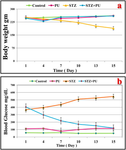
The body weight of the STZ-treated rats significantly and gradually decreased compared to that of the normal rats. The diabetic rats treated with punicalagin (1 mg/kg; every other day) showed a significant increase in body weight during the experimental period compared with the diabetic group (Fig. 1a); however, they remained significantly lower than the control group.
3.2 Blood sugar, insulin and HbA-1c levels
The blood glucose of the diabetic rats was significantly increased during the experimental period compared to that of the control group. The diabetic rats treated with punicalagin exhibited a marked amelioration over the experimental period (Fig. 1b) and at the end of the experimental period () compared with the diabetic group, but the level was significantly lower than that of the control group.
Table 1 Effects of streptozotocin (STZ) and punicalagin (PU) on the serum glucose (mg/dl) and insulin (pg/ml) levels and the percentages of HbA1c and HOMA-IR in rats in different groups at the end of the experimental period.
The STZ injection significantly elevated the serum glucose and HbA-1c levels two weeks after diabetic induction (). In addition these rats had significantly lower insulin levels and higher HOMA-IR than the controls (). Punicalagin treatment for 15 days after diabetic induction significantly ameliorated the serum glucose and HbA-1c levels compared to the STZ-treated rats. The diabetic rats treated with punicalagin exhibited a significant increase in the insulin levels and a decrease in the HOMA-IR values compared to the diabetic rats, but the results were similar to the control levels.
3.3 Biochemical observations
3.3.1 Serum lipid profiles
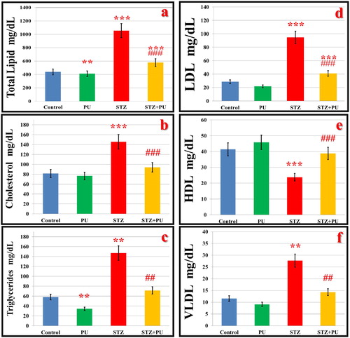
The lipid profile, including total lipids, TG, total cholesterol, LDL and VLDL, were significantly higher and HDL was significantly lower in the STZ-treated rats than in the control rats. Punicalagin treatment significantly ameliorated these lipid fractions at 15 days after diabetic induction in the rats (Fig. 2).
3.3.2 Serum markers of heart function
The concentration of troponin T and the activity of CK-MB and LDH in heart were significantly increased in STZ-induced diabetes compared to the normal control animals (). When diabetic rats were treated with punicalagin, the activity of both enzymes and troponin T level was normalized to a level comparable to that of the control rats and was significantly lower than that of the diabetic animals.
Table 2 Effects of streptozotocin (STZ) and punicalagin (PU) on the serum troponin T concentration (pg/ml) and CK-MB and LDH activities (U/ml) rats in different groups.
3.3.3 Oxidative stress and antioxidants
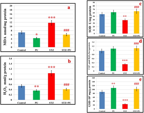
The levels of the lipid peroxidation product and H2O2 were significantly increased in the hearts of the STZ-induced diabetic rats (Fig. 3). In addition, significant decreases were demonstrated in the SOD and CAT activities and the GSH content in the hearts of STZ-treated rats. These changes appeared similar to control levels when punicalagin was administered for 15 days after diabetic induction and were significantly better than those of the diabetic rats (Fig. 3).
3.3.4 Proinflammatory cytokines
The ability of punicalagin to influence the production of IL-1b, IL-6, TNF-α was measured in the serum of the different rat groups using enzyme immunoassays. Animals that received STZ had significantly higher levels of IL-1b, IL-6 and TNF-α compared to the control values (). Punicalagin administration after diabetic induction ameliorated the increase in these inflammatory proteins in the serum and displayed insignificant changes when compared to the diabetic rats.
Table 3 Effect of streptozotocin (STZ) and punicalagin (PU) on the serum IL-6, IL-1b, and TNF-α levels, expressed as pg/ml, in rats in different groups.
3.4 Flow cytometry of apoptosis and apoptotic proteins
3.4.1 Annexin V/PI
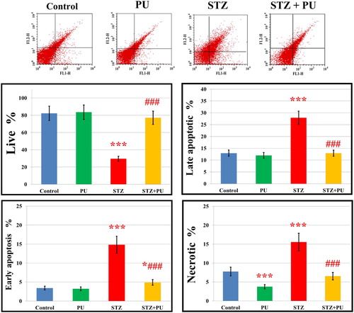
Flow cytometry was used to assess apoptosis in the heart using Annexin V-FITC staining (Fig. 4). The percentages of early and late apoptotic cells and necrotic cells were significantly higher in the hearts of diabetic rats than in those of the controls. The punicalagin therapy considerably minimized the number of apoptotic cells compared to the diabetic rats.
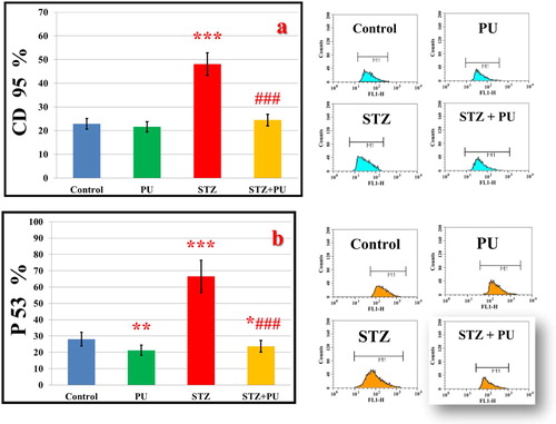
3.4.2 CD95 and P53
Similarly, CD95 and P53 expressions were significantly increased in the hearts of STZ-treated rats (Fig. 5a,b). Punicalagin administration after diabetic induction significantly ameliorated the increase in CD95 and P53 in the heart compared to the diabetic rats. The effects of punicalagin along with diabetes for particular apoptotic proteins have been evaluated.
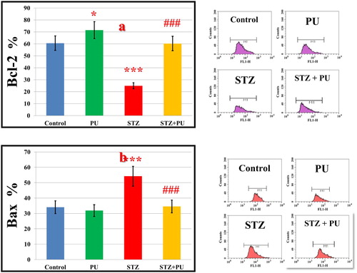
3.4.3 Bcl-2 and Bax
The analysis of the flow cytometry data revealed that the anti-apoptotic protein Bcl-2 was significantly reduced, while the apoptotic protein Bax was augmented in the hearts of STZ-induced diabetic rats (Fig. 6a,b). Punicalagin treatment considerably normalized the STZ-induced changes in the expression of these proteins.
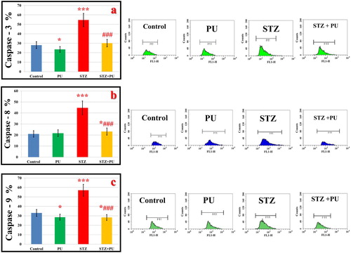
3.4.4 Caspases-3, 8 and 9
In Fig. 7, the expression levels of caspases-3, 8 and 9 were significantly up-regulated in the hearts of the STZ-induced diabetic rats. Punicalagin administration significantly normalized these apoptotic proteins (Figs. 6c,7a,b).
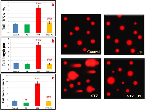
3.5 Comet assay
The changes in the levels of the comet parameters are displayed in Fig. 8. The STZ-treated rats showed an increase in the levels of all comet attributes, including percent DNA in tail, tail length and tail moment, suggesting that DNA damage occurred in the diabetic rats. Meanwhile, the treatment of diabetic rats with punicalagin significantly repressed the increase in the comet parameters.
3.6 Histological observations
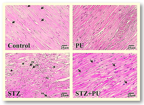
The histological examination of heart sections from the control and punicalagin-treated animals demonstrated the uniform size and regular arrangement of the cardiac muscle fibers, with centrally located round or oval nuclei. The heart sections of STZ-diabetic rats showed a disorganized array of the myocardial structure, myofibrillar discontinuation, myocyte degeneration, and pyknotic nuclei. Punicalagin treatment of STZ-induced diabetic rats reduced these changes in the STZ-injected rat hearts and revealed markedly less disorganization of the architecture of most of the cardiac muscle fibers, with centrally located vesicular nuclei (Fig. 9).
4 Discussion
The actual prevention and management of diabetic cardiomyopathy continues to be an essential health issue. To our knowledge, this is the first study demonstrating that punicalagin has cardioprotective effects in STZ-induced diabetes and that punicalagin reduces the risk of diabetes-induced cardiac apoptosis. Oxidative stress is a core player in the cardiac pathophysiology of hyperglycemia-induced cardiac injury; therefore, it is hypothesized that treatment with bioactive antioxidants is a promising approach to block the pathological changes and protect the heart function.
In the diabetic rats, the body weights were significantly decreased, which may be attributed to increased metabolism of the muscle tissue, fats and proteins [Citation32]. In punicalagin-treated diabetic rats, the body weights were markedly maintained within control levels, indicating a role of punicalagin in protecting against muscle damage, possibly by influencing disturbed muscle protein turnover and the subsequent loss of muscle mass after diabetic induction.
Punicalagin treatment significantly lowered fasting serum glucose, HbA1c and HOMA-IR levels and increased the insulin level in STZ-treated rats at the end of the experimental period. The mechanism underlying the glucose-lowering effect of punicalagin may be due to increased insulin release from the protected and/or remaining β-cells, restored insulin sensitivity [Citation33], changes in the absorption of dietary carbohydrates in the small intestine and facilitated utilization of glucose by the peripheral tissues that is mediated by an insulin-dependent glucose transporter [Citation34]. The present results are consistent with findings from cell culture and animal studies as well as clinical human research that pomegranates rich in punicalagin exhibited anti-diabetic effect [Citation12]. A pomegranate seed methanol extract that is rich in punicalagin showed hypoglycemic activity in STZ-induced diabetes and improved insulin sensitivity in rodent animals [Citation35].
It is well known that maintaining normal serum lipid levels through nutrition programs is an effective approach to decrease the major risk of cardiovascular disease and related disease complications [Citation36]. In the current study, the total cholesterol, LDL-cholesterol, VLDL-cholesterol and triglyceride levels in the serum were significantly reduced after diabetic rats were treated with punicalagin, indicating its beneficial effect on the lipid profile. In Zucker fatty diabetic rats, the pomegranate extract improved abnormal cardiac lipid metabolism by activating peroxisome proliferator-activated receptor-alpha (PPAR-α), thereby decreasing the circulating lipid levels and inhibiting their cardiac uptake [Citation37]. PPAR-α is a cardiac transcription factor involved in myocardial energy production via fatty acid uptake and oxidation. The augmented fatty acid oxidation improves insulin sensitivity by reducing lipid accumulation in the heart [Citation38]. The beneficial effect of punicalagin on the lipid profile in the present study explains the clinical study in diabetic patients with hyperlipidemia [Citation16,Citation39]. The authors claimed that pomegranate reduced cholesterol absorption, increased cholesterol excretion in feces, exerted positive effects on cholesterol metabolizing enzymes, markedly decreased total and LDL-cholesterol and improved the total/HDL and LDL/HDL-cholesterol ratios [Citation16]. Moreover, the pomegranate-mediated reduction of oxidized-LDL cellular uptake and cellular cholesterol biosynthesis was associated with a significant reduction in cellular oxidative stress [Citation40] [Citation41]. In accord with these results, PPARγ modulation by punicalagin resulted in reduced oxidative stress in macrophages in vitro [Citation42]. Thus, punicalagin treatment reduces lipid risk factors, confirming its anti-hyperlipidemic effects in diabetic rats.
A number of publications have reported the involvement of lipid peroxidation in the pathogenesis of diabetes-induced cardiac injury. The increased levels of H2O2 and MDA with decreased levels of SOD, CAT and GSH established excessive oxidative stress in STZ-induced diabetic rats. The increase in oxidative stress in diabetic rats is a consequence of higher ROS generation, which are produced by metabolic disturbances [Citation1,Citation43]. Punicalagin treatment significantly decreased the oxidative stress parameters, increased the GSH levels and normalized the activities of antioxidant enzymes in the hearts of STZ-treated rats, demonstrating the anti-peroxidation effect of punicalagin. It is worth mentioning that punicalagin treatment significantly augmented the GSH levels in the rat heart, revealing its potential ability to improve the antioxidant mechanisms in the heart. Similarly, there was a significant increase in the GSH content in the heart of the pomegranate-treated rats compared to the doxorubicin-treated group, indicating an antioxidant effect against doxorubicin-induced oxidative cardiotoxicity in rats [Citation44]. These results indicate that punicalagin possesses potent antioxidant capacity that is responsible for cardioprotection against oxidative stress.
Moreover, punicalagin administration significantly ameliorated the elevated levels of troponin T levels and LDH and CK-MB activities toward normal levels, indicating that punicalagin is an efficient cardioprotectant in diabetic conditions and can protect cardiomyocyte membrane integrity. Histopathological examination of the heart confirmed this finding and showed that punicalagin effectively prevented STZ-induced cardiac damage. The current data coincide with the ability of the punicalagin-rich pomegranate to protect against I/R injury, doxorubicin and isoproterenol-induced cardiotoxicity in rats, likely because of its actions in enhancing oxygen free radical scavenging activity and decreasing lipid peroxidative damage [Citation10,Citation44,Citation45]. These results indicate that punicalagin can defend the heart against oxidative stress and the associated pathology and dysfunction.
The increase in ROS resulting from sustained hyperglycemia can lead to myocardial inflammation, which is characterized by the production of large number of cytokines [Citation46]. Previous studies reported that the levels of proinflammatory cytokines and ROS were markedly increased and were associated with impaired glucose tolerance [Citation47,Citation48]. Therefore, it has been hypothesized that punicalagin can modulate cellular activity during inflammation due to its antioxidant properties. The current study demonstrated that punicalagin attenuated IL-1b, IL-6 and TNF-α in STZ-induced diabetes in rats. Several recent experimental studies suggest anti-inflammatory effects of pomegranate polyphenols in other settings that are characterized by increased oxidative stress. It has been reported that punicalagin treatment attenuated the elevation of TNF-alpha, IL-6 and IL-1b after LPS-induced acute respiratory distress syndrome in mice [Citation49], in rat primary microglia [Citation22], in cerebral I/R in rats [Citation50] and in an in vitro model of human intestinal epithelium [Citation51]. These data suggest that punicalagin could be an interesting nutritional source that contributes to preventing myocardial inflammation in diseases characterized by excessive oxidative stress, such as diabetes.
To understand the mechanism underlying the cardioprotective action of punicalagin on cardiac apoptosis in diabetes, we investigated the involvement of the STZ-mediated extrinsic and intrinsic apoptotic cell death pathway in cardiac tissues. Flow cytometry analysis demonstrated that punicalagin treatment ameliorated STZ-induced programmed cell death in cardiac tissues by regulating pro-apoptotic proteins, such as CD95; caspases-3, 8, and 9; Bax; and P53, and anti-apoptotic proteins, such as Bcl-2. These results indicate a marked modulation of both mitochondrial and death receptor apoptotic pathways in the hearts of punicalagin-treated diabetic rats and provide evidence that punicalagin has the ability to modulate apoptotic pathways in diabetes. Punicalagin significantly decreased trophoblast death as shown by the reduced levels of cleaved-PARP and LDH release [Citation52]. Punicalagin-rich pomegranate peel extract protected the liver and kidneys by stimulating antioxidant activities and elevating the anti-apoptotic protein Bcl-2 [Citation53]. Punicalagin reduced cerebral ischemia-reperfusion effects via attenuation of proinflammatory cytokines, up-regulation of Bcl-2 and down-regulation of Bax and caspase-3 [Citation50]. In contrast to the induction of apoptosis reported in cancer cells [Citation54], punicalagin protects against heart cell death in STZ-induced diabetes, reflecting the cell type-specific nature of the responses to punicalagin.
In addition, STZ-induced cardiac apoptosis was also evident from the DNA damage. A significant correlation between oxidative DNA damage and hyperglycemia was previously reported [Citation55]. Punicalagin treatment protected cardiac tissue from STZ-induced DNA damage, as evidenced by the inhibition of the increase of comet parameters compared to the diabetic rats. This implies that punicalagin prevented DNA strand breaks and maintained its supercoiled loops in an appropriately firm status. The precise mechanism is unidentified. However, this may be attributed to the effective antioxidant capacity of punicalagin. Therefore, punicalagin can decrease the DNA damage by scavenging the ROS in diabetic cases.
Significantly, the current results display, for the first time, that punicalagin has biological activities in addition to being an antioxidant in the diabetic heart. Punicalagin showed anti-apoptotic effects and protected DNA. It can trigger a variety of anti-apoptotic pathways due to its inherited antioxidant property. Punicalagin treatment effectively down-regulates P53 and up-regulates Bcl-2 and intracellular GSH levels in the STZ-treated rats. P53 and its regulated genes can be activated by hyperglycemia, leading to cardiomyocyte death [Citation56]. It is reported that P53 has the ability to induce apoptosis by an ROS-dependent pathway [Citation57,Citation58]. In contrast, Bcl-2 exhibits an anti-apoptotic and anti-necrotic influence via its antioxidant effect on intracellular ROS [Citation43,Citation59]. Bcl-2 can decrease lipid peroxidation by increasing cell resistance to ROS and blocking ROS production [Citation58]. Accordingly, it is suggested that the anti-apoptotic effect of punicalagin may occur via down-regulating P53 induction and up-regulating Bcl-2, which is related to the enhanced GSH and antioxidant levels in the heart. It also seems that punicalagin decreases apoptosis by blocking DNA damage via the suppression of caspases and pro-inflammatory cytokines that may be related to its anti-glycemic and anti-hyperlipidemic actions.
In conclusion, the underlying mechanisms of punicalagin-induced protection against diabetic cardiomyopathy are pleiotropic and include anti-hyperglycemic, anti-hyperlipidemic, antioxidant and anti-inflammatory activities. It can play an important role in the modulation of inflammatory and apoptotic cell signaling in the diabetic heart.
Acknowledgments
The facilities provided by the Faculty of Science, Mansoura University, Egypt, are greatly acknowledged and appreciated.
References
- T.V.FiorentinoA.PriolettaZuoP.F.FolliHyperglycemia-induced oxidative stress and its role in diabetes mellitus related cardiovascular diseasesCurr Pharm Des19201356955703
- M.F.ChowdhryH.A.VohraM.GalinanesDiabetes increases apoptosis and necrosis in both ischemic and nonischemic human myocardium: role of caspases and poly-adenosine diphosphate-ribose polymeraseJ Thorac Cardiovasc Surg1342007124131 131 e121–3
- WuT.G.LiW.H.LinZ.Q.WangL.X.Effects of folic acid on cardiac myocyte apoptosis in rats with streptozotocin-induced diabetes mellitusCardiovasc Drugs Ther222008299304
- DuY.GuoH.LouH.Grape seed polyphenols protect cardiac cells from apoptosis via induction of endogenous antioxidant enzymesJ Agric Food Chem55200716951701
- E.PeuchantJ.L.BrunV.RigalleauL.DubourgM.J.ThomasJ.Y.Danielet alOxidative and antioxidative status in pregnant women with either gestational or type 1 diabetesClin Biochem372004293298
- M.A.El-MissiryEnhanced testicular antioxidant system by ascorbic acid in alloxan diabetic ratsComp Biochem Physiol C Pharmacol Toxicol Endocrinol1241999233237
- M.I.GilF.A.Tomas-BarberanB.Hess-PierceD.M.HolcroftA.A.KaderAntioxidant activity of pomegranate juice and its relationship with phenolic composition and processingJ Agric Food Chem48200045814589
- C.M.MatthaiouN.GoutzourelasD.StagosE.SarafoglouA.JamurtasS.D.Koulocheriet alPomegranate juice consumption increases GSH levels and reduces lipid and protein oxidation in human bloodFood Chem Toxicol73201416
- S.GozlekciO.SaracogluE.OnursalM.OzgenTotal phenolic distribution of juice, peel, and seed extracts of four pomegranate cultivarsPharmacogn Mag72011161164
- R.N.JadejaM.C.ThounaojamD.K.PatelR.V.DevkarA.V.RamachandranPomegranate (Punica granatum L.) juice supplementation attenuates isoproterenol-induced cardiac necrosis in ratsCardiovasc Toxicol102010174180
- N.P.SeeramL.S.AdamsS.M.HenningNiuY.ZhangY.M.G.Nairet alIn vitro antiproliferative, apoptotic and antioxidant activities of punicalagin, ellagic acid and a total pomegranate tannin extract are enhanced in combination with other polyphenols as found in pomegranate juiceJ Nutr Biochem162005360367
- S.BanihaniS.SwedanZ.AlguraanPomegranate and type 2 diabetesNutr Res332013341348
- S.K.MiddhaT.UshaV.PandeHPLC evaluation of phenolic profile, nutritive content, and antioxidant capacity of extracts obtained from Punica granatum fruit peelAdv Pharmacol Sci20132013296236
- A.BasuK.PenugondaPomegranate juice: a heart-healthy fruit juiceNutr Rev6720094956
- M.HashemiR.KelishadiM.HashemipourA.ZakerameliN.KhavarianS.Ghatrehsamaniet alAcute and long-term effects of grape and pomegranate juice consumption on vascular reactivity in paediatric metabolic syndromeCardiol Young2020107377
- A.EsmaillzadehF.TahbazI.GaieniH.Alavi-MajdL.AzadbakhtCholesterol-lowering effect of concentrated pomegranate juice consumption in type II diabetic patients with hyperlipidemiaInt J Vitam Nutr Res762006147151
- ZouX.YanC.ShiY.CaoK.XuJ.WangX.et alMitochondrial dysfunction in obesity-associated nonalcoholic fatty liver disease: the protective effects of pomegranate with its active component punicalaginAntioxid Redox Signal21201415571570
- LiuW.MaH.L.FrostYuanT.J.A.DainN.P.SeeramPomegranate phenolics inhibit formation of advanced glycation endproducts by scavenging reactive carbonyl speciesFood Funct5201429963004
- P.G.DorseyP.GreenspanInhibition of nonenzymatic protein glycation by pomegranate and other fruit juicesJ Med Food172014447454
- M.N.Al-MuammarF.KhanObesity: the preventive role of the pomegranate (Punica granatum)Nutrition282012595604
- M.RosenblatN.VolkovaM.Roqueta-RiveraM.T.NakamuraM.AviramIncreased macrophage cholesterol biosynthesis and decreased cellular paraoxonase 2 (PON2) expression in Delta6-desaturase knockout (6-DS KO) mice: beneficial effects of arachidonic acidAtherosclerosis2102010414421
- O.A.OlajideA.KumarR.VelagapudiU.P.OkorjiB.L.FiebichPunicalagin inhibits neuroinflammation in LPS-activated rat primary microgliaMol Nutr Food Res58201418431851
- L.YaidikarB.BynaS.R.ThakurNeuroprotective effect of punicalagin against cerebral ischemia reperfusion-induced oxidative brain injury in ratsJ Stroke Cerebrovasc Dis23201428692878
- B.CerdaJ.J.CeronF.A.Tomas-BarberanJ.C.EspinRepeated oral administration of high doses of the pomegranate ellagitannin punicalagin to rats for 37 days is not toxicJ Agric Food Chem51200334933501
- B.RameshK.V.PugalendiAntihyperglycemic effect of umbelliferone in streptozotocin-diabetic ratsJ Med Food92006562566
- YangY.J.XiuZhangL.QinC.LiuJ.Antiviral activity of punicalagin toward human enterovirus 71 in vitro and in vivoPhytomedicine2020126770
- O.H.LowryN.J.RosebroughA.L.FarrR.J.RandallProtein measurement with the Folin phenol reagentJ Biol Chem1931951265275
- GongJ.QianL.KongX.YangR.ZhouL.ShengY.et alCardiomyocyte apoptosis in the right auricle of patients with ostium secundum atrial septal defect diseasesLife Sci80200711431151
- W.S.JuanLinH.W.ChenY.H.ChenH.Y.Y.C.HungTaiS.H.et alOptimal Percoll concentration facilitates flow cytometric analysis for annexin V/propidium iodine-stained ischemic brain tissuesCytometry A812012400408
- N.P.SinghM.T.McCoyR.R.TiceE.L.SchneiderA simple technique for quantitation of low levels of DNA damage in individual cellsExp Cell Res1751988184191
- R.R.TiceP.W.AndrewsN.P.SinghThe single cell gel assay: a sensitive technique for evaluating intercellular differences in DNA damage and repairBasic Life Sci531990291301
- J.DasV.VasanP.C.SilTaurine exerts hypoglycemic effect in alloxan-induced diabetic rats, improves insulin-mediated glucose transport signaling pathway in heart and ameliorates cardiac oxidative stress and apoptosisToxicol Appl Pharmacol2582012296308
- G.JelodarM.MohsenS.ShahramEffect of walnut leaf, coriander and pomegranate on blood glucose and histopathology of pancreas of alloxan induced diabetic ratsAfr J Tradit Complement Altern Med42007299305
- D.Del RioA.Rodriguez-MateosJ.P.SpencerM.TognoliniG.BorgesA.CrozierDietary (poly)phenolics in human health: structures, bioavailability, and evidence of protective effects against chronic diseasesAntioxid Redox Signal18201318181892
- M.ViladomiuR.HontecillasLuP.J.Bassaganya-RieraPreventive and prophylactic mechanisms of action of pomegranate bioactive constituentsEvid Based Complement Alternat Med20132013789764
- TanB.K.TanC.H.P.N.PushparajAnti-diabetic activity of the semi-purified fractions of Averrhoa bilimbi in high fat diet fed-streptozotocin-induced diabetic ratsLife Sci76200528272839
- HuangT.H.PengG.B.P.KotaLiG.Q.J.YamaharaB.D.Roufogaliset alPomegranate flower improves cardiac lipid metabolism in a diabetic rat model: role of lowering circulating lipidsBr J Pharmacol1452005767774
- P.FerreThe biology of peroxisome proliferator-activated receptors: relationship with lipid metabolism and insulin sensitivityDiabetes53Suppl. 12004S4350
- A.EsmaillzadehF.TahbazI.GaieniH.Alavi-MajdL.AzadbakhtConcentrated pomegranate juice improves lipid profiles in diabetic patients with hyperlipidemiaJ Med Food72004305308
- B.CerdaR.LlorachJ.J.CeronJ.C.EspinF.A.Tomas-BarberanEvaluation of the bioavailability and metabolism in the rat of punicalagin, an antioxidant polyphenol from pomegranate juiceEur J Nutr4220031828
- M.RosenblatN.VolkovaM.AviramPomegranate juice (PJ) consumption antioxidative properties on mouse macrophages, but not PJ beneficial effects on macrophage cholesterol and triglyceride metabolism, are mediated via PJ-induced stimulation of macrophage PON2Atherosclerosis21220108692
- M.ShinerB.FuhrmanM.AviramMacrophage paraoxonase 2 (PON2) expression is up-regulated by pomegranate juice phenolic anti-oxidants via PPAR gamma and AP-1 pathway activationAtherosclerosis1952007313321
- A.H.AminM.A.El-MissiryA.I.OthmanMelatonin ameliorates metabolic risk factors, modulates apoptotic proteins, and protects the rat heart against diabetes-induced apoptosisEur J Pharmacol7472015166173
- M.Hassanpour FardA.E.GhuleS.L.BodhankarM.DikshitCardioprotective effect of whole fruit extract of pomegranate on doxorubicin-induced toxicity in ratPharm Biol492011377382
- DongS.TongX.LiuH.GaoQ.Protective effects of pomegranate polyphenols on cardiac function in rats with myocardial ischemia/reperfusion injury]Nan Fang Yi Ke Da Xue Xue Bao322012924927
- S.A.HayatB.PatelR.S.KhattarR.A.MalikDiabetic cardiomyopathy: mechanisms, diagnosis and treatmentClin Sci (Lond)1072004539557
- S.KhuranaK.VenkataramanA.HollingsworthM.PicheTaiT.C.Polyphenols: benefits to the cardiovascular system in health and in agingNutrients5201337793827
- S.KhuranaM.PicheA.HollingsworthK.VenkataramanTaiT.C.Oxidative stress and cardiovascular health: therapeutic potential of polyphenolsCan J Physiol Pharmacol912013198212
- PengJ.WeiD.FuZ.LiD.TanY.XuT.et alPunicalagin ameliorates lipopolysaccharide-induced acute respiratory distress syndrome in miceInflammation382015493499
- L.YaidikarS.ThakurPunicalagin attenuated cerebral ischemia-reperfusion insult via inhibition of proinflammatory cytokines, up-regulation of Bcl-2, down-regulation of Bax, and caspase-3Mol Cell Biochem4022015141148
- S.HollebeeckJ.WinandM.F.HerentA.DuringJ.LeclercqY.Larondelleet alAnti-inflammatory effects of pomegranate (Punica granatum L.) husk ellagitannins in Caco-2 cells, an in vitro model of human intestineFood Funct32012875885
- ChenB.M.G.TuuliM.S.LongtineJ.S.ShinR.LawrenceT.Inderet alPomegranate juice and punicalagin attenuate oxidative stress and apoptosis in human placenta and in human placental trophoblastsAm J Physiol Endocrinol Metab3022012E114252
- A.E.Abdel MoneimM.S.OthmanS.M.MohmoudK.M.El-DeibPomegranate peel attenuates aluminum-induced hepatorenal toxicityToxicol Mech Methods232013624633
- WangS.G.HuangM.H.LiJ.H.LaiF.I.H.M.LeeY.N.HsuPunicalagin induces apoptotic and autophagic cell death in human U87MG glioma cellsActa Pharmacol Sin34201314111419
- SongF.JiaW.YaoY.HuY.LeiL.LinJ.et alOxidative stress, antioxidant status and DNA damage in patients with impaired glucose regulation and newly diagnosed Type 2 diabetesClin Sci (Lond)1122007599606
- F.FiordalisoA.LeriD.CesselliF.LimanaB.SafaiB.Nadal-Ginardet alHyperglycemia activates p53 and p53-regulated genes leading to myocyte cell deathDiabetes50200123632375
- D.CesselliI.JakoniukL.BarlucchiA.P.BeltramiT.H.HintzeB.Nadal-Ginardet alOxidative stress-mediated cardiac cell death is a major determinant of ventricular dysfunction and failure in dog dilated cardiomyopathyCirc Res892001279286
- ShengR.GuZ.L.XieM.L.ZhouW.X.GuoC.Y.EGCG inhibits cardiomyocyte apoptosis in pressure overload-induced cardiac hypertrophy and protects cardiomyocytes from oxidative stress in ratsActa Pharmacol Sin282007191201
- A.J.KowaltowskiG.FiskumRedox mechanisms of cytoprotection by Bcl-2Antioxid Redox Signal72005508514