
Abstract
Background
Intravenous thrombolysis (IVT) for acute brain infarctions caused by aortic dissection (AD) may lead to fatal outcomes; thus, it should be ruled out, especially if hypofibrinogenemia occurs after IVT. Successful management of AD-related acute brain infarction with hypofibrinogenemia after IVT has not been reported previously.
Case report
An 84-year-old woman developed sudden left limb weakness and aphasia for almost 4 h. Alteplase was administered intravenously immediately after cerebral hemorrhage was ruled out by emergent head computed tomography (CT). An anomaly suspected to be AD was detected during subsequent routine chest CT, which was confirmed by CT angiography to be a thoracoabdominal aortic dissecting aneurysm (DeBakey type I). Severe hypofibrinogenemia was also noted. After effective blood pressure control, intramuscular injection of vitamin K, and rehydration therapy, her brain cell metabolism improved, hemiplegia improved slightly, and hypofibrinogenemia recovered gradually. The patient’s cerebral hemorrhage did not progress, there was no chest pain or no aggravation of hemiplegia, and the fibrinogen level gradually returned to normal. The condition was stable during hospitalization. At 1.5 months after discharge, the patient showed minimal change in condition.
Conclusion
The symptoms of AD may be nonspecific and latent. IVT may be allowed to perform for some patients with AD related ischemical stroke, And IVT can improve the neural symptoms of AD-related ischemic stroke, but close monitoring is needed to avoid aneurysm rupture. Fibrinogen levels should also be monitored periodically after IVT for early detection of hypofibrinogenemia.
Introduction
Intravenous thrombolysis (IVT) is the first-line treatment for acute ischemic stroke (AIS) within the thrombolytic time window [Citation1]. Although aortic dissection (AD) rarely causes cerebral infarction [Citation2], IVT is contraindicated in AD, owing to its high risk of AD rupture [Citation3, Citation4]. Hypofibrinogenemia, a rare and severe complication of IVT, can increase the risk of symptomatic intracerebral hemorrhage [Citation5]. We report a rare case of successful IVT in a patient with AD-related AIS with severe hypofibrinogenemia.
Case presentation
An 84-year-old woman was admitted to our hospital due to sudden left limb weakness and aphasia for almost 4 h, without abdominal pain or chest pain. According to the information supplied by her family, she had no hereditary diseases, hypertension, or other chronic diseases, and she had no history of surgery. On admission, her blood pressure (BP) was 132/69 mmHg and her heart rate was 77 beats per minute; she was emaciated, had muscle strength of 2/5 in the left upper and lower extremities, had clear consciousness, and had motor aphasia, but there were no other obvious positive neurological signs. The National Institutes of Health Stroke Scale (NIHSS) score was 7. Emergency head computed tomography (CT) showed old lacunar infarctions bilaterally in the area under the cortex of the head (). Red blood cell count, platelet counts, random blood glucose, blood electrolytes, and blood coagulation test () showed no apparent abnormality. A total of 36 mg of recombinant tissue plasminogen activator (rt-PA; alteplase) was administered, initially with a 0.9 mg/kg intravenous bolus injection of 10% of the total amount, and the rest was administered intravenously within 1 h. The fibrinogen level sharply decreased, and fibrinogen could not be detected anymore () after IVT. Intramuscular injection of vitamin K was administered, but no antiplatelet treatment was administered. Her coagulation indicators gradually returned to normal within 48 h. The serum creatinine, glycated hemoglobin, troponin I, and creatine kinase-MB were normal; the hemoglobin concentration (85 g/L) decreased; the percentage of neutrophils (86.9%) increased; the B-type natriuretic peptide (BNP, 3091 pg/mL), plasma D-dimers (1.85 mg/L), C-reactive protein (92 mg/L), lactate dehydrogenase (272 U/L), and serum homocysteine levels (19 µmol/L) increased; BNP decreased to 1734 pg/mL within 3 days after diuretic therapy. The fecal occult blood test was weakly positive. One day after IVT, the muscle strength of the left limb was 3/5 (NIHSS score, 6), and repeat head CT showed a minor hematoma within the focus of the AIS (). On the third day after IVT, conventional chest CT showed an anomaly suspected to be AD (), and prompt thoracoabdominal CT angiography confirmed a thoracoabdominal AD (DeBakey type I) (). The patient’s guardians refused endovascular intervention for the AD, and she received medications to control the BP and improve the metabolism of brain cells as well as rehydration therapy. The patient was confined in the hospital for 3 days and then transferred to another hospital for treatment. At discharge, the changes in her condition were not significant, and she had no headache or chest/abdominal pain.
Figure 1. (a) Computed tomography (CT) performed before intravenous thrombolysis (IVT) shows old lacunar infarctions bilaterally in the area under the cortex of the head. (b) Head CT shows low-density foci in the right frontal and temporal lobes with minor hemorrhaging (no space-occupying effect) on the second day after IVT. (c) Chest CT shows inward displacement of the calcified plaques in the thoracic aortic arch (arrow) and suspicious double lumens from the aortic arch to the descending aorta, suggesting aortic dissection (AD). (d) CT angiography of the thoracoabdominal aorta shows a thoracoabdominal AD (DeBakey type I), with the AD extending from the aortic root to the brachiocephalic trunk (e) and then to the right common carotid artery. (f) Intimal slices split the lumen into two cavities in the aortic arch.
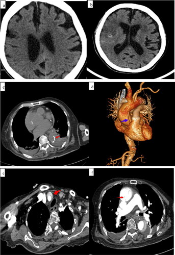
Table 1. Coagulation function before and after intravenous thrombolysis.
During the patient’s follow-up at 1.5 months, she appeared to have been treated by the conservative therapy administered in the gerontological hospital, and her condition was the same as that during discharge and was stable.
Discussion and conclusion
Causes and dangers of AD
Hypertension is the most common cause of AD, especially in younger patients with hypertension [Citation2, Citation6], but patients without hypertension who have AD, like our case, are not rare. Mortality in patients with Stanford type A AD may be as high as 40%–50% if appropriate emergency medical treatment is not obtained promptly [Citation7].
Signs and symptoms of AD
Patients with AD are prone to unstable hemodynamics [Citation8], such as BP difference >20 mmHg between both arms and nonpalpable weak pulse on one side [Citation9]. Typical patients with AD often have chest/back/abdominal pain, but our patient had no chest/back pain during hospitalization. Only 48% (11/23) of patients complained of chest/back pain [Citation10]. The above-mentioned signs and symptoms could deteriorate during IVT [Citation2]. Details of cases involving IVT with rt-PA for AD-related AIS are shown in . Neurological symptoms such as unconsciousness and dysphrasia can obscure the symptoms of AD, resulting in misdiagnosis of AD [Citation11].
Table 2. Details of cases that underwent IVT with rt-PA for AD-related acute ischemic stroke.
Methods of AD screening
Routine chest CT is the recommended screening, followed by carotid ultrasound and plasma D-dimers. The examining doctor should perform these screenings before IVT. Most chest X-rays obtained on admission do not show AD-specific clues, but follow-up chest X-ray is useful in AD diagnosis [Citation12]. Positive findings indicating acute AD include occlusion or intimal flap of the common carotid artery on carotid ultrasound, elevated serum D-dimer values, left hemiparesis, systolic BP difference >20 mmHg between the arms, and mediastinal widening on chest radiograph [Citation10]. The combination of physical, radiological, ultrasonographic, and laboratory findings in rapid screening of acute AD as a cause of AIS can help improve the diagnostic rate.
IVT for AD-related ischemic stroke
A few case reports showed that IVT can improve the neural symptoms of patients with AD-related AIS [Citation12–14] (); however, the greatest risk in IVT is aneurysm rupture induced by IVT. This case involving full-dose successful IVT for AD-related AIS that was successfully treated without operation is exceedingly rare.
In previous reports, AD was found opportunely and IVT was discontinued [Citation9, Citation14] () or emergency surgery was performed to ameliorate the effect of full-dose IVT [Citation12, Citation13] (). Fortunately, AD rupture did not occur after full-dose IVT in our patient. It should be noted that IVT is absolutely contraindicated for AD-related AIS.
Hypofibrinogenemia after IVT
Severe hypofibrinogenemia is observed during IVT with rt-PA in nearly 5% of stroke cases [Citation15]. Hypofibrinogenemia is a risk factor for major bleeding such as intracerebral hemorrhage during rt-PA infusions [Citation16], and fibrinogen assessment could be a rapid, inexpensive tool to identify patients at high risk of bleeding [Citation17]. An ∼80% decrease in fibrinogen level was observed 2 h after IVT, reaching ∼80% at 24 h and returning to normal by >2 day [Citation15, Citation18]. The minor non-space-occupying cerebral hemorrhage in this patient may be related to hypofibrinogenemia, but thankfully there was no AD rupture.
In conclusion, symptoms of AD may be non-specific and latent, and rapidly ruling out AD is extremely important for patients with AIS. Routine chest CT should be performed before IVT. IVT may be allowed to perform for some patients with AD related ischemical stroke, and IVT can improve the neural symptoms of patients with AD-related ischemic stroke, but close monitoring is needed to avoid aneurysm rupture. Fibrinogen levels should also be monitored periodically after IVT for early detection of hypofibrinogenemia.
Ethics approval and consent to participate
The study design was approved by the ethics review board of the 3rd Affiliated Hospital of Shenzhen University (no. 2019-SZLH-LW-003).
Disclosure statement
No potential conflict of interest was reported by the author(s).
Funding
The author received no financial support for the research, authorship, and/or publication of this article.
References
- Yang ZS, Mu J. Co-administration of tissue plasminogen activator and hyperbaric oxygen in ischemic stroke: a continued promise for neuroprotection. Med Gas Res. 2017;7(1):68–73.
- Sukockienė E, Laučkaitė K, Jankauskas A, et al. Crucial role of carotid ultrasound for the rapid diagnosis of hyperacute aortic dissection complicated by cerebral infarction: a case report and literature review. Medicina (Kaunas). 2016;52(6):378–388.
- Folgoas E, Toulgoat F, Sévin M, et al. [Ischemic stroke related to pauci-symptomatic acute aortic dissection. Risks of intravenous thrombolysis]. Rev Neurol (Paris)). 2012;168(4):357–362.
- Jo KW, Park IS, Kim YD, et al. Unexpected complication of intravenous recombinant tissue plasminogen activator thrombolysis in a patient with acute ischemic stroke: aortic dissection. Korean J Cerebrovasc Surg. 2009;11:204–206.
- Xu X, Li C, Wan T, et al. Risk factors for hemorrhagic transformation after intravenous thrombolysis in acute cerebral infarction: a retrospective single-center study. World Neurosurg. 2017;101:155–160.
- Tsivgoulis G, Vadikolias K, Heliopoulos I, et al. Aortic arch dissection causing acute cerebral ischemia: an uncommon contraindication for intravenous thrombolysis. Circulation. 2011;124(5):657–658.
- Hagan PG, Nienaber CA, Isselbacher EM, et al. The international registry of acute aortic dissection (IRAD): new insights into an old disease. JAMA. 2000;283(7):897–903.
- Choi N, Yoon J-E, Park B-W, et al. Delayed surgery for aortic dissection after intravenous thrombolysis in acute ischemic stroke. Korean J Thorac Cardiovasc Surg. 2016;49(5):392–396.
- Rodrıíguez-Luna D, Vilar RM, Peinazo M, et al. Intravenous thrombolysis in an elderly patient with acute ischemic stroke masking aortic dissection. J Stroke Cerebrovasc Dis. 2011;20(6):559–561.
- Ohara T, Koga M, Tokuda N, et al. Rapid identification of type a aortic dissection as a cause of acute ischemic stroke. J Stroke Cerebrovasc Dis. 2016;25(8):1901–1906.
- Lantos J, Nagy A, Hegedűs Z, et al. [Thrombolysis in case of ischemic stroke caused by aortic dissection]. Ideggyogy Sz. 2017;70(1-2):69–72.
- Do YR. Aortic dissection after intravenous thrombolysis in acute cerebral infarction. Kosin Med J. 2017;32(1):127–132.
- Kazmi SO, Achi O, Damani R. Full-dose thrombolysis for a right middle cerebral artery stroke after an acute aortic dissection. Ann Indian Acad Neurol. 2018;21(3):223–224.
- Hong KS, Park SY, Whang S, et al. Intravenous recombinant tissue plasminogen activator thrombolysis in a patient with acute ischemic stroke secondary to aortic dissection. J Clin Neurol. 2009;5(1):49–52.
- Matrat A, De Mazancourt P, Derex L, et al. Characterization of a severe hypofibrinogenemia induced by alteplase in two patients thrombolysed for stroke. Thromb Res. 2013;131(1):e45–e48.
- Skeik N, Gits CC, Ehrenwald E, et al. Fibrinogen level as a surrogate for the outcome of thrombolytic therapy using tissue plasminogen activator for acute lower extremity intravascular thrombosis. Vasc Endovascular Surg. 2013;47(7):519–523.
- Vandelli L, Marietta M, Gambini M, et al. Fibrinogen decrease after intravenous thrombolysis in ischemic stroke patients is a risk factor for intracerebral hemorrhage. J Stroke Cerebrovasc Dis. 2015;24(2):394–400.
- Zhang YL, Cui Y, Cai L, et al. The influence of ateplase on blood coagulation function in patients treated with intravenous thrombolysis within 24 hours. Shanxi Med J. 2015;44:1591–1594.