
Abstract
PsoP27 is an antigen expressed in psoriatic lesions. It plays an inflammatory role in psoriasis. This study objective was to characterize antibodies (Abs) against PsoP27 in patients with psoriatic arthritis (PsA) and rheumatoid arthritis (RA). Levels of Abs against native and citrullinated PsoP27 in PsA and RA patients’ synovial fluid (SF) and sera were determined by ELISA. SF of osteoarthritis (OA) patients and sera of healthy donors were used as controls. Levels of Abs against PsoP27 were correlated with disease activity scores. Abs against native and citrullinated PsoP27 levels in SF of PsA (n = 48; 0.38 ± 0.03 and 0.44 ± 0.04, respectively) and RA (n = 22; 0.57 ± 0.1 and 0.62 ± 0.09, respectively) were significantly higher than in OA patients (n = 23; 0.14 ± 0.01 and 0.15 ± 0.01, respectively) (p < .0001). For both Abs, there were no significant differences between their level in PsA and RA patients. There was no difference in the level of Abs against citrullinated PsoP27 in SF of seronegative versus seropositive RA patients. Levels of Abs against both native and citrullinated PsoP27 in the SF and level of systemic C-reactive protein in PsA correlated positively, while in RA there were no significant correlations with disease activity scores. No differences in level of Abs against PsoP27 were found in the sera of all three study groups. Abs against native and citrullinated PsoP27 are present in PsA and RA SF but not in those of OA patients, suggesting a potential role of those Abs in inflammatory joint diseases.
Introduction
Psoriatic arthritis (PsA) and rheumatoid arthritis (RA) are chronic inflammatory diseases, characterized by joint pain and swelling along with systemic manifestations [Citation1]. PsA is characterized by inflammation of the entheses and synovium, leading to joint erosions [Citation2]. It affects approximately 30% of psoriasis (PsO) patients, with 0.5% prevalence worldwide [Citation3]. PsA diagnosis is mostly clinical, based upon the classification criteria for PsA (CASPAR) [Citation4]. Laboratory diagnostic markers specific to PsA are lacking, leading to its definition as a ‘seronegative disease’. PsA and PsO share common pathogenic mechanisms [Citation5]. Specifically, keratinocytes and fibroblast-like synoviocytes exhibit similar proliferative activity which can lead to articular cartilage destruction and bone damage [Citation6]. As such, it can be speculated that PsO and PsA share common autoantigens.
PsoP27 is a protein generated by N- and C-terminal cleavage of SERPINB3/B4 by mast cells and it was designated as an autoantigen in PsO [Citation7]. PsoP27 is present in psoriatic skin plaques, but not in healthy skin. It is expressed in mast cells in psoriatic skin lesions [Citation8,Citation9] and participates in immune complexes generation [Citation10,Citation11]. SERPINB3/B4 was found to be involved in skin damage and disorders, such as atopic dermatitis, in a mouse model [Citation12] and in human keratinocytes [Citation13,Citation14]. Antibodies (Abs) against PsoP27 were found in PsO patients sera [Citation15], and after cyclosporine A therapy, PsoP27 expression was reduced [Citation16].
Synovial fluid (SF) autoantibodies (autoAbs) are highly prevalent in RA disease. Rheumatoid factor (RF) and anti-cyclic citrullinated peptide (anti-CCP) Abs are used as serological markers for RA diagnosis based on the American College of Rheumatology (ACR) criteria [Citation17]. Citrullination is a post-transcription modification caused by arginine amino acid conversion to citrulline by peptidyl arginine deaminase enzymes found in macrophages and neutrophils [Citation18]. Anti-citrullinated Abs recognize a variety of citrullinated antigens, including citrullinated forms of fibrinogen, vimentin, type II collagen and α-enolase [Citation19]. The fact that these Abs appear before disease onset [Citation20], their association with erosion development, further radiographic progression [Citation21], and their association with a more aggressive disease course [Citation22] supports the likelihood of their involvement in disease pathophysiology. Anti-CCP could be detected in 60–80% of RA patients’ sera with 85–99% specificity [Citation23]. However, up to one-third of RA patients are RF and/or anti-CCP seronegative [Citation24]. Seronegative RA remains a source of debate on whether they are truly autoAbs-negative or whether they harbor yet undefined RA-related autoAbs. Although citrullination is commonly associated with RA, it was shown to be present in other diseases, such as PsA and systemic lupus erythematosus (SLE) [Citation25,Citation26]. Anti-CCP are considered to be RA-specific although they have been found in 6% of PsA patients as well [Citation27]. In this study, Abs against PsoP27 were investigated in SF of PsA and RA and were compared to osteoarthritis (OA) patients. In addition, those Abs were analyzed in sera of PsA and RA patients and were compared to healthy donors. Correlation between levels of those Abs and clinical measures were determined.
Methods
Patients and samples
The study was conducted at the Rheumatology Department, Tel Aviv Medical Center during the years 2019–2022. The study was approved by the Center’s Institutional Review Board (Helsinki Committee – approval number 0394-19-TLV). All participants signed their informed consent prior to study entrance. SF samples from patients with PsA (n = 48), RA (n = 28) and OA (n = 23) were obtained via knee aspiration. Patients were classified according to the standard criteria: PsA according to the CASPAR criteria [Citation28], RA according to the ACR/EULAR criteria [Citation17] and OA according to the 1986 ACR criteria [Citation29]. Clinical indices were collected from the patients’ files on the date of enrollment. Disease activity was measured by the Simple Disease Activity Index (SDAI), with an SDAI score of 0 to ≤11 scored as 1, between >11 to ≤26 scored as 2, and >26 scored as 3.
The SF samples were centrifuged at 2500 rpm for 10 min to obtain cell-free supernatants, aliquoted and immediately stored at −80 °C until use. Blood samples were obtained from patients with PsA (n = 31), RA (n = 32) and healthy donors (n = 31) and centrifuged at 2500 rpm for 10 min, and the separated serum was stored at −80 °C.
Synthetic peptides
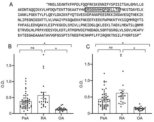
Two synthetic peptides (GL Biochem group, Shanghai, China) were used, and the sequences of those peptides are native PsoP27: SVDRSGNVHHQFQKLTLE, and citrullinated (cit) PsoP27: SVDCitSGNVHHQFQKLTLE, in which citrulline (cit) replaces arginine (R) position. The native peptide sequence was based on previous publication [Citation30] and the citrullinated peptide was designed as new antigenic substrate. The schematic PsoP27 peptide representation on the SERPINB3 protein is shown in .
Figure 1. PsoP27 peptide sequence and the detection of Abs against native and citrullinated PsoP27 in SF of PsA and RA patients. (A) Illustration of the PsoP27 peptide sequence (marked in black box) on the SERPINB3 protein. SFs were analyzed for levels of Abs against native (B) and citrullinated (C) PsoP27 in samples of PsA (n = 48), RA (n = 22) and OA (n = 23) patients. Levels of Abs against PsoP27 are shown as the value of optical density (O.D.). The p values were calculated by the non-parametric one-way ANOVA Kruskal–Wallis test and Dunn’s multiple comparison test, *p < .0001, ns: non-significant.
ELISA
Peptides were diluted to 10 μg/mL in phosphate-buffered saline (PBS), and microtiter plates were coated with 100 μL of each peptide and incubated overnight at 4 °C. After the plates were washed three times with Tween 0.05% PBS solution, blocking was performed with bovine serum albumin (BSA) 2% in PBS for 2 h and washed three times more. The plates then underwent incubation for 2 h with the SF diluted 1:10 or the serum samples diluted 1:50, after which they were washed three times with Tween 0.05% PBS solution. After the washes, the plates underwent incubation with anti-human IgG horseradish peroxidase conjugated followed by five washes. Reactions were induced with 3,3′,5,5′-tetramethylbenzidine for 30 min and stopped with 1 M sulfuric acid. The plates were read on an ELISA plate reader BioTek 800TS microplate reader (BioTek, Winooski, VT) using GEN5 version 2.05 software. Optical density (O.D.) units for comparison of wavelength absorbance measurement were determined at a wavelength of 450 nm with a reference wavelength of 630 nm.
Statistical analysis
Non-parametric methods were used for statistical comparisons since the data showed a non-normal distribution. Statistical differences with respect to levels of Abs against PsoP27 between three independent groups were calculated using the Kruskal–Wallis test followed by Dunn’s post hoc test. Analyses for comparison of independent data between two groups were performed with the Mann–Whitney U test. The Pearson rank test was used to analyze the correlation between levels of Abs against PsoP27 and disease activity parameters. All statistical analyses were performed with the statistical program GraphPad Prism 8.0 (GraphPad Software, San Diego, CA). All values are expressed as the mean ± standard error, and p < .05 was considered statistically significant.
Results
Detection of Abs against native and citrullinated PsoP27 in the SF but not the sera of PsA and RA patients
The demographic and clinical parameters of patients that donated SF and sera are summarized in , respectively. The mean levels of Abs against native PsoP27 in the SF of the PsA group (O.D. ± SE) (0.38 ± 0.03) and the RA group (0.57 ± 0.1) were both higher than that of OA group (0.14 ± 0.01). This difference of PsA and RA as compared to the control OA group was significant (p < .0001) but no significant differences in level of Abs against native PsoP27 in SF were detected between the PsA and RA groups (). Similar results were observed with the level of Abs against citrullinated PsoP27 () in PsA (0.44 ± 0.04) and RA (0.62 ± 0.09) as compared to OA (0.15 ± 0.01). Here, again, the difference was similarly significant for PsA and RA compared to OA (p < .0001), with no significant differences between PsA and RA. There were also no significant differences in the level of Abs against citrullinated PsoP27 in SF of seropositive (CCP+) (n = 14; 0.46 ± 0.04) compared to seronegative (CCP–) RA patients (n = 14; 0.68 ± 0.14) (). Interestingly, the level of Abs against citrullinated PsoP27 in CCP seronegative RA subgroup in this cohort was higher than their level in the seropositive RA subgroup, but with no statistical significance. Analysis of Abs against PsoP27 in sera showed no difference in the mean O.D. between the levels of Abs against native and citrullinated PsoP27 in the groups tested; PsA (0.22 ± 0.02 and 0.39 ± 0.05, respectively), RA patients (0.24 ± 0.017 and 0.27 ± 0.02) and healthy controls (0.27 ± 0.02 and 0.33 ± 0.02) ().
Figure 2. Level of Abs against citrullinated PsoP27 in SF of seropositive and seronegative RA patients. SF were analyzed for level of Abs against citrullinated PsoP27 in samples of seropositive (n = 14) and seronegative (n = 14) RA patients. Level of Abs against citrullinated PsoP27 is shown as the value of optical density (O.D.). The p values were calculated using the Mann–Whitney U test. ns: non-significant.
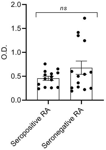
Figure 3. Sera levels of Abs against native and citrullinated PsoP27. Sera were analyzed for levels of Abs against native (A) and citrullinated (B) PsoP27. Samples were derived from PsA (n = 32) and RA (n = 32) patients and healthy controls (n = 31). Levels of Abs against PsoP27 are shown as the value of optical density (O.D.). The p values were calculated using the one-way ANOVA Kruskal–Wallis test and Dunn’s multiple comparison test. ns: non-significant.
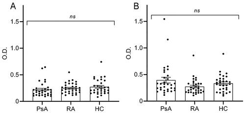
Table 1. Demographic and clinical characteristics of (A) PsA, RA and OA patients in the SF section and (B) PsA and RA patients and healthy controls in the serum section.
Correlation between levels of Abs against PsoP27 in SF and PsA and RA clinical measures
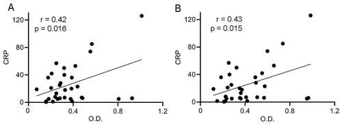
In order to determine whether the level of Abs against PsoP27 could correlate with PsA and RA disease activity, we correlated the laboratory and clinical indices including C-reactive protein (CRP) level, tender and swollen joints counts (TJ and SJ) and SDAI for both PsA and RA patients. The PASI score was analyzed for PsA only. There was a significant correlation between the level of Abs against native PsoP27 and systemic CRP level r = 0.42, p = .016 in the SF of PsA patients (). Similarly, the level of Abs against citrullinated PsoP27 in the SF of PsA patients was correlated also with CRP level r = 0.43, p = .015 (). Other disease activity scores showed no correlation with level of Abs against PsoP27. The results for RA showed no significant correlations with disease activity scores of CRP, TJ, SJ and SDAI.
Figure 4. Correlation between level of Abs against PsoP27 in SF of PsA patients and systemic CRP. Both levels of Abs against native and citrullinated PsoP27 in SF correlated positively with CRP level in the PsA cohort. Each point represents single SF sample of PsA patient. The x-axis reflects the levels of Abs against native PsoP27 (A) and citrullinated PsoP27 (B) in optical density (O.D.). The y-axis reflects the systemic CRP level (mg/L). The relationship between variables was evaluated using the Pearson rank correlation test. Trend lines indicate linear correlation. r: Pearson’s rank correlation coefficient, p value indicates statistical significance.
Diagnostic value of Abs against PsoP27 in SF of PsA and RA patients
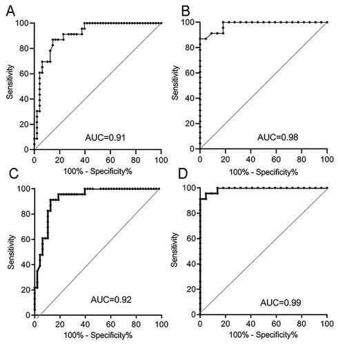
ROC analysis was used to determine the diagnostic performance of Abs against PsoP27 in SF for their potential to discriminate PsA and RA from OA (). It emerged that to distinguish PsA from OA, level of Abs against native PsoP27 showed diagnostic capability at cut-off value of 0.2 with a sensitivity of 91% and a specificity of 77% (95% confidence interval) (). To distinguish RA from OA, level of Abs against native PsoP27 showed diagnostic capability at cut-off value of 0.23 with a sensitivity of 91% and a specificity of 86% (95% confidence interval) (). Levels of Abs against citrullinated PsoP27 were showing slightly higher diagnostic capability to distinguish PsA from OA at a cut-off value of 0.19 with a sensitivity of 91% and a specificity of 87.5% (95% confidence interval) (). Even higher values were found for level of Abs against citrullinated PsoP27 to distinguish RA from OA a cut-off value of 0.25 yields and a sensitivity and specificity of 95% ().
Figure 5. Receiver operating characteristic (ROC) curve analysis to assess the capacity of Abs against citrullinated PsoP27 in SF to discriminate between PsA and RA and OA patients. Abs against native PsoP27 to discriminate between (A) PsA versus OA patients, and (B) RA versus OA patients. Abs against citrullinated PsoP27 used to discriminate between (C) PsA versus OA patients and (D) RA versus OA patients.
Discussion
The findings of this study demonstrated presence of Abs against PsoP27 in the SF of PsA and RA patients. Both Abs against native and citrullinated PsoP27 in the SF were found in higher levels as compared with that of OA. Moreover, the levels of Abs against native and citrullinated PsoP27 in the SF of PsA patients correlated significantly with the systemic CRP level. In RA, there were no significant correlations between the level of Abs against PsoP27 in the SF and disease activity scores.
Abs against PsoP27 were reported to be present in psoriatic skin lesions and absent in healthy skin. PsoP27 antigen was studied by Iversen and coworker who found that it participated in inflammatory reactions not only in psoriatic skin lesions but in other inflammatory diseases as well [Citation31], most probably due to its role in forming complement activating immune-complexes, as shown by Asbakk et al. [Citation10]. We hypothesized that PsO and PsA share common inflammatory pathways, and identified presence of Abs against PsoP27 in two inflammatory arthropathies prototypes, PsA and RA, in comparison with a non-inflammatory arthropathy, OA. Our findings suggest that Abs against PsoP27 may participate in inflammatory process in affected joints. Moreover, Abs against citrullinated PsoP27 were present in SF of both RA subgroups, regardless their CCP status.
Several theories have been proposed to explain the pathogenesis of joint and skin damage in PsA. Chimenti et al. [Citation32] suggested that cytochrome C and tryptase may have a role in skin and joint inflammation, indeed, PsoP27 is expressed in tryptase-positive cells [Citation8]. We found that the level of Abs against PsoP27 correlated with CRP in PsA but not in RA. This observation is in line with Iversen’s study that showed PsoP27 antigen levels in psoriatic skin lesions correlate with disease activity and that they declined after treatment with cyclosporine A [Citation16], which has been used for PsA treatment in the past. In accordance to our findings, a different study identified a particular immune cell subset IL-17+CD8+ T cells in SF that their level correlated with systemic CRP level in PsA but not in RA [Citation33]. Both studies describe difference in synovial immunopathology features between PsA and RA.
RA is characterized by autoAbs that bind citrullinated proteins, those autoAbs although in small amount, can be detected in other auto-inflammatory diseases: we reported earlier on presence of Abs against citrullinated proteins in the SF of patients PsA [Citation34]. Abs against citrullinated proteins were also detected in the sera of a small subset of PsA patients [Citation35,Citation36], and their presence was indicative of a more aggressive disease [Citation26].
We analyzed SF and sera aiming to determine the presence of Abs against PsoP27 in PsA and RA patients. Although these two compartments may share similarities, the lack of Abs against PsoP27 in sera might indicate that those Abs are produced locally in the joints. SF is enriched with neutrophils that release genomic DNA–protein complexes known as neutrophil extracellular traps (NETs) in a process termed NETosis [Citation37]. The resulting network of DNA and associated proteins (e.g. histones, myeloperoxidase and neutrophil elastase) is present in the synovium of both PsA [Citation38] and RA [Citation39]. NETosis is a source of citrullinated antigens [Citation39]. We therefore hypothesize that PsoP27 citrullination is a product of NETs, but further studies are needed to determine the source of Abs against PsoP27 in SF.
Our study has several limitations, starting with the unequal numbers of men and women and age differences of the patients in each group. The numbers of men and women with PsA were approximately equal, whereas the patients with RA and OA were mostly women. The patients with OA were older, while those with PsA and RA were younger and relatively similar in age. In addition, comparison of sera from PsA and RA to older healthy subjects or OA patients control groups could reduce the age-related heterogeneity.
Second, part of the patients in the cohort had been treated with steroids (prednisone), synthetic or biologic DMARDs that could have decreased their inflammatory status as well as the level of Abs against PsoP27 to a degree that cannot be assessed. Third, we could not detect specific Abs against PsoP27 in the sera of PsA and RA patients. Including a control group of PsO, might detect those Abs, since there are evidence that PsoP27 autoantigen present in skin plaques of PsO patients.
To our knowledge, this is the first study to demonstrate Abs against native and citrullinated PsoP27 that are present in SF of both PsA and RA but not in OA. We demonstrated a positive correlation between the level of Abs against PsoP27 and systemic CRP in PsA but not in RA, indicating that Abs against PsoP27 can be a possible inflammatory marker in PsA disease.
Disclosure statement
No potential conflict of interest was reported by the author(s).
References
- Verheul MK, Fearon U, Trouw LA, et al. Biomarkers for rheumatoid and psoriatic arthritis. Clin Immunol. 2015;161(1):2–10. doi: 10.1016/j.clim.2015.04.005.
- Eder L, Gladman DD. Psoriatic arthritis: phenotypic variance and nosology. Curr Rheumatol Rep. 2013;15(3):316. doi: 10.1007/s11926-013-0316-4.
- Stolwijk C, Boonen A, van Tubergen A, et al. Epidemiology of spondyloarthritis. Rheum Dis Clin North Am. 2012;38(3):441–476. doi: 10.1016/j.rdc.2012.09.003.
- Mease P. Psoriatic arthritis and spondyloarthritis assessment and management update. Curr Opin Rheumatol. 2013;25(3):287–296. doi: 10.1097/BOR.0b013e32835fd8d5.
- Haroon M, Winchester R, Giles JT, et al. Certain class I HLA alleles and haplotypes implicated in susceptibility play a role in determining specific features of the psoriatic arthritis phenotype. Ann Rheum Dis. 2016;75(1):155–162. doi: 10.1136/annrheumdis-2014-205461.
- McGonagle D, Conaghan PG, Emery P. Psoriatic arthritis: a unified concept twenty years on. Arthritis Rheum. 1999;42(6):1080–1086. doi: 10.1002/1529-0131(199906)42:6<1080::AID-ANR2>3.0.CO;2-7.
- Iversen OJ, Lysvand H, Slupphaug G. Pso p27, a SERPINB3/B4-derived protein, is most likely a common autoantigen in chronic inflammatory diseases. Clin Immunol. 2017;174:10–17. doi: 10.1016/j.clim.2016.11.006.
- Iversen OJ, Lysvand H, Jacobsen T, et al. The psoriasis-associated antigen, Pso p27, is expressed by tryptase-positive cells in psoriatic lesions. Arch Dermatol Res. 1995;287(5):503–505. doi: 10.1007/BF00373437.
- Lysvand H, Hagen L, Klubicka L, et al. Psoriasis pathogenesis – Pso p27 is generated from SCCA1 with chymase. Biochim Biophys Acta. 2014;1842(5):734–738. doi: 10.1016/j.bbadis.2014.02.005.
- Asbakk K, Bergh K, Iversen OJ. The psoriasis-associated antigen, Pso p27, participates in the formation of complement activating immune-complexes in psoriatic scale. APMIS. 1990;98(2):143–149. doi: 10.1111/j.1699-0463.1990.tb01014.x.
- Lysvand H, Helland R, Hagen L, et al. Psoriasis pathogenesis – Pso p27 constitutes a compact structure forming large aggregates. Biochem Biophys Rep. 2015;2:132–136. doi: 10.1016/j.bbrep.2015.06.001.
- Sivaprasad U, Kinker KG, Ericksen MB, et al. SERPINB3/B4 contributes to early inflammation and barrier dysfunction in an experimental murine model of atopic dermatitis. J Invest Dermatol. 2015;135(1):160–169. doi: 10.1038/jid.2014.353.
- Kantaputra P, Daroontum T, Chuamanochan M, et al. SERPINB3, adult-onset immunodeficiency, and generalized pustular psoriasis. Genes. 2023;14(2):266. doi: 10.3390/genes14020266.
- Titapiwatanakun B, Miyahira Y, Mayuzumi N, et al. SCCA2-transfected human keratinocytes show increased secretion of IL-1alpha and IL-6, but not of TNF-alpha. Arch Dermatol Res. 2005;297(6):274–277. doi: 10.1007/s00403-005-0609-1.
- Iversen OJ, Rødahl E. The major internal protein, p27, of a retrovirus-like particle participates in immune complex formation in psoriasis. Arch Virol. 1985;86(1–2):37–45. doi: 10.1007/BF01314112.
- Dalaker M, Jacobsen T, Lysvand H. Expression of the psoriasis-associated antigen, Pso p27, is inhibited by cyclosporin A. Acta Derm Venereol. 1999;79(4):281–284.
- Aletaha D, Neogi T, Silman AJ, et al. 2010 rheumatoid arthritis classification criteria: an American College of Rheumatology/European League against rheumatism collaborative initiative. Ann Rheum Dis. 2010;69(9):1580–1588. doi: 10.1136/ard.2010.138461.
- Spengler J, Lugonja B, Ytterberg AJ, et al. Release of active peptidyl arginine deiminases by neutrophils can explain production of extracellular citrullinated autoantigens in rheumatoid arthritis synovial fluid. Arthritis Rheumatol. 2015;67(12):3135–3145. doi: 10.1002/art.39313.
- Van Steendam K, Tilleman K, Deforce D. The relevance of citrullinated vimentin in the production of antibodies against citrullinated proteins and the pathogenesis of rheumatoid arthritis. Rheumatology. 2011;50(5):830–837. doi: 10.1093/rheumatology/keq419.
- Rantapää-Dahlqvist S, de Jong BAW, Berglin E, et al. Antibodies against cyclic citrullinated peptide and IgA rheumatoid factor predict the development of rheumatoid arthritis. Arthritis Rheum. 2003;48(10):2741–2749. doi: 10.1002/art.11223.
- Boman A, Brink M, Lundquist A, et al. Antibodies against citrullinated peptides are associated with clinical and radiological outcomes in patients with early rheumatoid arthritis: a prospective longitudinal inception cohort study. RMD Open. 2019;5(2):e000946. doi: 10.1136/rmdopen-2019-000946.
- Lundberg K, Nijenhuis S, Vossenaar ER, et al. Citrullinated proteins have increased immunogenicity and arthritogenicity and their presence in arthritic joints correlates with disease severity. Arthritis Res Ther. 2005;7(3):R458–R467. doi: 10.1186/ar1697.
- van Venrooij WJ, van Beers JJ, Pruijn GJ. Anti-CCP antibody, a marker for the early detection of rheumatoid arthritis. Ann N Y Acad Sci. 2008;1143(1):268–285. doi: 10.1196/annals.1443.013.
- Avouac J, Gossec L, Dougados M. Diagnostic and predictive value of anti-cyclic citrullinated protein antibodies in rheumatoid arthritis: a systematic literature review. Ann Rheum Dis. 2006;65(7):845–851. doi: 10.1136/ard.2006.051391.
- Kakumanu P, Sobel ES, Narain S, et al. Citrulline dependence of anti-cyclic citrullinated peptide antibodies in systemic lupus erythematosus as a marker of deforming/erosive arthritis. J Rheumatol. 2009;36(12):2682–2690. doi: 10.3899/jrheum.090338.
- Perez-Alamino R, Garcia-Valladares I, Cuchacovich R, et al. Are anti-CCP antibodies in psoriatic arthritis patients a biomarker of erosive disease? Rheumatol Int. 2014;34(9):1211–1216. doi: 10.1007/s00296-014-2956-8.
- Eker YO, Pamuk ON, Pamuk GE, et al. The frequency of anti-CCP antibodies in patients with rheumatoid arthritis and psoriatic arthritis and their relationship with clinical features and parameters of angiogenesis: a comparative study. Eur J Rheumatol. 2014;1(2):67–71. doi: 10.5152/eurjrheumatol.2014.022.
- Taylor W, Gladman D, Helliwell P, et al. Classification criteria for psoriatic arthritis: development of new criteria from a large international study. Arthritis Rheum. 2006;54(8):2665–2673. doi: 10.1002/art.21972.
- Altman R, Asch E, Bloch D, et al. Development of criteria for the classification and reporting of osteoarthritis. Classification of osteoarthritis of the knee. Diagnostic and therapeutic criteria committee of the American Rheumatism Association. Arthritis Rheum. 1986;29(8):1039–1049. doi: 10.1002/art.1780290816.
- Iversen OJ, Lysvand H, Bergh K, et al. The N-terminal amino acid sequence of the psoriasis-associated antigen, Pso p27. Arch Dermatol Res. 1995;287(8):761–763. doi: 10.1007/BF01105802.
- Rødahl E, Iversen OJ. Antigens related to the major internal protein, p27, of a psoriasis associated retrovirus-like particle are expressed in patients with chronic arthritis. Ann Rheum Dis. 1985;44(11):761–765. doi: 10.1136/ard.44.11.761.
- Chimenti MS, Sunzini F, Fiorucci L, et al. Potential role of cytochrome c and tryptase in psoriasis and psoriatic arthritis pathogenesis: focus on resistance to apoptosis and oxidative stress. Front Immunol. 2018;9:2363. doi: 10.3389/fimmu.2018.02363.
- Menon B, Gullick NJ, Walter GJ, et al. Interleukin-17 + CD8+ T cells are enriched in the joints of patients with psoriatic arthritis and correlate with disease activity and joint damage progression. Arthritis Rheumatol. 2014;66(5):1272–1281. doi: 10.1002/art.38376.
- Caspi D, Anouk M, Golan I, et al. Synovial fluid levels of anti-cyclic citrullinated peptide antibodies and IgA rheumatoid factor in rheumatoid arthritis, psoriatic arthritis, and osteoarthritis. Arthritis Rheum. 2006;55(1):53–56. doi: 10.1002/art.21691.
- Alenius GM, Berglin E, Rantapää Dahlqvist S. Antibodies against cyclic citrullinated peptide (CCP) in psoriatic patients with or without joint inflammation. Ann Rheum Dis. 2006;65(3):398–400. doi: 10.1136/ard.2005.040998.
- Shibata S, Tada Y, Komine M, et al. Anti-cyclic citrullinated peptide antibodies and IL-23p19 in psoriatic arthritis. J Dermatol Sci. 2009;53(1):34–39. doi: 10.1016/j.jdermsci.2008.06.008.
- Brinkmann V, Reichard U, Goosmann C, et al. Neutrophil extracellular traps kill bacteria. Science. 2004;303(5663):1532–1535. doi: 10.1126/science.1092385.
- Baeten D, Kruithof E, De Rycke L, et al. Infiltration of the synovial membrane with macrophage subsets and polymorphonuclear cells reflects global disease activity in spondyloarthropathy. Arthritis Res Ther. 2005;7(2):R359–R369. doi: 10.1186/ar1501.
- Khandpur R, Carmona-Rivera C, Vivekanandan-Giri A, et al. NETs are a source of citrullinated autoantigens and stimulate inflammatory responses in rheumatoid arthritis. Sci Transl Med. 2013;5(178):178ra40. doi: 10.1126/scitranslmed.3005580.