Abstract
Background: Healthcare students have difficulties achieving a conceptual understanding of 3D anatomy and misconceptions about physiological phenomena are persistent and hard to address. 3D visualization has improved the possibilities of facilitating understanding of complex phenomena. A project was carried out in which high quality 3D visualizations using high-resolution CT and MR images from clinical research were developed for educational use. Instead of standard stacks of slices (original or multiplanar reformatted) volume-rendering images in the quicktime VR format that enables students to interact intuitively were included. Based on learning theories underpinning problem based learning, 3D visualizations were implemented in the existing curricula of the medical and physiotherapy programs. The images/films were used in lectures, demonstrations and tutorial sessions. Self-study material was also developed.
Aims: To support learning efficacy by developing and using 3D datasets in regular health care curricula and enhancing the knowledge about possible educational value of 3D visualizations in learning anatomy and physiology.
Method: Questionnaires were used to investigate the medical and physiotherapy students’ opinions about the different formats of visualizations and their learning experiences.
Results: The 3D images/films stimulated the students will to understand more and helped them to get insights about biological variations and different organs size, space extent and relation to each other. The virtual dissections gave a clearer picture than ordinary dissections and the possibility to turn structures around was instructive.
Conclusions: 3D visualizations based on authentic, viable material point out a new dimension of learning material in anatomy, physiology and probably also pathophysiology. It was successful to implement 3D images in already existing themes in the educational programs. The results show that deeper knowledge is required about students’ interpretation of images/films in relation to learning outcomes. There is also a need for preparations and facilitation principles connected to the use of 3D visualizations.
Introduction
There are several educational problems related to the understanding of anatomical structures and their spatial relationships, and complex physiological processes. Facilitating understanding of e.g., the (anatomical and physiological) relationship between different heart chambers is a demanding teaching issue. Traditionally, this will be learnt from simple instruction, traditional lectures, dissections and books. Better understanding of spatial anatomic relations would help students draw rational conclusions in more complex situations. Thus, they would be able to understand deviations derived from, e.g. enlargement of the heart.
Functional anatomy is traditionally learned by reading books and by studying anatomical models. Because of the dynamic nature of movement, many concepts of functional anatomy are not well portrayed in standard textbooks and figures. Students have difficulties understanding the effects of axial load during weight bearing, or the change of muscle force direction due to transition of a flexor into an extensor depending on how the joint is angled.
To address such problems, a project aimed at supporting learning efficacy by using computer visualizations of three-dimensional (3D) datasets as a means in the students’ learning processes was carried out at the Faculty of Health Sciences, Linköping University. The design of the project was based on learning theories underpinning student-centred learning and the development of dedicated 3D visualizations. The medicine and physiotherapy programmes at the faculty were involved in the project with a focus on anatomy and physiology of the cardiovascular and musculoskeletal systems.
A general implementation problem when involving IT in education is the failure to integrate, e.g., 3D visualizations as a regular part of educational programmes, partly due to lacking access to the 3D presentation software. Also the learning issues and the actual educational context have to be considered.
The aim of the educational development project was twofold.
To develop different presentation formats of high quality 3D visualizations for educational use and integrate the 3D visualizations in various learning situations in regular health care curricula.
To enhance the knowledge about students’ views and attitudes concerning the educational value of 3D visualizations in learning anatomy and physiology.
Background
Specific educational problems concerning anatomy and physiology
Recent studies support what is commonly experienced: students have difficulties achieving a conceptual understanding of 3D anatomy based on abstract teaching (Cottam Citation1999; Miller Citation2000; Garg et al. Citation2001; Dev et al. Citation2002). Misconceptions about physiological phenomena are persistent and hard to address in education (Michael Citation1998, Citation2002). Pictures, models, dissections and physical examinations have long been used to meet the students’ need of visible representation. More recently, information technology (IT), and in particular 3D visualization, has improved the possibilities for students to imagine hidden structures and functions, thereby facilitating understanding of complex phenomena. The usefulness of 3D images is well documented (Rosse Citation1995; Zirkel & Zinkel Citation1997; Garg et al. Citation2001; Dev 2002; McLachlan et al. Citation2004).
Simplification, in order to support student learning, might lead to misconceptions and misunderstandings that are hard to let go (Spiro et al. Citation1989). Concept maps (Novak Citation2003), active learning environments in which students can discuss their understanding (Modell et al. Citation2000, Citation2004; Fyrenius et al. Citation2005) have been used to promote meaningful learning, and different approaches to address physiological phenomena have been shown among medical students (Fyrenius et al. Citation2007). A “moving-approach”, where students actively use various learning forms to expose themselves to challenge and new inputs, supports the idea of learning environments that offer a variety of learning modalities.
Visualization
The development of 3D visualization of the body has moved from physical representations, such as natural skeletons and models, to virtual representations, such as animated models (e.g. 3D Brain and ADAM), and assembled digital photos of sliced cadavers (the visible human project) with more or less realistic views.
Within clinical medical imaging, there has been a development from simple 2D projections (projection radiography) to 3D datasets, with either three spatial dimensions, common in computed tomography (CT) and magnetic resonance imaging (MRI), or two spatial and one temporal dimension, as in ultrasound. In order to present the 3D (or higher) datasets on available 2D devices (monitors or printouts), techniques from computer graphics, such as volume rendering (Drebin & Carpenter Citation1988; Calhoun & Kuszyk Citation1999), are utilized. The quality of 3D image presentation has improved lately and may provide the basis for 3D visualization for educational purpose. A technical solution that has been proposed for educational purposes is to convert volume-rendering images to the QuickTime VR (QTVR) format, which gives the user an impression of turning an object by moving the mouse in the image (Nieder et al. Citation2000; Trelease et al. Citation2000; Friedl 2002).
Visualization of processes in the human body involving movement (e.g. the heart, and blood flow in vessels) requires techniques that present variations in velocity. New techniques to visualize dynamic processes in a more accurate and intuitive way, including the direction of the movement, are being developed for diagnostic and therapeutic purposes. Thus, complex patterns can be visualized and displayed in an intuitive way (Wigström et al. Citation1999; Fyrenius et al. Citation2001).
Another advantage of using CT- or MRI- or ultrasound-generated images is that they offer access to clinical cases. In the clinical routine, though, such image presentations are usually accessed with high-end dedicated computers. The extraction of sequences for use on low-end computers may be time-consuming and is currently done only to a limited extent.
Student-centred education
Problem-based learning (PBL) has been the basic pedagogical approach in all programmes at our faculty since it was established in 1986. A broad theoretical base (pragmatism, meaningful learning, cognitive psychology, social constructivism) is underpinning assumptions about student-centred learning in contrast to teacher-centred education (Maudsley Citation1999; Savin-Baden Citation2000; Silén Citation2004). The students work with scenarios in small groups (6–9 students and a tutor) identifying problems and learning needs. Other strategies and forums for learning, such as resource sessions, seminars, lectures, skills training, practice within the professional domain and self-studies are regarded as parts of the PBL approach (Hammar et al. Citation2006).
Several researches regard the experience of meaningfulness as a prerequisite and a driving force for learning. It might mean that the content is understandable (comprehensible) but also that it means something to the learner. Other dimensions of meaningfulness are experiences of usefulness and something that is valued as important. In PBL, scenarios based on authentic situations serve as a meaningful context for learning and creating a relationship to the actual profession. In this context, understanding involves the ability to grasp wholes and relationships within the body. The importance of meaningful learning is emphasized within PBL and has been especially noted in learning within physiology (Michael Citation1998, Citation2002; Richardson & Speck Citation2004) and in relation to learning IT and learning in higher education (Lockyer et al. Citation2001). The learner's processing of information to become ones own understanding is emphasised. These processes involve posing questions, looking for possible answers, analysing and reflecting. Processing includes application of knowledge, such as expressing something, to use it and to appraise and judge. Feedback as well as experiences of fun and excitement has been shown to improve learning.
The students’ ownership of their own learning involves collaboration and interaction between the student and the teacher (Silén Citation2003). The teacher expertise in different subjects and knowledge to facilitate learning processes, create opportunities for meaningful design of learning situations.
The educational project
Existing courses related to anatomy and physiology concerning cardiovascular and musculoskeletal phenomena in the medical and physiotherapy programmes were engaged in the project. All implementation steps were applied to the regular teaching. Not only the project group, which included student representatives, but all teachers were involved in the planning and the implementation. Different formats, suitable for presentation of 3D images, were identified. New learning material was produced according to the need in different educational settings.
Design
3D visualization formats for certain educational needs
At Linköping University, research and development concerning visual interactive technology for diagnostic use has lead to the formation of a cross-faculty Center for Medical Image Science and Visualization (CMIV) (http://www.cmiv.liu.se). The images are mostly produced from clinical or research examinations on high-end CT and MRI equipment. In this project, such images were adapted for use in educational programmes.
Already available 3D images were explored and developed from two perspectives: (1) to offer suitable formats for storing and presentation of the images and (2) the potential to use 3D visualization to enhance understanding of structures and functions of the body related to topics students find difficult.
For lectures and demonstrations for medical students, CT images of the heart and great vessels were transferred to a radiological workstation (Siemens Leonardo), which was connected, either to a conventional computer projector or to an advanced system for stereoscopic projection (Barco Infitec). For lectures in the physiotherapy programme, cineradiographic image sequences of the knee joint were recorded during various motions and displayed in the classroom with an ordinary projector.
For self-studies, clinical CT examinations of both the thorax and the knee joint were selected. The datasets were transferred to a General Electric Advantage Workstation, where volume-rendering images were produced and stored in QTVR format. These images were then transferred to a web server so that students could access them on their own computers or on several on-campus sites.
Interventions in the medical and physiotherapy programmes
The well-implemented strategy for PBL implies a sustainable environment for educational development and integration of IT in the curricula. Instead of teaching anatomy as a separate study topic, it is presented in relation to dynamics and processes within the body during a specific task aiming at future professional use.
Since 2001, web-based scenarios for PBL using hypertext and multimedia have been produced within the educational development using information technology (EDIT) project. The database used for the web-based scenarios was well suited for 3D visualizations in scenarios and in self-study material. The EDIT project has prepared students to use computer-based resources.
The following interventions were carried out in the medical and physiotherapy programme. The interventions represent different image formats and vary with respect to the degree of interactivity and complexity. All interventions were part of the themes the students were studying, thus providing comprehensible contexts.
Learning situations in the medical programme
The interventions in the medical programme were carried through twice, a pilot project in autumn 2005 and the main project in spring 2006. Students in the second and third semester (of 11) were involved. In both semesters the students were studying the cardiovascular field.
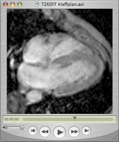
In the second semester, the students were introduced to different images and films presented within an internet-based scenario. The images and films consisted of a rotating CT image of the heart, an MRI movie of the pumping heart (), and a dynamic colour Doppler echocardiograph. The pedagogical aim was to challenge the students’ interpretation of the visualizations, trigger their formulation of learning needs and provide possibilities for them to apply their understanding after studies of the heart.
Figure 1. Image-based scenario example. Frame from an MRI movie illustrating the motion of the cardiac chambers.
In the third semester, the students attended a lecture entitled “Functional anatomy of the thoracic organs”, given by a radiologist and a clinical physiologist interacting. Using an advanced rendering workstation in the classroom, various 3D and planar images were shown, mostly from CT but also from echocardiography, and the students’ attention was drawn to clinically important anatomical relations, examples of normality, normal variation and pathology such as aortic aneurysms and their consequences.
In the third semester, the students were also offered a demonstration in the virtual reality theatre and the self-study material. At the demonstration, 4D MR images of the pumping cardiac ventricles as well as flow, both intracardiac and in the aorta, were presented stereoscopically, using novel software to a great extent developed at CMIV. The stereo display demonstrates the potential of reconstructed 3D anatomy and the display of 4D flow datasets.
Both the lecture and the demonstration aimed at explaining complex and difficult phenomena using advanced technology not possible for students to handle alone. The aim was also to stimulate the students’ curiosity and will to learn more.
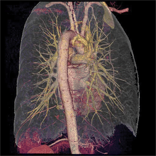
A collection of QuickTime VR images of CT datasets of the heart and great vessels, which can be interactively rotated by the students on their own computers, was introduced as self-study material in the medical programme. The anatomical structures have been stained to facilitate differentiation of structures from each other. The images produced include ():
thoracic organs in a semitransparent rib-cage, which can be rotated in any direction;
isolated heart, with the coronary arteries and their branches down to minute dimensions;
semitransparent heart muscle wall with the blood-filled chambers opaque.
Learning situations in the physiotherapy programme
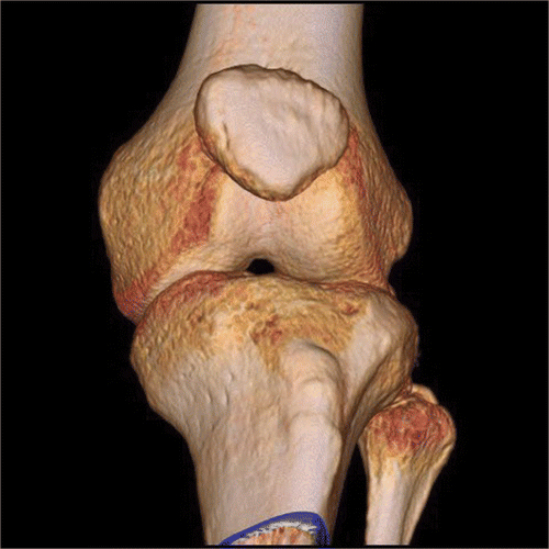
One group of students, the first semester, in the physiotherapy programme was engaged in the project. They attended a lecture entitled “motion pattern in the knee joint”. A low-end computer was used and the 3D images were demonstrated in order to visualize the complex motion pattern of the knee joint in unloaded and loaded situations. The images produced included:
lower limb CT images in 3D (QuickTime VR), which can be rotated in all directions, with semitransparent bones (femur and/or tibia alternatively), allow for thorough and detailed inspection of the complex anatomy of the joint and may help understand the relationship between the bones;
fluoroscopic (gray-scale) x-ray video sequences of the knee of a person suffering from an anterior cruciate ligament injury were produced demonstrating how the bones (tibia, femur and patella) move in relation to each other during weight bearing, knee flexion – extension, squat and both isolated and combined muscle contractions.
Evaluation of the students’ experience and attitudes
The main concern evaluating the project was to understand the possible educational value of introducing 3D visualizations in the students’ learning process related to anatomy and physiology.
Methods
Data collection
Questionnaires were used to gather information from the students about the different interventions in the project related to the basic ideas about student-centred learning. In a pilot study, autumn 2005, mainly open questions were used. The second time the interventions were carried through, spring 2006, the questionnaires were developed based on analyses from previous results. A set of questions to be answered on a 5-graded Likert scale ranging from “Agree completely” to “Don’t agree at all” and a number of open questions with room for comments was used in the medical programme. In the physiotherapy programme a set of open questions similar to the pilot study questionnaires was used. The questionnaires had the same core but were adapted to relate to each specific intervention and educational programme. The basic core of questions concerned:
students’ experience of and attitudes towards visualization;
their learning processes and their own activity connected to the use of the 3D material, which had been made available in the project;
what the students consider difficult to understand and the role of 3D images/films in the theme where the intervention was carried out;
availability related to the self-study material.
Participants
One set of inquires has been performed within the medical programme in the main study. The set comprises three questionnaires, two from the second semester students and one set from the third semester students. One questionnaire with open questions was used close to the given lecture in the physiotherapy programme. The answering rate from all questionnaires is presented in .
Table 1. The answering rate from the questionnaires used in the project
Results
Learning situations
Scenarios used in tutorial sessions
The opinion of medical students regarding images and films included in scenarios and tutorial sessions is displayed in .
Figure 4. Questionnaire answers of medical students after scenarios concerning the anatomy and physiology of the heart, (n = 62). Asterisks denote questions with a significant difference (p < 0.05) between agreeing answers (1–2) and disagreeing answers (4–5). Degree signs denote questions with borderline significance (0.05¾p < 0.10) of the difference.
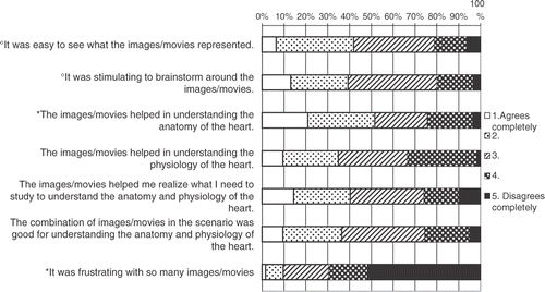
The students found it easy to see what the images represent and they thought they were stimulating for their brainstorming. They found the images of a greater help to understand the anatomy than physiology. The images/films seem to have been to some help in realizing the need for further study. Many students thought that the combination of images used in the scenario was helpful for their understanding.
The students’ comments indicate that various kinds of images (ultrasound, MRI cine loops and 3D animations) gave rise to different difficulties in the interpretation. One volume-rendered CT image clearly showing the heart and great vessels as a rotating sequence was most appreciated, and the students spent more time discussing it compared to the others. Some groups hardly used the echocardiographic cineloops with color-coded velocity at all. The students commented that they find it too difficult to understand and not worthwhile spending time on.
Lectures – Demonstrations
The medical students in the third semester attended the lecture “Functional anatomy of the thoracic organs”. Their answers on the Likert scale are shown in .
Figure 5. Questionnaire answers of medical students after the lecture “Functional anatomy of the thoracic organs” (n = 26). Asterisks denote questions with a significant difference (p < 0.05) between agreeing answers (1–2) and disagreeing answers (4–5).
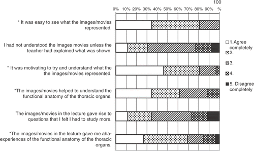
They thought it was easy to understand the images/films, but some of them believed they needed support from the teacher. The students found the lecture showing various 3D and planar images, very stimulating. They believed the images/films helped them to understand the functional anatomy of the thoracic organs. It is significant that the lecture gave them aha-experiences of the functional anatomy. In the comments, the students bring to the fore that the visualizations are especially helpful to understanding topographic anatomy. Compared to their earlier studies, the 3D dimensions gave them aha-experiences of the size and space of different thoracic organs and vessels related to the skeleton and other structures. Some students comment that they realize individual variations related to anatomic structures.
When the students were asked to compare the lecture with a dissection of ovine heart-lung specimen they performed in the second semester, they preferred the “virtual dissection” which gave a clearer picture; it was easier to discern different structures and the possibilities to turn around and look from different angles were very useful. Still, they do not want to replace the “real” dissection. The dimensions of touch and close experiences of a “real” organ are much appreciated.
The medical students were also offered the demonstration in the Virtual Reality theatre. Their opinions are displayed in .
Figure 6. Questionnaire answers of medical students after the demonstration of 4D MR images of the pumping cardiac ventricles and flow (n = 23). Asterisks denote questions with a significant difference (p < 0.05) between agreeing answers (1–2) and disagreeing answers (4–5).
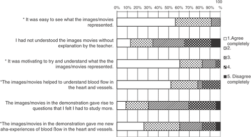
The answers are very similar compared to the lecture. Their comments show that the 4D demonstration of the blood flow and pumping cardiac ventricles was much appreciated and regarded as a help to understand the physiology of the heart and vessels. The students were thrilled and stimulated imagining the possibilities for learning about other areas of the body but also about pathological phenomena.
In the physiotherapy programme, the students were overall very positive about the use of the images and films. The fluoroscopic films helped them to understand the movements between the bones in the knee joint and how the motion pattern is guided by joint compression and muscle action, a topic that had been difficult to understand by reading books and looking at anatomical models. In their free comments about the lecture, the students wrote that “… we could confirm our knowledge on knee anatomy”, “enhanced my knowledge of knee function”, “interesting to see in reality what happens in the knee joint during movement” etc. Most of the students wanted to have more images/films like these demonstrating motions in other joints and visualization of how muscle contraction influence the motion pattern in the joints.
Self-study material
The students on the whole, both from the medical and physiotherapy programme, express great interest for the self-study material. Still, in questionnaires distributed close to the end of the circulation theme in the medical programme, many students reported that they had not used it.
The students’ answers indicate that they were interested but the main reason why they had not used it was lack of time. The students who used the material were very satisfied and found a great help in their studies. They appreciated especially that they could rotate the images and watch the organs from different angles. They could look at it many times and at their own pace. The difference from only looking at pictures in a book is that they realize the spatial extent and location of organs. The images of the knee and joints were also much appreciated by the physiotherapy students who used it.
Difficult topics
The medical students comment that a combination of different teaching modalities is important for achieving understanding. 3D images provide a good option to create new perspectives on cardiovascular anatomy and physiology. They rate a dissection of the heart and lungs very high related to their understanding of the anatomy of the heart. They comment that this gives them a 3D visualization that really contributes to their understanding of the anatomy of the heart. When the students study on their own, they mainly use books, and there seems to be little use of any kind of 3D visualizations. The elements considered most difficult to understand are the conduction system of the heart, blood pressure regulation and cardiac flow physiology. It is also difficult to understand topographic relationships: where organs are situated and relations between different organs.
Supervision and availability
General comments indicate that some students want more help to interpret the images used in the scenario. They want help from the tutor, others ask for written explanations, labels and arrows. Other students comment that the images in the scenario were used again during the second tutorial meeting and they could understand them better when they had studied more.
Concerning the lectures most of the students answer that they can follow and interpret the images with the help of the lecturer. There are students who think they need more help to understand 3D images. Some medical students thought the lecture and the demonstration had too much emphasis on technical aspects.
Only few students report that they had difficulties getting hold of the self-study material in the EDIT database and that it took a long time to load. Most of the students who had used the material used computers in the campus library, but some of them also used their own computers at home. They did not seem to find it difficult to handle the images. Some of them wanted more instructions related to what is shown in the pictures, while others thought it was a challenge and a learning experience to try to find this out on their own.
Discussion
The experience from the project concerning more easily available formats for presenting high quality 3D images point in a practicable direction. In addition to the standard stacks of slices (original or multiplanar reformatted), we have included volume rendering images in the QuickTime VR format that enables students to interact much more intuitively by turning the anatomical objects around two axes with the mouse rather than sliding through them or starting and stopping a rotating sequence. Images in this interactive format can easily be produced with modern medical imaging equipment. This means that images of high quality can become available for the students, within reasonable costs, covering all parts of the body. They can also easily be made available for all medical and health care educational programmes. The technical difficulties in producing the images were not greater than expected.
With access to advanced medical imaging workstations, it also seems easy to introduce lectures and demonstrations using modern rendering techniques in the curriculum with promising results. This, however, presupposes that diagnostic clinicians with a high level of competence in this area are involved in the teaching.
Why bother to produce new images and films and not use material available in the market? We would argue that material produced in collaboration with the needs expressed by teachers and students carry a special potential for successful implementation. The images and films can also be related to the actual context, the content and educational level. The 3D visualizations used in the project are developed from authentic material and not specially adjusted to “average normal”. That might help the students to understand that anatomic structures inherit individual biological differences. Some students have noticed and commented on that and a significant factor that drives learning is the experiences of variation and differences (Marton & Booth Citation1997).
An important aim was to ensure that visual interactive technology becomes an integral and accessible part of the regular curriculum. The idea of implementing the images directly integrated in the existing curricula turned out to be successful. All interventions implied through the project have now become a regular part of the courses that were engaged. The produced images for self-study of thoracic and knee anatomy will be available for future medical and physiotherapy students as well as for the other students at the faculty (nurses, occupational therapists etc.). The lecture and demonstration for medical students is now part of the regular curriculum.
One explanation of the successful integration might be the involvement of teachers in both the planning and implementation phase of the project. Also, the common base of educational philosophy, PBL, provides learning situations that are already well known by students and teachers and are suitable for introducing 3D images and films. The use of the already existing EDIT internet database for presentation and storing of self-study material seems to be a good alternative for the students to access and use the images and films.
However, the need for work to introduce the newly produced media to be actually used by the students should not be underestimated. The least successful part of the project was the fact that the students did not use the self-study material to the extent that was possible. The reason given by the students was lack of time. We believe the appropriate point of time in the course to introduce the material had not been found. The self-study material was probably introduced too late and a new theme insisting on attention started immediately after the circulation theme.
The impact of the 3D images and films related to the students’ understanding of anatomy and physiology could not be thoroughly judged from the evaluation. Several issues need to be further investigated. Still there are some indications worth taking into account. The images and films implemented in the different learning situations all add certain values related to their understanding compared to pictures in books, models and ordinary dissections. Images and films in the scenarios are not appreciated as much as the lectures and the self-study material. Students discuss the interactive 3D images of anatomy in the groups whereas the films of echocardiographic cineloops with colour-coded velocity are almost neglected. One explanation might be the early stage of their education and it may be more useful to introduce that kind of visualizations at a later stage in the curriculum. Another explanation might be that tutors are not being prepared to support the students.
Compared to their earlier studies, the 3D aspect presented in the lectures and the self-study material gave the students new insights about size and space of different organs and the relation to other functional structures. From a learning perspective, this excitement and experiences of differences are important motivational factors stimulating the students’ will to explore their own understanding. The use of 3D images and films seems to contribute to the students’ understanding of especially anatomy. Although the medical students rated ordinary dissections high, the virtual dissection added educational value by giving a clearer picture. The possibility to turn structures around was also pointed out as instructive. Considering the difficulties of offering enough ordinary dissections in education, the rendering workstation used in the classroom presents a great opportunity to improve the learning. On the other hand, a low-end computer, e.g. the student's own, can be used to visualize complex phenomena like motion pattern of the knee joint in unloaded and loaded situations.
The students report that they were stimulated and motivated to understand and study when they took part in the visualizations in scenarios, lectures and the self-study material. This is a very important sign related to student-centred learning. If the images stimulate the curiosity and the will to learn more that is very important for the students taking responsibility for their own learning (Silén Citation2003).
Along with some encouraging results the evaluation of the project points at several issues that need to be further explored. One important further step, which we are currently working on, is to carry out studies that attempt to measure whether better learning results are indeed achieved with the new technique. Deeper knowledge is also required about how the students interpret and reason about the images/films and if they enhance their understanding. 3D visualizations based on authentic material might add special values in the students’ learning process. This issue needs to be further investigated related to learning outcomes. Further development of advanced 3D visualizations should also be built on deeper knowledge about what students find difficult to understand. Other urgent questions relate to preparation and facilitation needed for different formats and meaningful learning like the need for more information and training related to the use of the self-study material.
In conclusion, visualizations with varying degrees of interactivity, produced by modern medical imaging equipment, are a promising resource in student-centred medical education. An optimal implementation is achieved by integrating the new modalities in the curricula, understanding that visualizations for self-studies, tutorial groups or lectures pose different demands on the material, and by involving and educating teachers and tutors. A continuing evaluation by feedback from students and teachers is also essential for the efficient use of modern imaging technology in medical education.
Additional information
Notes on contributors
Charlotte Silén
CHARLOTTE SILÉN, RN, PhD, Assistant Professor in Higher Education and Head of the Division for Learning and Teaching Research in Medicine and Care. She is senior lecturer in the nursing program. Her research field is student centred learning in higher education, especially PBL, self directed learning, the tutors role, assessment and e-learning.
Staffan Wirell
STAFFAN WIRELL, MD, PhD, Assistant Professor in Medical Radiology and senior lecturer in the medical program. He is a member of the undergraduate medical program committee and deeply involved in educational development concerning the medical program. He is also active in pedagogical courses for faculty.
Joanna Kvist
JOANNA KVIST, RPT, PhD, Associated Professor in Physical Therapy and senior lecturer in the physical therapy program. Her research area of interest includes rehabilitation in sports medicine with specific focus in knee injuries.
Eva Nylander
EVA NYLANDER, MD, PhD, Professor in Clinical Physiology. She is responsible for the cardiovascular field in the medical program.
Örjan Smedby
ÖRJAN SMEDBY, MD, PhD, Professor of Medical Radiology and involved in the medical program. He was one of the founders of the Centre for Medical Image Science and Visualization, CMIV. He has been the project director for the 3D visualisation project that is described and analyzed in this article.
References
- Calhoun PS, Kuszyk BS. Three-dimensional volume rendering of spiral CT data: theory and method. Radiographics 1999; 19(3)745–64
- Cottam WW. Adequacy of medical school gross anatomy education as perceived by certain postgraduate residency programmes and anatomy course directors. Clin Anat 1999; 12(1)55–65
- Dev P, Montgomery K, Senger S, Leroy Heinrichs W, Srivastava S, Waldron K. Simulated medical learning environments on the Internet. J Am Med Informatics Assoc 2002; 9(5)437–447
- Drebin RA, Carpenter L. Volume rendering. Comput Graph 1988; 22(4)65–74
- Friedl R, Preisack MB, Klas W, Rose T, Stracke S, Quast KJ. Virtual reality and 3D visualizations in heart surgery education. Heart Surg Forum 2002; 5(3)17–21
- Fyrenius A, Bergdahl B, Silén C. Lectures in problem-based learning-why, when and how? An example of interactive lecturing that stimulates meaningful learning. Med Teach 2005; 27: 61–65
- Fyrenius A, Wirell S, Silén C. Student approaches to achieving understanding Approaches to learning revisited. Stud Higher Educ 2007; 32(2)149–165
- Fyrenius A, Wigström L, Ebbers T, Karlsson M, Engvall J, Bolger AF. Three-dimensional flow in the human left atrium. Heart 2001; 86(4)448–55
- Garg AX, Norman G, Sperotable L. How medical students learn spatial anatomy. Lancet 2001; 3; 357(9253)363–4
- Hammar M, Bergdahl B, Öhman L, (Eds). Celebrating the Past by Expanding the Future, The Faculty of Health Sciences, Linköpings Universitet 1986–2006. Medical Faculty Report 3. Linköping University Electronic press, LinköpingSweden 2006
- Lockyer L, Patterson J, Harper B. ICT in higher education: evaluating outcomes for health education. J Comp Ass Learning 2001; 17(3)275–83
- Marton F, Booth S. Learning and Awareness. Lawrence Erlbaum Associates, Publishers, Mahwah, NJ 1997
- Maudsley G. Do we all mean the same thing by “problem-based learning”? A review of the concepts and a formulation of the ground rules. Acad Med 1999; 74(2)178–185
- McLachlan JC, Bligh J, Bradley P, Searle J. Teaching anatomy without cadavers. Med Educ 2004; 38: 418–24
- Michael J. Students’ misconceptions about perceived physiological responses. Advan Physiol Educ 1998; 19(1)90–98
- Michael J. Undergraduates’ understanding of cardiovascular phenomena. Advan Physiol Educ 2002; 26(2)72–84
- Miller R. Approaches to learning spatial relationships in gross anatomy: perspective from wider principles of learning. Clin Anat 2000; 13: 439–43
- Modell HI, Michael JA, Adamson T, Goldberg J, Horwitz BA, Bruce DS, Hudson ML, Whitescarver SA, Williams S. Helping undergraduates repair faulty mental models in the student laboratory. Advan Physiol Educ 2000; 23: 82–90
- Modell HI, Michael JA, Adamson T, Horwitz BA. Enhancing active learning in the student laboratory. Advan Physiol Educ 2004; 28: 107–111
- Nieder GL, Scott JN, Anderson MD. Using QuickTime virtual reality objects in computer-assisted instruction of gross anatomy: Yorick–the VR Skull. Clin Anat 2000; 13(4)287–93
- Novak JD. The promise of new ideas and new technology for improving teaching and learning. Cell Biol Educ 2003; 2: 122–132
- Richardson D, Speck D. Addressing students’ misconceptions of renal clearance. Advan Physiol Educ 2004; 28(4)210–212
- Rosse C. The potential of computerized representations of anatomy in the training of health care providers. Acad Med 1995; 70: 499–505
- Savin–Baden M. Problem-based Learning in Higher Education: Untold Stories. The Society for Research into Higher Education & Open University Press, Buckingham 2000
- Silén C. Responsibility and independence in learning – what is the role of the educators and the framework of the educational programme?. Improving Student Learning: Improving Student Learning – Theory, Research and Practice, C Rust. The Oxford Centre for Staff and Learning Development, Oxford 2003
- Silén C. Does Problem-based Learning make students go meta?. Challenging research in Problem-based Learning, M Savin-Baden, K Wilkie. SRHE & Open University Press, Maidenhead 2004
- Spiro RJ, Feltovich PJ, Coulson RL, Anderson DK. Multiple analogies for complex concepts: antidotes for analogy induced misconception in advanced knowledge acquisition. Similarity and Analogical Reasoning, S Vosniadou, A Ortony. Cambridge University Press, Cambridge 1989; 498–531
- Trelease RB, Nieder GL, Dorup J, Hansen MS. Going virtual with Quicktime VR: new methods and standardized tools for interactive dynamic visualization of anatomical structures. Anat Rec 2000; 261(2)64–77
- Wigström L, Ebbers T, Fyrenius A, Karlsson M, Engvall J, Wranne B, Bolger AF. Particle trace visualization of intracardiac flow using time-resolved 3D phase contrast MRI. Mag Res Med 1999; 41: 793–799
- Zirkel JB, Zirkel PA. Technological alternatives to actual dissection in anatomy instruction: a review of the research. Educ Technol 1997; 37(6)52–56

