
Dear Sir,
Here we describe a 41-year-old female who was referred in 2004 with a fibroid uterus initially noted during pregnancy. On examination she had an 18-week sized uterus, which subsequent radiological investigation confirmed as two uterine leiomyomas. The masses continued to enlarge and were unresponsive to hormonal therapy. Embolisation was unsuitable, and so a hysterectomy was performed, which revealed a single large mass arising from the left round ligament.
Macroscopically, received separately from the main hysterectomy specimen, was a firm, well-circumscribed mass measuring 80 × 60 × 40 mm, with a uniform, white/yellow, whorled cut surface, typical of a leiomyoma, labelled mass from round ligament. The uterus was otherwise unremarkable.
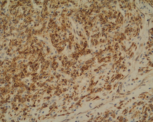
Histologically, the tumour was composed of haphazardly arranged round to spindle-shaped cells with vesicular nuclei and little cytoplasm, forming a well-circumscribed, encapsulated mass. There were alternating hypercellular and hypocellular areas, with intervening small, branching vascular channels (). Areas of hyalinisation and myxoid change were also noted, as well as occasional interstitial mast cells and lymphocytes. There was no cytological atypia, necrosis or significant mitotic activity. Immunohistochemistry showed strong expression of CD34, CD99, bcl-2 () and MIC2; desmin and S100 were negative. The vascular channels showed positive expression of SMA and caldesmon, but tumour cells were negative.
Figure 1. Solitary fibrous tumour H&E.
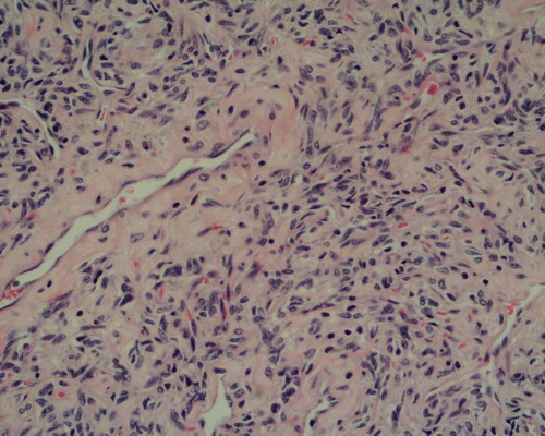
Figure 2. Immunohistochemistry for bcl 2.
Solitary fibrous tumour is a rare tumour of the female genital tract typically found in the lung, and also reported in many sites including the peritoneum, mediastinum and retroperitoneum (Fletcher Citation2000). As this case demonstrates, these tumours can clinically and radiologically, with increased high definition scanning of the female genital tract, mimic a leiomyoma. In an era of increasing sub-specialisation, it is important to consider tumours not typical for a given site.
Extrapleural solitary fibrous tumours are uncommon mesenchymal tumours of (myo)fibroblastic type, which may be found at any location (Guillou et al. Citation2002). Many extrapleural manifestations have been described, including the uterus and fallopian tube (Berzal-Cantalejo et al. Citation2005; Wakami et al. Citation2005). No literature however, has documented involvement of the round ligament.
References
- Berzal-Cantalejo F, Montesinos-Carbonell M, Montesinos-Carbonell M L, et al. Solitary fibrous tumour arising in the fallopian tube. Gynecologic Oncology 2005; 96: 880–882
- Fletcher C DM. Soft tissue tumours. Diagnostic histopathology of tumors2nd ed., C DM Fletcher. Churchill Livingstone, China 2000; 2: 1496–1497
- Guillou L, Fletcher J A, Fletcher C DM, . Fibroblastic/myofibroblastic tumours Extrapleural solitary fibrous tumour and haemangiopericytoma. World Health Organisation classification of tumours, pathology and genetics: tumours of soft tissue and bone, C DM Fletcher, K Krishnan Uwi, F Musters, et al. IARC Press, London 2002; 86–90
- Wakami K, Tateyama H, Kawashima H, et al. Solitary fibrous tumour of the uterus producing high-molecular-weight insulin-like growth factor II and associated with hypoglycaemia. International Journal of Gynecological Pathology 2005; 24: 79–84