

Abstract
Taxanes are important chemotherapeutic agents used to manage breast cancer and gynaecological malignancies. However, ovarian toxicity induced by the taxane docetaxel (DOC) is of great concern. We investigated DOC-induced toxicity in the ovaries of female CD1 strain mice. The mice were divided into control (saline), DOC-5 (5 mg/kg DOC), and DOC-10 (10 mg/kg DOC) groups and administered saline or DOC on the first day of the study and two weeks later. Two weeks after the second dose, the ovaries were removed for analysis after inducing superovulation. Ovary weight, the number of secondary follicles, and the total number of follicles were reduced after DOC administration. Additionally, the expression levels of caspase-3 and the pro-apoptotic protein Bcl-2 interacting mediator of cell death (BIM) increased. Our findings suggest that high-dose DOC induces damage to growing follicles; however, it may not affect primordial follicles.
What is already known on this subject? Docetaxel (DOC) is one of the most effective chemotherapeutic agents used to manage various cancers. Some in-vitro studies have examined paclitaxel-induced ovarian toxicity; however, limited research on DOC is available.
What do the results of this study add? We investigated DOC-induced ovarian toxicity in female CD1 strain mice at 5 mg/kg and 10 mg/kg. We found that DOC reduced ovary weight, the number of secondary follicles, and the total number of follicles, with the higher dose having a higher effect.
What are the implications of these findings for clinical practice and/or further research? We believe that our study makes a significant contribution to the knowledge about the effect of DOC on ovarian function.
Impact statement
Introduction
Recently, breast cancer prevalence among young women has been increasing. The lifetime incidence rate of breast cancer development among females was found to be 12.3% (approximately 1 in 8 cases) between 2010 and 2012. Breast cancer is the most common malignancy among women worldwide (Rojas and Stuckey Citation2016, Ghoncheh et al. Citation2016). The incidence rate of breast cancer ascended gradually by 0.3% per year from 2012 to 2016. This was largely due to the increase in early stage and hormone receptor-positive breast cancers. Contrastingly, the death rate due to breast cancer is declining. Specifically, a 40% decrease in the rate was observed from 1989 to 2017 (DeSantis et al. Citation2019). Ovarian damage due to chemotherapy for breast cancer degrades the quality of life of women and can lead to infertility and premature menopause (Koyama et al. Citation1977, Meirow et al. Citation2007, Nicosia et al. Citation1985, Reichman and Green Citation1994).
The risk of chemotherapy-induced amenorrhoea (CIA) differs and is dependent on patient age as well as the dose and type of chemotherapeutic agent used (Gadducci et al. Citation2007). Cyclophosphamide (CTX) is a material agent used for breast cancer therapy; however, it can result in CIA. It has been indicated that 68% of patients experience amenorrhoea after undergoing combination treatment with CTX, methotrexate, and 5-fluorouracil (Bines et al. Citation1996). In the past, standard chemotherapy for breast cancer was based on anthracycline and CTX. Anthracycline-based chemotherapy has a high efficacy. Additionally, it may protect ovarian function compared to other treatments based on the use of alkylating agents (Gadducci et al. Citation2007, Bines et al. Citation1996, Pérez-Fidalgo et al. Citation2010). Nevertheless, 75.5% women still experience amenorrhoea after therapy with anthracyclines and CTX (Pérez-Fidalgo et al. Citation2010). There are a few reports on the effects of taxane-based chemotherapy on ovarian function in young women (Fornier et al. Citation2005, Tham et al. Citation2007).
Docetaxel (DOC) is a second-generation taxane and one of the most effective chemotherapeutic agents used to manage early to advanced-stage breast cancer, non-small cell lung cancer, and gynaecological malignancies. It also enhances the antitumor efficacy of radiotherapy (Gligorov and Lotz Citation2004). Fornier et al. (Citation2005) have reported that the risk of CIA is minimal in taxane-based chemotherapy. Conversely, Tham et al. (Citation2007) showed that premenopausal women receiving chemotherapy with anthracyclines (doxorubicin and CTX) followed by a taxane frequently experienced amenorrhoea compared with the women receiving chemotherapy without a taxane. The incidence of amenorrhoea among such patients was 55% after treatment with anthracycline alone and 64% after treatment with anthracycline and additional taxane (Tham et al. Citation2007). Thus, it is unclear why ovarian toxicity is induced by taxane-based chemotherapy and whether the toxicity is dose-dependent. This is partly because studies have not been performed to assess these issues in experimental animals. The clinical outcomes obtained with preoperative DOC chemotherapy for breast cancer are satisfactory. This is because the treatment induces a few severe side effects, although it has the potential to induce ovarian toxicity (Gligorov and Lotz Citation2004). DOC is a beneficial drug for the management of both breast cancer and gynaecological malignancies. Neoadjuvant chemotherapy with DOC is also an important treatment strategy. DOC-induced CIA is the most important side effect to be considered in young patients. Thus, the present study aimed to investigate DOC-induced ovarian toxicity in female mice.
Materials and methods
Animals
Female CD1 strain mice, aged 8 weeks, were obtained from Charles River Laboratories Japan, Inc. (Yokohama, Japan) and housed at the animal laboratory of Tokushima University (Tokushima, Japan). The animals were kept under a 12/12 h light/dark cycle (light turned on at 8:00 am and turned off at 8:00 pm) at a controlled temperature of 24 °C. They had free access to sterilised food and enough fresh water. All experimental and animal care procedures were approved by the animal experiment ethic committee of Tokushima University (registration code: T29-33, Kanako Yoshida registered) and carried out according to the National Animal Experiment Guidelines.
A total of 25 mice were used in this study. Sixteen mice were divided into the following three groups to evaluate the effects of DOC on oocytes and follicles: control, DOC-5 (5 mg/kg DOC), and DOC-10 (10 mg/kg DOC). The remaining nine mice were similarly divided into three groups for the evaluation of apoptosis in their ovaries. DOC (FUJIFILM Wako Pure Chemical Co., Osaka, Japan) was dissolved in phosphate-buffered saline (PBS) and injected intraperitoneally. The control group was administered PBS alone. The mice were injected with the same dose of DOC or PBS 14 days after the first dose administration. Twelve days after the second dose administration, the animals were intraperitoneally injected with 10 IU of pregnant mare serum gonadotropin (Funakoshi Co., Ltd., Tokyo, Japan), followed by 10 IU of human chorionic gonadotropin (hCG; Sigma Aldrich Japan, Inc., Tokyo, Japan) 48 h after the induction of superovulation. The ovaries and oviducts were removed 15 h after hCG administration for analysis.
Oocytes counting
Oviduct and cumulus-oocyte samples were placed in G-MOPS media and then in HEPES-buffered medium, containing 80 IUmL−1 hyaluronidase, for denuding of cumulus-oocyte complexes. Metaphase II oocytes and germinal vesicles were then counted under a stereomicroscope.
Follicle classification and quantification
The removed ovaries were fixed in 10% formalin for 24 h, embedded in paraffin, and cut into serial sections (5 μm thick). The sections were laid in order on microscope slides, and every tenth section was stained with haematoxylin and eosin. Follicles were counted under an optical microscope and classified as previously described (Morgan et al. Citation2013). Specifically, follicles were classified as primordial when the oocyte was surrounded by flat-shaped pre-granulosa cells, primary when the oocyte was surrounded by one layer of cuboidal granulosa cell, and secondary when the granulosa cells form multiple layers. Tertiary follicles were defined as follicles with fluid-filled cavities. Follicular health was evaluated based on standard morphological criteria. For instance, follicles were considered unhealthy when they had any of the following features: an oocyte with concentrated or non-uniform eosin staining, shrunken cytoplasm, numerous pyknotic granulosa cells, or no attachment between oocyte and granulosa cells. Only healthy primordial, primary, secondary, and tertiary follicles were counted.
Polyacrylamide gel electrophoresis and Western blotting
The possibility of DOC activating the intracellular apoptotic pathway in the ovaries of mice was investigated in this study. The test was performed by measuring the expression of the apoptosis detection marker cleaved caspase-3 via western blotting. It has been shown previously that the B-cell lymphoma (BCL)-2 family activates the apoptotic pathway by promoting the permeability of the outer mitochondrial membrane when cells are exposed to paclitaxel (Youle and Strasser Citation2008, Chipuk et al. Citation2010). Members of the BCL-2 family are divided into the following three groups based on BCL-2 domain homology and action mechanism: anti-apoptotic BCL-2 family proteins (BCL-2, myeloid cell leukaemia 1, BCL-extra-large), BCL-2 family effector proteins (BCL-2-associated X protein [BAX], BCL-2 antagonist killer [BAK]), and BH3-only proteins (direct activators: BCL-2-interacting mediator of cell death [BIM] and BCL-2-interacting domain death agonist [BID], “sensitizer” or “derepressor”: BCL-2 antagonist of cell death [BAD], PUMA and Noxa) (Chipuk et al. Citation2010). To investigate the apoptotic pathway induced by DOC, western blotting was carried out to detect BCL-2 and BIM levels. The removed ovaries were washed with cold PBS and snap-frozen for protein extraction. T-PERTM Tissue Protein Extraction Reagent (Thermo Fisher Scientific, Tokyo, Japan), containing HaltTM Protease Inhibitor Cocktail and HaltTM Phosphatase Inhibitor Cocktail (Thermo Fisher Scientific), was added to two ovaries. The ovaries and reagents were homogenised and then centrifuged. Protein concentration was measured using PierceTM BCA Protein Assay Kit (Thermo Fisher Scientific). The same amounts of protein samples were separated on 12% Mini-PROTEAN TGX Gels (Bio-Rad Laboratories, Inc., Tokyo, Japan) and transferred onto a nitrocellulose membrane. After blocking with 3% bovine serum albumin blocking buffer for 2 h, the membrane was probed overnight at 4 °C with cleaved caspase-3 rabbit monoclonal antibody (1:1000; Cell Signalling Technology, Inc., Beverly, MA, USA), anti-Bcl-2 rabbit monoclonal antibody (1:2000; Abcam, Tokyo, Japan), or anti-Bim rabbit monoclonal antibody (1:500, Abcam) and β-actin rabbit polyclonal antibody (1:1000). After washing in Tris-buffered saline with Tween 20 (Takara Bio Inc., Shiga, Japan), the membrane was incubated with goat anti-rabbit IgG antibody (1:1000, Cell Signalling Technology, Inc.) for 1 h at 24 °C. Fluorescence was detected using an ECL Plus system (GE Healthcare, Amersham, UK). The membrane was reprobed after incubation in 15 mL stripping buffer (Takara Bio Inc.) for 1 h at 24 °C. Band intensities were quantified using the ImageJ software (version 1.53k; National Institutes of Health, Bethesda, MA, USA).
Statistical analysis
All data were analysed using SPSS Statistics (version 20.0; IBM, Armonk, NY, USA). A one-way analysis of variance, followed by Bonferroni post hoc tests, was used to analyse normally distributed data to investigate differences between the control and treatment groups. Kruskal–Wallis test, followed by Dunn’s post hoc tests, was used to analyse whether the data were not normally distributed, whereas paired t-test was used. All results are presented as mean ± standard error of the mean (SEM). P < 0.05 was considered to represent statistical significance.
Results
Effects of DOC on the weight of ovaries and number of oocytes
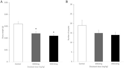
The weights of ovaries in the DOC-5 and DOC-10 groups were significantly lower than those in the control group (P = 0.024 and P = 0.022, respectively) (). However, the number of oocytes was not different among the three groups ).
Figure 1. DOC affects ovary weight but not the number of oocytes after superovulation is induced in mice. (A) Ovary weight. (B) Number of oocytes after superovulation was induced in the control (PBS, n = 12), DOC-5 (5 mg/kg DOC, n = 10), and DOC-10 (10 mg/kg DOC, n = 10) groups. Data are expressed as mean ± SEM. * indicates P < 0.05 when data for the DOC-treated groups are compared to those for the control group. PBS: phosphate-buffered saline; SEM: standard error of the mean.
Effects of DOC on the number of follicles
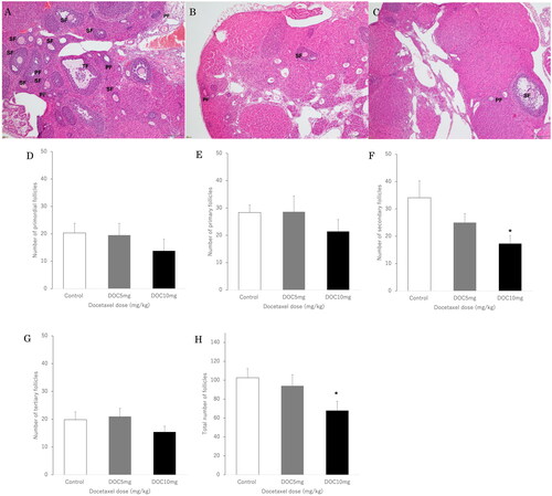
Histological evaluation of the ovaries in the control group showed primary, secondary, and atretic follicles with normal morphology (). However, the number of follicles was significantly reduced in the DOC-5 and DOC-10 groups ().
The number of secondary follicles in the DOC-10 group was significantly lower than that in the control group (P = 0.045). However, the number of secondary follicles in the DOC-5 group was not different from that in the control group (). Similarly, the total number of follicles in the DOC-10 group was significantly lower than that in the control group (P = 0.025); however, there was no difference in total number of follicles between the DOC-5 and control groups (). The numbers of primordial, primary, and tertiary follicles did not differ among the three groups ()).
Figure 2. Hematoxylin and eosin staining of ovaries (×40 magnification). (A) Control group of ovaries showing normal morphology. (B) DOC-5 (5 mg/kg DOC) and (C) DOC-10 (10 mg/kg DOC) groups demonstrating reduced follicular numbers at each stage. PF; primary follicle, SF; secondary follicle, TF; tertiary follicle. DOC affects the number of secondary follicles and the total number of follicles but not primordial, primary, or tertiary follicles. Number of (D) primordial, (E) primary, (F) secondary, and (G) tertiary follicles. (H) Total number of follicles in the control (n = 12), DOC-5 (5 mg/kg DOC, n = 10), and DOC-10 (10 mg/kg DOC, n = 10) groups. The data show significant decreases in the number of secondary follicles and the total number of follicles in the DOC-10 group. Data are expressed as mean ± SEM. * indicates P < 0.05 when the data for the DOC-treated groups are compared to those for the control group.
DOC-induced apoptosis is BIM-dependent in the mouse ovary
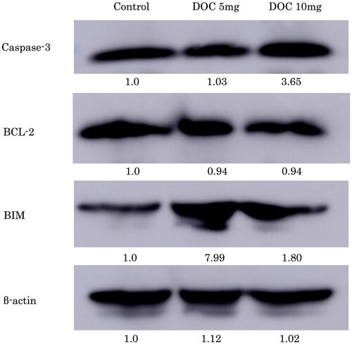
Cleaved caspase-3 expression in the DOC-10 group was 3.65-fold higher than that in the control group (). Additionally, BIM expression in the DOC-5 group was 7.99-fold higher than that in the control group, whereas BCL-2 expression was not different among the DOC-5, DOC-10, and control groups.
Figure 3. DOC increases caspase-3 and BIM expression but not BCL-2 expression in mice. Representative images showing western blotting of whole ovary proteins for caspase-3, BCL-2, and BIM expression in the control, DOC-5 (5 mg/kg DOC) and DOC-10 (10 mg/kg DOC) groups. Six ovaries were analysed for each treatment group. The data presented were normalised against those for the PBS-treated group.
Discussion
Taxanes affect the cytoskeleton; they bind to the beta subunit of tubulin, thereby causing cell cycle arrest at the G2-M phase. However, evidence of the potential gonadotoxicity of taxanes may limit their use. Some clinical studies have shown that treatment with taxanes does not result in an additional increase in amenorrhoea rate, although it may cause a slight increase in the incidence of reversible amenorrhoea (Pérez-Fidalgo et al. Citation2010, Davis et al. Citation2005, Berliere et al. Citation2008, Reh et al. Citation2008, Abusief et al. Citation2010). Nevertheless, the findings from several follow-up studies indicate that the probability of CIA associated with taxane-based chemotherapy is higher than that associated with anthracycline-based chemotherapy (Fornier et al. Citation2005, Han et al. Citation2009, Hortu et al. Citation2020). In a previous study on rats, treatment with paclitaxel reduced the number of primordial follicles (Yucebilgin et al. Citation2004). This is similar to what occurs in humans (Kai et al. Citation1994), which indicates that paclitaxel is gonadotoxic. It was also found in another study that rats treated with high-dose paclitaxel exhibit a decrease in the number of foetuses and implantations due to the inhibition of ovulation. However, these adverse effects were not seen in a later follow-up period during the study, suggesting that the effects of paclitaxel on the ovaries may be reversible (Tarumi et al. Citation2009).
In the present study, the outcomes of the in vivo experiment showed that DOC treatment resulted in reduced ovary weight, indicating that it can impair ovarian function. This alteration in ovary weight was more evident in mice that were administered the higher DOC dose. We also found that DOC directly affected the number of secondary follicles but did not affect the number of stored primordial follicles. These effects might be due to either a decrease in the number of follicles reaching the secondary stage or an increase in the number of follicles that die or burn out as soon as they reach maturity. It was shown in a previous in vitro study that granulosa cells of developing follicles are exposed to be injured firstly. This indicates that DOC does not directly damage oocytes but activates the apoptotic pathway by promoting the permeability of the outer mitochondrial membrane, and consequently somatic cells are damaged (Lopes et al. Citation2014). Our results showed that due to the initial damage to the granulosa cells, the number of secondary follicles decreased.
The little available information on the effects of DOC on fertility in humans is mostly in relation to the periods of temporary or permanent amenorrhoea occurrence, which do not accurately indicate ovarian toxicity (Berliere et al. Citation2008, Zhou et al. Citation2010). Furthermore, there are conflicting reports on the impact of DOC on the severity of amenorrhoea after therapy; some results have indicated a considerable impact (Han et al. Citation2009, Martin et al. Citation2005), whereas others have indicated otherwise (Zhou et al. Citation2010, Minisini et al. Citation2009). However, a more detailed analysis has revealed that combination therapy with a taxane and other chemotherapeutic agents does worsen ovarian toxicity (Anderson et al. Citation2006). To our best knowledge, there is insufficient information on the toxic effects of DOC on fertility in animals. In vivo studies have been carried out to reveal the effects of paclitaxel on ovarian function in adult mice; however, the results are highly incompatible. It was found in a previous study that high-dose paclitaxel (injection, 7.5 mg/kg), whether administered as a single dose or as multiple doses, can cause a reduction in primordial follicle count one week after treatment (Yucebilgin et al. Citation2004, Gücer et al. Citation2001). In contrast, multiple doses of paclitaxel (5 mg/kg) in adult rats do not result in a reduction in follicle count at any pre-antral growth stage, although there could be some effects on antral follicles (Tarumi et al. Citation2009).
Patients who require chemotherapy generally undergo plural doses over several months. In the human ovary, 200 days or more are required to transition from primordial follicles to antral follicles (McGee and Hsueh Citation2000); therefore, long-term treatment can damage follicles at various stages of development. The main effect on ovarian toxicity is the depletion of dormant primordial follicles or not, as this alteration can lead to premature ovarian insufficiency (POI). Depletion of primordial follicles can occur due to indirect effects as well as direct damage to primordial follicles. The loss of developing follicles can lead to the recruitment and growth of dormant primordial follicles, resulting in "burnout of follicle reserves" (Roness et al. Citation2013). Mature follicles are more likely to be damaged by chemotherapy than immature follicles are (Gücer et al. Citation2001, Nicosia et al. Citation1985b); however, specific damage to late antral follicles could have a minor effect on long-period reproduction function. The outcomes of the present study show that mature follicles can be severely affected by DOC, even at low doses. Additionally, the reduction in primordial follicle number was not significant; however, the high-dose DOC tended to have a negative effect. Oxytocin is reported to reduce methotrexate-induced ovarian toxicity in rats (Hortu et al. Citation2020); thus, it may be considered as a potential drug candidate for reducing chemotherapy-induced ovarian toxicity.
The observed ovarian toxicity caused by DOC administration was supposedly due to the main action mechanism of taxanes. Taxanes bind to the beta subunit of tubulin, providing stabilisation of tubulin polymerisation, resulting in cell cycle arrest and mitosis suppression (Gligorov and Lotz Citation2004). Therefore, cells with active mitosis such as granulosa cells are more sensitive to DOC than oocytes are. Moreover, even if the oocytes are intact, if the somatic cells are damaged by an extrinsic stress, oocyte-granulosa cell connection is lost, resulting in damage to the oocytes (Thomson et al. Citation2010). The outcomes of the present study show that DOC administration promotes the mitochondrial apoptotic pathway of granulosa cells, thereby promoting expression of the apoptosis detection marker cleaved caspase-3. It has been previously shown that the BCL-2 family activates the apoptotic pathway when cells are exposed to paclitaxel (Youle and Strasser Citation2008, Chipuk et al. Citation2010). BH3-only proteins operate upstream of BAX and BAK because they cannot promote the apoptosis of cells in which both BAX and BAK are deleted. Furthermore, cytochrome c release via BAX and/or BAK activation is promoted by BH3-only proteins but inhibited by the anti-apoptotic BCL-2 family of proteins (Youle and Strasser Citation2008, Chipuk et al. Citation2010). BIM, a BH3-only protein, acts as a trigger for paclitaxel-induced apoptosis. Downregulation of BIM expression by gene silencing experiments results in inhibited paclitaxel-induced cytotoxicity in cell-based models (Sunters et al. Citation2003, Li et al. Citation2005, Janssen et al. Citation2007, Hortu et al. Citation2020), Kutuk and Letai Citation2010). Additionally, previous studies have revealed that paclitaxel causes BIM accumulation and BIM-dependent apoptosis (Tan et al. Citation2005). In the present study, we found that DOC-induced apoptosis is BIM-dependent in the mouse ovary.
In conclusion, the findings of our in vivo study indicate a certain evidence of DOC-induced ovarian damage. Activation of resting primordial follicles following depletion of secondary follicles can affect the number of pooling primordial follicles and lead to POI.
Acknowledgement
The authors would like to express our deep gratitude to all those who assisted us in conducting this research.
Disclosure statement
No potential conflict of interest was reported by the authors.
References
- Abusief, M.E., et al. 2010. The effects of paclitaxel, dose density, and trastuzumab on treatment-related amenorrhea in premenopausal women with breast cancer. Cancer, 116 (4), 791–798.
- Anderson, R.A., et al. 2006. The effects of chemotherapy and long-term gonadotrophin suppression on the ovarian reserve in premenopausal women with breast cancer. Human Reproduction (Oxford, England), 21 (10), 2583–2592.
- Berliere, M., et al. 2008. Incidence of reversible amenorrhea in women with breast cancer undergoing adjuvant anthracycline-based chemotherapy with or without docetaxel. BMC Cancer, 8, 56.
- Bines, J., Oleske, D.M. and Cobleigh, M.A., 1996. Ovarian function in premenopausal women treated with adjuvant chemotherapy for breast cancer. Journal of Clinical Oncology : official Journal of the American Society of Clinical Oncology, 14 (5), 1718–1729.
- Chipuk, J.E., et al. 2010. The BCL-2 family reunion. Molecular Cell, 37 (3), 299–310.
- Davis, A.L., Klitus, M. and Mintzer, D.M., 2005. Chemotherapy-induced amenorrhea from adjuvant breast cancer treatment: the effect of the addition of taxanes. Clinical Breast Cancer, 6 (5), 421–424.
- DeSantis, C.E., et al. 2019. Breast cancer statistics, 2019. CA: A Cancer Journal for Clinicians, 69 (6), 438–451.
- Fornier, M.N., et al. 2005. Incidence of chemotherapy-induced, long-term amenorrhea in patients with breast carcinoma age 40 years and younger after adjuvant anthracycline and taxane. Cancer, 104 (8), 1575–1579.
- Gadducci, A., Cosio, S. and Genazzani, A.R., 2007. Ovarian function and childbearing issues in breast cancer survivors. Gynecological Endocrinology : The Official Journal of the International Society of Gynecological Endocrinology, 23 (11), 625–631.
- Ghoncheh, M., Pournamdar, Z. and Salehiniya, H., 2016. Incidence and mortality and epidemiology of breast cancer in the world. Asian Pacific Journal of Cancer Prevention : APJCP, 17 (S3), 43–46.
- Gligorov, J. and Lotz, J.P., 2004. Preclinical pharmacology of the taxanes: implications of the differences. The Oncologist, 9 (S2), 3–8.
- Gücer, F., et al. 2001. Effect of paclitaxel on primordial follicular reserve in mice. Fertility and Sterility, 76 (3), 628–629.
- Hortu, I., et al. 2020. Protective effect of oxytocin on a methotrexate-induced ovarian toxicity model. Archives of Gynecology and Obstetrics, 301 (5), 1317–1324.
- Han, H.S., et al. 2009. Analysis of chemotherapy-induced amenorrhea rates by three different anthracycline and taxane containing regimens for early breast cancer. Breast Cancer Research and Treatment, 115 (2), 335–342.
- Janssen, K., et al. 2007. Apaf-1 and caspase-9 deficiency prevents apoptosis in a Bax-controlled pathway and promotes clonogenic survival during paclitaxel treatment. Blood, 110 (10), 3662–3672.
- Kai, S., et al. 1994. Reproductive and developmental toxicity studies of paclitaxel. (I)–Intravenous administration to rats prior to and in the early stages of pregnancy. The Journal of Toxicological Sciences, 19 (SupplementI), 57–67.
- Koyama, H., et al. 1977. Cyclophosphamide-induced ovarian failure and its therapeutic significance in patients with breast cancer. Cancer, 39 (4), 1403–1409.
- Kutuk, O. and Letai, A., 2010. Displacement of Bim by Bmf and Puma rather than increase in Bim level mediates paclitaxel-induced apoptosis in breast cancer cells. Cell Death and Differentiation, 17 (10), 1624–1635.
- Li, R., et al. 2005. Apoptosis of non-small-cell lung cancer cell lines after paclitaxel treatment involves the BH3-only proapoptotic protein Bim. Cell Death and Differentiation, 12 (3), 292–303.
- Lopes, F., et al. 2014. Docetaxel induces moderate ovarian toxicity in mice, primarily affecting granulosa cells of early growing follicles. Molecular Human Reproduction, 20 (10), 948–959.
- Martin, M., Breast Cancer International Research Group 001 Investigators., et al. 2005. Adjuvant docetaxel for node-positive breast cancer. The New England Journal of Medicine, 352 (22), 2302–2313.
- McGee, E.A. and Hsueh, A.J.W., 2000. Initial and cyclic recruitment of ovarian follicles. Endocrine Reviews, 21 (2), 200–214.
- Meirow, D., et al. 2007. Cortical fibrosis and blood-vessels damage in human ovaries exposed to chemotherapy. Potential mechanisms of ovarian injury. Human Reproduction (Oxford, England), 22 (6), 1626–1633.
- Minisini, A.M., et al. 2009. Determinants of recovery from amenorrhea in premenopausal breast cancer patients receiving adjuvant chemotherapy in the taxane era. Anti-Cancer Drugs, 20 (6), 503–507.
- Morgan, S., et al. 2013. Cisplatin and doxorubicin induce distinct mechanisms of ovarian follicle loss; imatinib provides selective protection only against cisplatin. PLOS One, 8 (7), e70117.
- Nicosia, S.V., Matus-Ridley, M. and Meadows, A.T., 1985. Gonadal effects of cancer therapy in girls. Cancer, 55 (10), 2364–2372.
- Pérez-Fidalgo, J.A., et al. 2010. Incidence of chemotherapy-induced amenorrhea in hormone-sensitive breast cancer patients: the impact of addition of taxanes to anthracycline-based regimens. Breast Cancer Research and Treatment, 120 (1), 245–251.
- Reh, A., Oktem, O. and Oktay, K., 2008. Impact of breast cancer chemotherapy on ovarian reserve: a prospective observational analysis by menstrual history and ovarian reserve markers. Fertility and Sterility, 90 (5), 1635–1639.
- Reichman, B.S. and Green, K.B., 1994. Breast cancer in young women: effect of chemotherapy on ovarian function, fertility, and birth defects. Journal of National Cancer Institute Monographs, 16, 125–129.
- Rojas, K. and Stuckey, A., 2016. Breast cancer epidemiology and risk factors. Clinical Obstetrics and Gynecology, 59 (4), 651–672.
- Roness, H., et al. 2013. Ovarian follicle burnout: a universal phenomenon? Cell Cycle (Georgetown, Tex.), 12 (20), 3245–3246.
- Sunters, A., et al. 2003. FoxO3a transcriptional regulation of Bim controls apoptosis in paclitaxel-treated breast cancer cell lines. The Journal of Biological Chemistry, 278 (50), 49795–49805.
- Tan, T.T., et al. 2005. Key roles of BIM-driven apoptosis in epithelial tumors and rational chemotherapy. Cancer Cell, 7 (3), 227–238.
- Tarumi, W., et al. 2009. Ovarian toxicity of paclitaxel and effect on fertility in the rat. The Journal of Obstetrics and Gynaecology Research, 35 (3), 414–420.
- Tham, Y.L., et al. 2007. The rates of chemotherapy-induced amenorrhea in patients treated with adjuvant doxorubicin and cyclophosphamide followed by a taxane. American Journal of Clinical Oncology, 30 (2), 126–132.
- Thomson, T.C., Fitzpatrick, K.E. and Johnson, J., 2010. Intrinsic and extrinsic mechanisms of oocyte loss. Molecular Human Reproduction, 16 (12), 916–927.
- Youle, R.J. and Strasser, A., 2008. The BCL-2 protein family: opposing activities that mediate cell death. Nature Reviews. Molecular Cell Biology, 9 (1), 47–59.
- Yucebilgin, M.S., et al. 2004. Effect of chemotherapy on primordial follicular reserve of rat: an animal model of premature ovarian failure and infertility. The Australian & New Zealand Journal of Obstetrics & Gynaecology, 44 (1), 6–9.
- Zhou, W.B., et al. 2010. Incidence of chemotherapy-induced amenorrhea associated with epirubicin, docetaxel and Navelbine in younger breast cancer patients. BMC Cancer, 10, 281.