

Introduction
Hidradenoma papilliferum (HP) is a rare, benign apocrine tumour occurring mainly in the anogenital region of female patients between 20 and 90 years of age (Raiteb and Kouach Citation2017, Birge et al. Citation2021). It has also been reported in extragenital regions such as the breast (Kondo et al. Citation2018), head, chest, and abdomen (Jovic et al. Citation2021). An accurate diagnosis may be challenging since it is pathologically very similar to other dermatological benign tumours (Patel et al. Citation2020), therefore the actual prevalence and incidence are unknown.
The treatment approach for HP is complete excision either to remove the small risk of malignant transformation or to overcome cosmetic concerns (Kondo et al. Citation2018, Patel et al. Citation2020). Histopathological examination is essential for the accurate diagnosis of HP (Theodosiou et al. Citation2016). The differential diagnosis for this issue includes vulvar cancer as well as benign dermatological tumours. Malignant transformation is very rare.
In this paper, we reported hidradenoma papilliferum of the perineum in a virgin woman.
Case presentation
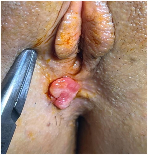
A 23-year-old virgin woman with a 4–5 year history of itching and bleeding due to irritation of a 2 × 2 cm mass lesion on the perineum, was referred to our tertiary hospital due to suspicion of vulvar cancer. There was no change in size in the last 3 years. She stated no changes occurred during her menstrual period. Her past medical history was unremarkable. Clinical examination revealed a single well-circumscribed, skin-colored, smooth, non-ulcerated, non-tender polypoid mass lesion arising from the perineum (). The rest of the gynecological examination was normal without any inguinal lymphadenopathy. Since our patient was a virgin, smear and human papillomavirus tests were not performed. We performed an excisional biopsy of the mass for diagnosis under local anaesthesia. Histopathological examination suggested the diagnosis of hidradenoma papilliferum. Immunohistochemical studies revealed CK 7 positivity in luminal cells and p63 positivity in the myoepithelial cells. The patient was discharged with no complaint following the procedure. Follow-up of the patient shows no actual recurrence. Written informed consent was obtained from the patient for publication of this case report and accompanying image. Ethics committee approval was unnecessary due to the nature of the study.
Figure 1. Clinical features of the vulvar lesion on the perineum.
Discussion
Although HP was generally described as asymptomatic in previous studies (Spindler et al. Citation2019, Patel et al. Citation2020), our patient had complaints such as itching and bleeding in the lesion due to irritation. This single, solitary nodule may cause tenderness (Spindler et al. Citation2019), dyspareunia (Birge et al. Citation2021), pain, bleeding, burning, and discharge if associated with ulceration (Seo et al. Citation2019), and increasing in diameter with the menstrual flow (Woodworth et al. Citation1971). HP is most commonly seen in the labia majora or labia minora, here we identified it in a rarer location, the perineum. It can mimic other vulvar malign neoplasms clinically so the final diagnosis needs to be confirmed histologically. Malignant cases were reported despite the rarity of malignant transformation (Shah, et al. Citation2008, Theodosiou et al. Citation2016, Kim et al. Citation2021). Several cases of ductal carcinoma in situ arising within a pre-existing hidradenoma papilliferum have been documented, and three cases have reported invasive carcinomas arising from HP (malignant perianal papillary hidradenoma, vulvar adenosquamous carcinoma and six cases of hidradenocarcinoma) (Vazmitel et al. Citation2008, Kondo et al. Citation2018, McGauran et al. Citation2021). One of these cases died from a disseminated tumour 2 months after local excision (Bannatyne et al. Citation1989). Paget’s disease associated with adenocarcinoma developing in a hidradenoma papilliferum in a 50-year-old lady was also described (Weilburg et al. Citation1967). However, in another case authors have failed to demonstrate continuity between the two lesions (Stefanato et al. Citation2000). These rare cases show that not all hidradenomas are innocent.
Baker et al. performed a retrospective review of a total of 189 vulvar adnexal lesions. Most of these lesions were benign, with hidradenoma papilliferum being the most common (Baker et al. Citation2013). It may appear with different clinical presentations including single bluish or red solitary nodule being the most common (Raiteb and Kouach Citation2017). However, some cases of multiple HP have been described (Veraldi et al. Citation1990). It occurs four times more frequently on the vulva than in the perianal area (Hama et al. Citation2013). The major sites of HP involvement include labia minora (50%), labia majora (40%), fourchette (7%), and clitoris (3%) (Scurry et al. Citation2009). One case of hymenal involvement was reported by Birge et al. (Citation2021). In another study HP locations were distributed as follows in order of descending frequency: the interlabial sulcus (59.6%), adjacent inner labia majora (25%), perineum (5.8%), clitoral hood (3.8%), and outer labia majora (1.9%) (El-Khoury et al. Citation2016). Less frequently reported locations are mons pubis, anus, and perineum (Meeker et al. Citation1962, Seo et al. Citation2019). The clinical appearance may be cystic, ulcerated, pedunculated, or solid (Scurry et al. Citation2009). The lesions range in size from 3 to 25 mm (Blind et al. Citation2019). It has been reported in much larger diameters in localizations other than the vulva. Bartholin’s abscess and HP can occur concomitantly if the tumour mass causes partial or complete obstruction of ductal drainage (Docimo et al. Citation2008).
The aetiology of HP is unclear (Seo et al. Citation2019). Although it has been reported in the literature that women with HP are usually of reproductive age and sexually active, indicating the sexually transmitted factors in the aetiology (Theodosiou et al. Citation2016, Birge et al. Citation2021), this possibility does not seem very likely because our patient is virgin. Moreover, a causal role for HPV in HP could not be confirmed (Kazakov et al. Citation2005). Previous studies have demonstrated the relationship between HP and hormone receptors and the lack of reported prepubertal cases (Stenchever et al. Citation2001). 90% of HP express oestrogen receptors and more weakly, for progesterone receptors explaining the predominancy of HP in reproductive females (Offidani and Campanati Citation1999). However, it has been suggested that the hormone prolactin may be responsible for the enlargement of the lesion since negative oestrogen and progesterone receptors had been demonstrated in another case (Hernández-Angeles et al. Citation2017). Hormonal stimulation may have a role in the pathogenesis and detection of HP in a pregnant woman and infertile patient undergoing injection in preparation for in vitro fertilisation (IVF) may confirm this (McClain et al. Citation2015, Olecki and Scow Citation2021). Tumourigenic alterations in several pathways have been demonstrated (Pfarr et al. Citation2019, Macagno et al. Citation2022).
Most examiners may think that the vulvar or perianal lesion is benign, based on the clinical appearance of HP. Many of the HP appear clinically as small, well-circumscribed, firm, non-ulcerated, predominantly unilobular, covered with normal skin, freely movable, slightly elevated, umbilicated, and slow-growing nodules in the skin. Others are soft and seemed to be cystic to the examiner (Meeker et al. Citation1962). HP may sometimes present with bleeding, pain, pruritus, and ulceration which raise doubts about malignancy. Histopathological examination reveals a characteristic, well-circumscribed dermal lesion with prominent elongated tubular and papillary structures lined by columnar cells without cellular atypia and mitoses (Jovic et al. Citation2021). Tumour epithelium is composed of a basal layer of cuboidal cells and a luminal layer of larger columnar cells on the basement membrane which show decapitation secretion. There are no mitotic figures, cytological atypia, prominent nucleoli, infiltrative growth pattern, or stromal desmoplasia as in the malignant tumours. The mean age of HP cases (52 years (range 31–91 years)) at presentation is lower than malignant cases (59.6 years (range 40–84 years)) (Scurry et al. Citation2009). These clinical and histological features can reassure us that the lesion is benign. However, the authors suggest that a correct diagnosis is rarely made clinically because clinically it mimics other cutaneous neoplasms so histopathological examination is necessary (Hama et al. Citation2013). Diagnosis of malignancy arising from HP is challenging due to the wide spectrum of histopathological features that have been described. Features consistent with a diagnosis of malignancy include loss of circumscription, infiltration, deep extension, nuclear pleomorphism, increased mitotic activity, necrosis, and perineural and vascular invasion (McGauran et al. Citation2021). Mitotic count in these lesions can be variable and often high, but it does not predict a more aggressive outcome (Sington et al. Citation2006). Besides these, recent immunohistochemical and molecular markers have been used for the diagnosis of HP. 50–75% of cases demonstrate MAML2 gene fusion. They express strongly and diffusely p40, p63, CK5/6, and AE1/AE3. Higher Ki67 and PHH3 labelling index and p53 expression have been reported in malignant cases (Macagno et al. Citation2022).
Tumours of the apocrine gland are closely related to each other histopathologically. Tubular apocrine adenoma, clear cell (apocrine) hidradenoma, and syringocystadenoma papilliferum should be included in the histopathological differential diagnosis of HP. Syringocystadenoma papilliferum is very rarely seen in the female genital tract. Cystic invaginations extending downward from the epidermis along with plasma cell infiltration are characteristic of histological findings of syringocystadenoma papilliferum and the absence of connection in the papillary structure distinguishes hidradenoma papilliferum from syringocystadenoma papilliferum. GCDFP-15, a sensitive marker for apocrine differentiation, is mostly positive in HP while in syringocystadenoma papilliferum lower GCDFP-15 staining has been reported (Nishie et al. Citation2004). In tubular apocrine adenoma, tubules have a dilated lumen with papillary projections extending into it. In clear cell hidradenoma, the lesions are constructed with apocrine-like tubular structures and clear cells can be seen. Hidradenoma papilliferum sometimes displays histopathology similar to these conditions (Minami et al. Citation2006, Lee et al. Citation2011).
Surgical excision of the lesion is curative regardless of the site and recurrence may occur due to incomplete excision (Kondo et al. Citation2018, Spindler et al. Citation2019).
Conclusions
Anogenital HP is a rare benign adnexal lesion that may be symptomatic. Differential diagnosis based on a clinical presentation should include both benign and malignant lesions. Histopathological examination confirms the diagnosis due to the inadequacy of specific clinical findings and rules out a malignant tumour.
Disclosure statement
No potential conflict of interest was reported by the author(s).
Additional information
Funding
References
- Baker, G.M., Selim, M.A. and Hoang, M.P., 2013. Vulvar adnexal lesions: a 32-year, single-institution review from Massachusetts General Hospital. Archives of Pathology & Laboratory Medicine, 137 (9), 1237–1246.
- Bannatyne, P., Elliott, P. and Russell, P., 1989. Vulvar adenosquamous carcinoma arising in a hidradenoma papilliferum, with rapidly fatal outcome: case report. Gynecologic Oncology, 35 (3), 395–398.
- Birge, O., et al., 2021. Hidradenoma papilliferum of the hymen: a case report. Journal of Medical Case Reports, 15 (1), 162.
- Blind, A., Weingertner, N., and Cribier, B., 2019. Étude anatomoclinique et immunohistochimique d’une tumeur dérivant des glandes ano-génitales de type mammaire: l’hidradénome papillifère [Anatomoclinical and immunohistochemical study of hidradenoma papilliferum, a tumor deriving from anogenital mammary-like glands. Annales de dermatologie et de venereologie, 146 (8–9), 528–536.
- Docimo, S., Jr., Shon, S., and Elkowitz, D.E., 2008. Bartholin’s abscess arising within hidradenoma papilliferum of the vulva: a case report. Cases Journal, 1 (1), 282.
- El-Khoury, J., et al., 2016. Vulvar hidradenoma papilliferum (HP) is located on the sites of mammary-like anogenital glands (MLAGs): analysis of the photographs of 52 tumors. Journal of the American Academy of Dermatology, 75 (2), 380–384.
- Hama, M., Oiso, N. and Kawada, A., 2013. Ulcerated hidradenoma papilliferum. International Journal of Dermatology, 52 (2), 198–199.
- Hernández-Angeles, C., Nadal, A. and Castelo-Branco, C., 2017. Hidradenoma papilliferum of the vulva in a postpartum woman: a case report. Journal of Obstetrics and Gynaecology, 37 (5), 683–684.
- Jovic, A., et al., 2021. Vulvar hidradenoma papilliferum dermoscopically mimicking basal cell carcinoma. Dermatology Practical & Conceptual, 11 (4), e2021070.
- Kazakov, D.V., et al., 2005. Hidradenoma papilliferum with oxyphilic metaplasia: a clinicopathological study of 18 cases, including detection of human papillomavirus. The American Journal of Dermatopathology, 27 (2), 102–110.
- Kim, G.Y., Solanki, M.H. and Guo, R., 2021. Vulvar apocrine hidradenocarcinoma arising in a hidradenoma papilliferum-a case report. Journal of Cutaneous Pathology, 48 (8), 1085–1087.
- Kondo, R.N., et al., 2018. Ectopic hidradenoma papilliferum. Anais brasileiros de dermatologia, 93 (3), 474–475.
- Lee, H.J., et al., 2011. Hidradenoma papilliferum occurring on the nasal skin. Annals of Dermatology, 23 (Suppl 2), S254–S257.
- Macagno, N., et al., 2022. Recent advances on immunohistochemistry and molecular biology for the diagnosis of adnexal sweat gland tumors. Cancers (Basel), 14 (3), 476.
- McClain, C.M., et al., 2015. Hidradenoma papilliferum associated with pregnancy: a case report. Journal of Cutaneous Pathology, 42 (12), 983–986.
- McGauran, M.F.G., et al., 2021. Poroid hidradenocarcinoma and atypical hidradenoma papilliferum of the vulva - two cases. Gynecologic Oncology Reports, 38, 100886.
- Meeker, J. H., Neubecker, R. D. and Helwıg, E. B., 1962. Hidradenoma papilliferum. American Journal of Clinical Pathology, 37, 182–195.
- Minami, S., et al., 2006. Non-anogenital (ectopic) hidradenoma papilliferum with sebaceous differentiation: a case report and review of reported cases. The Journal of Dermatology, 33 (4), 256–259.
- Nishie, W., et al., 2004. Hidradenoma papilliferum with mixed histopathologic features of syringocystadenoma papilliferum and anogenital mammary-like glands. Journal of Cutaneous Pathology, 31 (8), 561–564.
- Offidani, A. and Campanati, A., 1999. Papillary hidradenoma: immunohistochemical analysis of steroid receptor profile with a focus on apocrine differentiation. Journal of Clinical Pathology, 52 (11), 829–832.
- Olecki, E.J. and Scow, J.S., 2021. Hidradeonoma papilliferum of the anus: a case report about the relationship between neoplasms of the mammary-like-glands and hormones. Cureus, 13 (2), e13061. Feb 1
- Patel, S., 2020. Hidradenoma papilliferum: everyone else’s diagnosis. Indian Journal of Dermatology, 65 (2), 151–153.
- Pfarr, N., et al., 2019. Several genotypes, one phenotype: PIK3CA/AKT1 mutation-negative hidradenoma papilliferum show genetic lesions in other components of the signaling network. Pathology, 51 (4), 362–368.
- Raiteb, H. and Kouach, J., 2017. Hidradenoma papilliferum. Pan African Medical Journal, 26, 196.
- Scurry, J., et al., 2009. Mammary-like gland adenoma of the vulva: review of 46 cases. Pathology, 41 (4), 372–378.
- Seo, G.J., et al., 2019. Hidradenoma papilliferum of the anus: a report of 2 cases and review of the literature. Annals of Coloproctology, 35 (6), 361–363.
- Shah, S.S., Adelson, M. and Mazur, M.T., 2008. Adenocarcinoma in situ arising in vulvar papillary hidradenoma: report of 2 cases. The International Journal of Gynecological Pathology, 27 (3), 453–456.
- Sington, J., et al., 2006. Mitotic count is not predictive of clinical behavior in hidradenoma papilliferum of the vulva: a clinicopathologic study of 19 cases. The American Journal of Dermatopathology, 28 (4), 322–326.
- Spindler, L., et al., 2019. Anal and vulvar hidradenoma papilliferum are similar: a study of 14 cases. Annals of Dermatology and Venereology, 146 (8–9), 537–541.
- Stefanato, C.M., Finn, R. and Bhawan, J., 2000. Extramammary Paget disease with underlying hidradenoma papilliferum: guilt by association? The American Journal of Dermatopathology, 22 (5), 439–442.
- Stenchever, M.A., et al., 2001. Comprehensive gynecology. Benign gynecologic lesions. 4th ed., vol. 18. Mosby, Inc., 484–485.
- Theodosiou, G., et al., 2016. An unusual lesion in the right place. Dermatology Practical & Conceptual, 6 (3), 7–9.
- Vazmitel, M., et al., 2008. Hidradenoma papilliferum with a ductal carcinoma in situ component: case report and review of the literature. The American Journal of Dermatopathology, 30 (4), 392–394.
- Veraldi, S., Schianchi-Veraldi, R. and Marini, D., 1990. Hidradenoma papilliferum of the vulva: report of a case characterized by unusual clinical behavior. The Journal of dermatologic surgery and oncology, 16 (7), 674–676.
- Weilburg, R.D., Miller, G.V. and Von Pohle, K.C., 1967. Paget’s disease of the vulva associated with adenocarcinoma developing in a hidradenoma papilliferum. American Journal of Obstetrics and Gynecology, 98, 294–295.
- Woodworth, H., Jr., et al., 1971. Papillary hidradenoma of the vulva: a clinicopathologic study of 69 cases. American Journal of Obstetrics and Gynecology, 110 (4), 501–508.