

Abstract
Ovarian cancer is one of the most common malignant tumours affecting the female reproductive organs. CD147 (BSG) and CD98hc (SLC3A2) are oncogenes that form the CD98hc-CD147 complex, which regulates the proliferation, metastasis, metabolism, and cell cycle of cancer cells. The roles of the CD98hc-CD147 complex in ovarian cancer remain unclear. We analysed the expression and prognostic value of CD147 and CD98hc in ovarian cancer using the TCGA and ICGC databases. The effect of CD147 and CD98hc on the tumour immune response was analysed using the TIMER database. CD98hc was more highly expressed in normal tissues than primary tumour tissues, while CD147 was more highly expressed in primary tumour tissues than normal tissues. CD98hc expression was significantly associated with neutrophil and dendritic cell levels. CD147 and CD98hc were correlated with DNA repair, the cell cycle, and DNA replication. The CD98hc-CD147 complex could serve as a target for ovarian cancer treatment.
IMPACT STATEMENT
What is already known on this subject? CD98hc and CD147 are oncogenes that induce the proliferation and metastasis of cancer cells. The CD98hc-CD147 complex has been identified as a risk factor for cancer patients and causes resistance to cancer treatment.
What do the results of this study add? We confirmed the expression levels of CD98hc and CD147 in ovarian cancer tissues and the effects of these oncogenes on the tumour immune response.
What are the implications of these findings for clinical practice and/or further research? The CD98hc-CD147 complex may serve as a new target for ovarian cancer therapy.
Introduction
Ovarian cancer is one of the most common malignant tumours affecting the female reproductive organs, and its incidence rate is second only to cervical and uterine body cancer (Sung et al. Citation2021, Li et al. Citation2022). Epithelial ovarian cancer is the most common type of gynaecological tumour and poses a serious threat to women’s lives (Siegel et al. Citation2021). The recurrence rate of advanced epithelial ovarian cancer is very high, and the five-year survival rate is approximately 30% (Siegel et al. Citation2021, Upadhyay et al. Citation2022). Moreover, the treatment effect of patients with recurrent ovarian cancer often fails to reach the initial treatment, and the time to remission after treatment will decrease with the increasing number of times of recurrence (Marchetti et al. Citation2012, Musella et al. Citation2017, Cybula and Bieniasz Citation2022). Therefore, revealing the molecular mechanism of ovarian cancer cell proliferation and invasion is of great significance for the development of targeted drugs for the treatment of ovarian cancer.
CD98 is a type II transmembrane glycoprotein with a size of approximately 85 kDa (Ip and Sethi Citation2016). It consists of a heavy chain (CD98 heavy chain [CD98hc, SLC3A2]) and a light chain (LAT1, LAT2, XCT, y + LAT1, y + LAT2, or ASC1) covalently linked by disulphide bonds to form a heterodimer (Ip and Sethi Citation2016). The CD98 protein promotes tumorigenesis and metastasis by regulating cell adhesion and extracellular matrix synthesis (Cano-Crespo et al. Citation2019). Moreover, SLC3A2/LAT1 mediates the transport of most neutral and aromatic amino acids and provides nutrients for cell metabolism (Scalise et al. Citation2020). Previous studies have shown that CD98hc is highly expressed in tumour tissues and suggests a poor prognosis. Moreover, its high expression is related to tumour development, progression, and migration (Kaira et al. Citation2014, Satoh et al. Citation2017, El Ansari et al. Citation2018). CD147 (basigin, BSG) is a single transmembrane glycoprotein, belonging to the immunoglobulin superfamily (de la Cruz Concepción et al. Citation2022). This gene is highly expressed in a variety of malignancies, and its high expression is an indicator of poor clinical prognosis (Chu et al. Citation2014, Liu et al. Citation2017, Zhang et al. Citation2022). Moreover, CD147 can be used as both an inducer and an effector to promote the growth, metabolism, invasion, metastasis, angiogenesis, drug resistance, death resistance, and other malignant behaviours of tumour cells through various mechanisms, leading to tumour progression (de la Cruz Concepción et al. Citation2022). Together, CD147 and CD98hc form the CD98hc-CD147 complex to further regulate the sugar and amino acid metabolism of cells (Xu and Hemler Citation2005). In this study, we aimed to clarify the clinicopathological and prognostic significance of the CD98hc-CD147 complex in ovarian cancer using bioinformatics analysis.
Materials and methods
R programming and gene expression profiling interactive analysis (GEPIA)
R software v4.0.3 was used to analyse the expression of CD98hc and CD147 in ovarian cancer and normal tissues. The log-rank test was used to compare differences in survival between patients grouped according to the quartile of expression of these genes. The RNA-sequencing expression (level 3) profiles and corresponding clinical information of patients with ovarian cancer were downloaded from the TCGA (https://portal.gdc.com) and ICGC databases (https://dcc.icgc.org/releases/current/Projects). The ‘Forestplot’ package was used for multivariate Cox regression analysis, and the forest plot was generated to show the P-value, hazard ratio, and 95% confidence interval of the related variables, such as CD147, CD98hc, and age. The correlation between CD147 or CD98hc and the pathway scores was analysed by Spearman correlation analysis using the ‘GSVA’ package. Finally, GEPIA was used to analyse the correlation between CD98hc and CD147 expression in ovarian cancer and normal tissues.
TIMER database
The TIMER database (https://cistrome.shinyapps.io/timer/) was used to evaluate the association between CD147 or CD98hc expression and immune cell infiltrate levels in ovarian cancer. This database estimates the level of tumour infiltrates by determining the abundance of six immune infiltrates, including CD4+ T cells and CD8+ T cells. Furthermore, the somatic copy number alteration module was used to compare tumour infiltration levels in ovarian cancer with different somatic copy number alterations of CD147 or CD98hc, including deep deletion (-2), arm-level deletion (-1), diploid/normal (0), arm-level gain (+1), and high amplification (+2).
Results
Expression levels and prognostic roles of CD147 and CD98hc in ovarian cancer
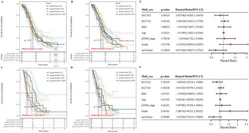
CD98hc expression was higher in normal tissues than in primary tumour tissues (p = 5.2e–05) () and recurrent tumour tissues (p = 9.1e–16) (), while CD147 expression was lower in normal tissues than in primary tumour tissues (p = 4.1e–15) (). However, no difference in CD147 expression was found between normal tissues and recurrent tumour tissues (p = 0.24) (). In addition, we found a positive correlation between CD147 and CD98hc in ovarian cancer tissues (R = 0.38, p = 8.9e–16) (), but not in normal tissues (R = 0.21, p = 0.055) (). Based on the TCGA database, Kaplan–Meier analysis revealed that CD147 and CD98hc were not correlated with the overall survival rate of patients with ovarian cancer (P > 0.05) (). However, based on the ICGC database, CD98hc was an unfavourable prognostic marker for patients with ovarian cancer (p = 0.026) (). The results of CD147 were consistent in both databases (P > 0.05) (). Additionally, the multivariate Cox regression analysis showed that CD147 and CD98hc were not associated with the overall survival and disease-free survival of patients with ovarian cancer (P > 0.05) (). However, age was associated with overall survival (CI, 1.01785; 95% CI, 1.0025–1.03344, p = 0.02251) and disease-free survival (CI, 1.02034; 95% CI, 1.00458–1.03634, p = 0.01122) of patients with ovarian cancer ().
Figure 1. Expression and correlation of CD98hc and CD147 in ovarian cancer. The expression of (A) CD98hc and (B) CD147 was evaluated in primary cancer tissues and matched normal tissues of patients with ovarian cancer using R software v4.0.3 (****P < 0.05). (C) CD98hc and (D) CD147 expression were evaluated in recurrent cancer tissues and matched normal tissues of patients with ovarian cancer using R software v4.0.3 (****P < 0.05). The correlation between CD98hc and CD147 was evaluated in normal tissue (E) and cancer tissue (F) using GEPIA.
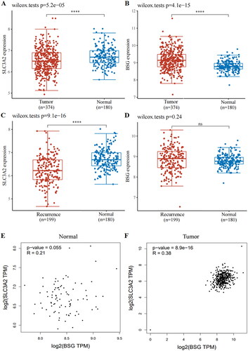
Figure 2. Prognostic roles of CD98hc and CD147 in patients with ovarian cancer. Kaplan–Meier analysis was used to analyse the effects of CD98hc (A, C) and CD147 (B, D) on the overall survival rate of patients with ovarian cancer based on TCGA and ICGC databases. Multivariate Cox regression analysis was used to analyse the risk factors for the overall survival (E) and disease-free survival of patients with ovarian cancer (F).
CD98hc expression was correlated with immune cell infiltration in ovarian cancer
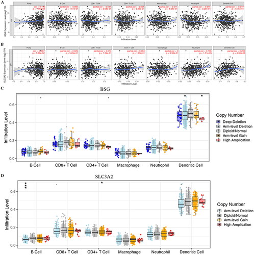
CD147 expression was correlated with ovarian cancer tumour purity (R = 0.106, p = 1.96e–02) (); however, no correlation was observed between CD98hc expression and tumour purity (R = −0.087, p = 5.66e–02) (). Moreover, CD98hc expression was significantly associated with levels of neutrophils (R = 0.196, p = 1.51e–05) and dendritic cells (R = 0.208, p = 4.30e–06) (). No such relationship between CD147 expression and immune cell infiltration in ovarian cancer was found (P > 0.05) (). In addition, the copy number of CD147 and CD98hc was significantly associated with the immune-infiltration level of dendritic cells in ovarian cancer (p < 0.05) ().
Figure 3. Correlation of CD98hc or CD147 expression with immune cell infiltration in ovarian cancer tissues. The association between CD147 (A) or CD98hc (B) expression and immune cell infiltrate levels in patients with ovarian cancer were evaluated using the TIMER database (https://cistrome.shinyapps.io/timer/). The somatic copy number alteration module was used to compare tumour infiltration levels in patients with ovarian cancer with different somatic copy number alterations of CD147 (C) or CD98hc (D).
Signalling pathways associated with CD147 or CD98hc in ovarian cancer
CD147 expression was correlated with tumour inflammation (p = 0.012) (Supplementary Figure 1(A)), DNA repair (p = 0.011) (Supplementary Figure 1(H)), the G2M checkpoint (p = 0.010) (Supplementary Figure 1(I)), MYC expression (p = 0.008) (Supplementary Figure 1(M), and DNA replication (p = 0.005) (Supplementary Figure 1(Q)) in ovarian cancer tissues. Moreover, CD98hc expression was correlated with tumour proliferation (p = 0.001) (Supplementary Figure 2C), epithelial-mesenchymal transition (EMT) markers (p = 4.62e–04) (Supplementary Figure 2(D)), angiogenesis (p = 8.36e–05) (Supplementary Figure 2(F), apoptosis (p = 0.006) (Supplementary Figure 2G), DNA repair (p = 0.007) (Supplementary Figure 2(H)), the G2M checkpoint (p = 8.32e–07) (Supplementary Figure 2(I)), inflammation (p = 0.012) (Supplementary Figure 2J), the PI3K-AKT-mTOR pathway (p = 0.003) (Supplementary Figure 2(K)), P53 pathway (p = 1.93e–05) (Supplementary Figure 2L), ROS level (p = 0.001) (Supplementary Figure 2P), DNA replication (p = 1.45e–05) (Supplementary Figure 2(Q)), extracellular matrix degradation (p = 0.022) (Supplementary Figure 2(S)), and ferroptosis (p = 1.11e–08) (Supplementary Figure 2T) in ovarian cancer.
Discussion
CD98hc has been demonstrated to be highly expressed in a variety of tumours and has become a marker of tumour diagnosis and prognosis (Kaira et al. Citation2014, Satoh et al. Citation2017, El Ansari et al. Citation2018). To the best of our knowledge, no previous study has shown the association between the expression of CD98hc and the prognosis of patients with ovarian cancer. In this study, we found that CD98hc expression was lower in ovarian cancer tissues than in normal tissues. According to the TCGA database, CD98hc was not a prognostic biomarker of ovarian cancer. However, according to the ICGC database, CD98hc was an unfavourable prognostic biomarker for patients with ovarian cancer. However, because the number of patients in the ICGC database is small, we cannot judge the true prognostic role of CD98hc. Moreover, multivariate Cox regression analysis showed that CD98hc was not a risk factor for patients with ovarian cancer. Therefore, based on current data, CD98hc cannot be used as a prognostic biomarker for patients with ovarian cancer. In the future, a larger number of patient samples will be needed to verify these results. CD98hc, as an oncogene, is closely related to the biological behaviour, including the proliferation, invasion, and migration of tumour cells (Cano-Crespo et al. Citation2019, Scalise et al. Citation2020). For example, upregulation of CD98hc has been found to increase cisplatin-induced apoptosis of ovarian cancer cells and decrease the volume and weight of implanted tumours (Cui et al. Citation2018). This evidence suggests that CD98hc does not function as an oncogene in ovarian cancer.
CD147 has been found to be overexpressed in ovarian cancers and associated with the survival of patients with ovarian cancer (Yang and Chen Citation2013). Consistent with previous studies, our results also showed higher CD147 expression in ovarian cancer tissues than in normal tissues. However, CD147 was not a prognostic biomarker of ovarian cancer according to the grouping of patients by quartile of CD147 expression. Moreover, CD147 and CD98hc were found to be co-expressed in drug-resistant SKOV3 cells (SKOV3/DDP cells) (Yang et al. Citation2007). Additionally, inhibition of CD98hc and CD147 separately or simultaneously was functionally related with drug resistance of SKOV3/DDP cells (Yang et al. Citation2007). We also confirmed the correlation of CD98hc and CD147 in ovarian cancer tissues but not in normal tissues.
Recurrence after surgery and first-line chemotherapy is a major problem of ovarian cancer (Fagotti et al. Citation2020) and is related to immune escape and treatment resistance (Laganà et al. Citation2015). The cardiac allograft rejection of CD98hc-deficient T cells was weakened for inhibiting lymphocyte migration and increasing the expansion of regulatory T cells (Liu et al. Citation2012). Moreover, CD98hc was found to facilitate the immunosuppressive function of regulatory cells and indirectly promote tumour immune escape (Geng et al. Citation2021). Additionally, the absence of CD98hc selectively impaired the activation of ERK1/2 after B cell receptor ligation, which is essential for B cell proliferation and adaptive humoral immunity (Cantor Citation2014). Therefore, CD98hc can regulate both humoral and cellular immunity. Another recent study showed that CD98hc could serve as a target for chimeric antigen receptor T therapy given its overexpression on the membrane of cancer cells (Köseer et al. Citation2022). In our study, we found that CD98hc expression was correlated with the infiltration levels of neutrophils and dendritic cells in ovarian cancer. However, the mechanism requires further experimental analysis. In contrast, the function of CD147 in the immune system has been widely studied (Hu et al. Citation2010, Shang et al. Citation2018), and it has been found to be a key factor for the formation of immune synapses (Hu et al. Citation2010). Downregulation of CD147 (BSG) on the surface of T cells can significantly inhibit immune synapse formation between T and B cells (Hu et al. Citation2010). However, we did not observe a correlation between CD147 expression and immune cell infiltration in ovarian cancer tissues. These findings indicate that CD98hc is the initial factor of ovarian cancer immunity, and CD147 is associated with CD98hc. Finally, Yang et al. (Citation2007) found that CD147 appeared sequentially after CD98hc expression in ovarian cancer cells.
Previous studies have confirmed that CD98hc and CD147 can regulate the invasion, metastasis, survival, angiogenesis, and drug resistance of ovarian cancer cells (Huang et al. Citation2005, Hu et al. Citation2016, Gao et al. Citation2021). Based on our bioinformatics analysis, we confirmed that CD147 and CD98hc were correlated with DNA repair, the cell cycle, and DNA replication. CD147 is also a regulator of MYC expression. Moreover, CD98hc expression is correlated with EMT, angiogenesis, apoptosis, the PI3K-AKT-mTOR pathway, P53 pathway, ROS level, and ferroptosis. Furthermore, an increased incidence of ovarian cancer has been demonstrated in patients with endometriosis in previous studies (Börner et al. Citation2018, Králíčková et al. Citation2020). Notably, CD147 may induce EMT in patients with endometriosis (Wang et al. Citation2018), and CD147 has been suggested to be the driving factor of ovarian cancer.
Our results provide an overview of the mechanisms of CD98hc and CD147 in ovarian cancer. This study will provide benefits to researchers for deep mechanism analysis. Moreover, these findings suggest that the CD98hc-CD147 complex may serve as a new target for tumour therapy. Although this bioinformatics analysis provides a clear target for ovarian cancer treatment, these results should be validated in vivo and in vitro.
Author contributions
All authors made substantial contributions to the conception and design of the study, acquisition of data, or analysis and interpretation of data; took part in drafting the article or revising it for important intellectual content; agreed to submit to the current journal; gave final approval of the version to be published; and agree to be accountable for all aspects of the work.
Supplemental Material
Download Zip (13.9 MB)Disclosure statement
No potential conflict of interest was reported by the author(s).
Data availability statement
There is no data needed to be deposited.
Additional information
Funding
References
- Börner, C., et al., 2018. Pain mechanisms in peritoneal diseases might be partially regulated by estrogen. Reproductive Sciences, 25 (3), 424–434.
- Cano-Crespo, S., et al., 2019. CD98hc (SLC3A2) sustains amino acid and nucleotide availability for cell cycle progression. Scientific Reports, 9 (1), 14065.
- Cantor, J.M., 2014. CD98 is a potential target for ablating B cell clonal expansion and autoantibody in multiple sclerosis. Journal of Neuroimmunology, 274 (1-2), 230–233.
- Chu, D., et al., 2014. CD147 expression in human gastric cancer is associated with tumor recurrence and prognosis. PLoS One, 9 (6), e101027.
- Cui, Y., et al., 2018. ZEB1 promotes chemoresistance to cisplatin in ovarian cancer cells by suppressing SLC3A2. Chemotherapy, 63 (5), 262–271.
- Cybula, M. and Bieniasz, M., 2022. Patient-derived tumor models are attractive tools to repurpose drugs for ovarian cancer treatment: pre-clinical updates. Oncotarget, 13, 553–575.
- de la Cruz Concepción, B., et al., 2022. EMMPRIN is an emerging protein capable of regulating cancer hallmarks. European Review for Medical and Pharmacological Sciences, 26 (18), 6700–6724.
- El Ansari, R., et al., 2018. The multifunctional solute carrier 3A2 (SLC3A2) confers a poor prognosis in the highly proliferative breast cancer subtypes. British Journal of Cancer, 118 (8), 1115–1122.
- Fagotti, A., et al., 2020. Randomized trial of primary debulking surgery versus neoadjuvant chemotherapy for advanced epithelial ovarian cancer (SCORPION-NCT01461850). International Journal of Gynecological Cancer, 30 (11), 1657–1664.
- Gao, L., et al., 2021. Interaction of CD147 and human epididymis protein 4 promotes invasion and metastasis of ovarian cancer. Journal of Cancer, 12 (24), 7422–7435.
- Geng, J., et al., 2021. CD98-induced CD147 signaling stabilizes the Foxp3 protein to maintain tissue homeostasis. Cellular & Molecular Immunology, 18 (12), 2618–2631.
- Hu, J., et al., 2010. Involvement of HAb18G/CD147 in T cell activation and immunological synapse formation. Journal of Cellular and Molecular Medicine, 14 (8), 2132–2143.
- Hu, Z., et al., 2016. The fucosylated CD147 enhances the autophagy in epithelial ovarian cancer cells. Oncotarget, 7 (50), 82921–82932.
- Huang, Y., et al., 2005. Cystine-glutamate transporter SLC7A11 in cancer chemosensitivity and chemoresistance. Cancer Research, 65 (16), 7446–7454.
- Ip, H. and Sethi, T., 2016. CD98 signals controlling tumorigenesis. The International Journal of Biochemistry & Cell Biology, 81 (Pt A), 148–150.
- Kaira, K., et al., 2014. CD98 is a promising prognostic biomarker in biliary tract cancer. Hepatobiliary & Pancreatic Diseases International : HBPD INT, 13 (6), 654–657.
- Köseer, A.S., et al., 2022. Validation of CD98hc as a therapeutic target for a combination of radiation and immunotherapies in head and neck squamous cell carcinoma. Cancers, 14 (7), 1677.
- Králíčková, M., et al., 2020. Endometriosis and risk of ovarian cancer: what do we know? Archives of Gynecology and Obstetrics, 301 (1), 1–10.
- Laganà, A.S., et al., 2015. Cytogenetic analysis of epithelial ovarian cancer’s stem cells: an overview on new diagnostic and therapeutic perspectives. European Journal of Gynaecological Oncology, 36 (5), 495–505.
- Li, S., et al., 2022. Clinical characteristics and survival outcomes in patients with ovarian strumal carcinoid. BMC Cancer, 22 (1), 1090.
- Liu, B., et al., 2017. Overexpression of EMMPRIN is associated with lymph node metastasis and advanced stage of non-small cell lung cancer: a retrospective study. BMC Pulmonary Medicine, 17 (1), 214.
- Liu, Z., et al., 2012. Deletion of CD98 heavy chain in T cells results in cardiac allograft acceptance by increasing regulatory T cells. Transplantation, 93 (11), 1116–1124.
- Marchetti, C., et al., 2012. Olaparib, PARP1 inhibitor in ovarian cancer. Expert Opinion on Investigational Drugs, 21 (10), 1575–1584.
- Musella, A., et al., 2017. Bevacizumab in ovarian cancer: state of the art and unanswered questions. Chemotherapy, 62 (2), 111–120.
- Satoh, T., et al., 2017. Prognostic significance of the expression of CD98 (4F2hc) in gastric cancer. Anticancer Research, 37 (2), 631–636.
- Scalise, M., et al., 2020. Membrane transporters for amino acids as players of cancer metabolic rewiring. Cells, 9 (9), 2028.
- Shang, Y.K., et al., 2018. System analysis of the regulation of the immune response by CD147 and FOXC1 in cancer cell lines. Oncotarget, 9 (16), 12918–12931.
- Siegel, R.L., et al., 2021. Cancer statistics, 2021. CA: A Cancer Journal for Clinicians, 71 (1), 7–33.
- Sung, H., et al., 2021. Global cancer statistics 2020: GLOBOCAN estimates of incidence and mortality worldwide for 36 cancers in 185 countries. CA: a Cancer Journal for Clinicians, 71 (3), 209–249.
- Upadhyay, A., et al., 2022. Early-Stage epithelial ovarian cancer: predictors of survival. Gynecologic Oncology Reports, 44, 101083.
- Wang, C., et al., 2018. CD147 induces epithelial-to-mesenchymal transition by disassembling cellular apoptosis susceptibility protein/E-cadherin/β-catenin complex in human endometriosis. The American Journal of Pathology, 188 (7), 1597–1607.
- Xu, D. and Hemler, M.E., 2005. Metabolic activation-related CD147-CD98 complex. Molecular & Cellular Proteomics : MCP, 4 (8), 1061–1071.
- Yang, H. and Chen, B., 2013. CD147 in ovarian and other cancers. International Journal of Gynecological Cancer, 23 (1), 2–8.
- Yang, H., et al., 2007. Bridge linkage role played by CD98hc of anti-tumor drug resistance and cancer metastasis on cisplatin-resistant ovarian cancer cells. Cancer Biology & Therapy, 6 (6), 942–947.
- Zhang, W., et al., 2022. Expression of CD147 after neoadjuvant chemotherapy and its relationship with prognosis in patients with triple negative breast cancer. American Journal of Translational Research, 14 (5), 2952–2961.