

Abstract
FAM64A is a mitotic regulator which promotes cell metaphase-anaphase transition and is highly expressed in a cell-cycle-dependent manner. In this study, we examined the clinicopathological and prognostic significance of FAM64A mRNA expression in gynecological cancers. We conducted a bioinformatics analysis of FAM64A mRNA expression using Gene Expression Omnibus (GEO), The Cancer Genome Atlas (TCGA), xiantao, The University of ALabama at Birmingham CANcer data analysis Portal (UALCAN), and Kaplan-Meier (KM) plotter databases. FAM64A expression was elevated in breast, cervical, endometrial, and ovarian cancers when compared with normal tissue. Expression was positively correlated with white race, low T stages, infiltrating ductal carcinoma, or favourable PAM50 classification in breast cancer patients, and with clinical stage, histological grade and TP53 mutation, and endometrial cancer serous subtype. FAM64A expression was negatively associated with overall and/or recurrence-free survival rates in breast and endometrial cancer patients, while the opposite was observed in cervical and ovarian cancer patients. FAM64A functioned as an independent predictor of overall and disease-specific survival in breast cancer patients. FAM64A-correlated genes were involved in ligand-receptor interactions, and chromosomal, cell cycle, and DNA replication processes in breast, cervical, endometrial and ovarian cancers. Top hub genes primarily included cell cycle-related proteins in breast cancer, mucins and acetylgalactosaminyl transferases in cervical cancer, kinesin family members in endometrial cancer, and synovial sarcoma X and the cancer/testis antigen in ovarian cancer. FAM64A mRNA expression was positively related to Th2 cell infiltration, but negatively associated with neutrophil and Th17 cell infiltration in breast, cervical, endometrial, and ovarian cancers. FAM64A expression may be considered a potential biomarker reflecting carcinogenesis, histogenesis, aggressive behaviour, and prognosis in gynecological cancers.
What is already known on this subject? FAM64A is located in cell nucleolar and nucleoplasmic regions, and during mitosis it putatively controls metaphase-to-anaphase transition. FAM64A appears to regulate different physiological processes, including apoptosis, tumorigenesis, neural differentiation, stress responses, and the cell cycle.
What the results of this study add? FAM64A expression was up-regulated in breast, cervical, endometrial, and ovarian cancers, and positively correlated with white race, low T stages, infiltrating ductal carcinoma, or favourable PAM50 classification in breast cancer patients, and with clinical stage, histological grade, and TP53 mutation, and a serous subtype in endometrial cancer. FAM64A expression was negatively associated with overall and/or recurrence-free survival rates in breast and endometrial cancer patients, while the opposite was observed in cervical and ovarian cancer patients. FAM64A functioned as an independent predictor of overall and disease-specific survival in breast cancer. FAM64A-correlated genes were involved in ligand-receptor interactions, chromosomal, cell cycle, and DNA replication processes, while FAM64A mRNA expression was positively related to Th2 cell infiltration but negatively correlated with neutrophil and Th17 cell infiltration in four gynecological cancers.
What the implications of these findings for clinical practice and/or further research? In the future, abnormal FAM64A mRNA expression may serve as a biomarker of carcinogenesis, histogenesis, aggressiveness, and prognosis in gynecological malignancies.
Impact statement
Introduction
CATS proteins (also called FAM64A and RCS1) were initially identified as novel CALM interaction factors which influenced subcellular leukemogenic fusion protein CALM/AF10 localisation. They were also known as phosphatidylinositol-binding clathrin assembler protein (PICALM) interacting mitotic regulatory factors (PIMREG). FAM64A is a highly expressed and cell-cycle-dependent gene induced by mitogens (Zhao et al. Citation2008, Archangelo et al. Citation2013, Zhou et al. Citation2021). Zhao et al. (Citation2008) showed that FAM64A levels peaked in mitosis, but dropped sharply as cells entered the G1 phase. FAM64A controlled the timing of anaphase onset, while FAM64A loss accelerated metaphase progression to anaphase, concomitant with securin and cyclin B degradation. Mitotic FAM64A interacted with the NuRD chromatin-remodeling complex and was involved in transcription regulation or chromatin structure. Indeed, FAM64A is efficiently ubiquitinated by anaphase-promoting complex/cyclosome (APC/C) via a unique D-box at its N terminus, and was degraded during mitotic withdrawal in a Cdh1-dependent manner. Zhou et al. (Citation2021) reported that dihydrotestosterone increased FAM64A expression via direct androgen receptor binding to the FAM64A promoter, and significantly promoted androgen-dependent prostate cancer cell proliferation, migration, invasion, and cell cycle progression. Archangelo et al. (Citation2013) identified FAM64A as a substrate for kinase interacting stathmin (KIS) and mapped phosphorylation sites to FAM64A serine 131, and KIS levels depended on cell cycle in the opposite direction to FAM64A level.
Reportedly, FAM64A was abundantly expressed in hypoxic foetal cardiomyocyte nuclei, but its expression was strongly inhibited by oxygen exposure. FAM64A knockdown enhanced cardiomyocyte proliferation, while foetal cardiomyocyte division required FAM64A and subsequent APC/C degradation (Hashimoto et al. Citation2017). Significantly increased FAM64A expression was observed in all-trans retinoic acid-induced granulocytic differentiation, while decreased expression was observed during erythroid, megakaryocytic, and monocytic differentiation. In lymphoma cells, FAM64A silencing reduced cell proliferation, blocked cell cycle progression, and lowered migratory capacity upon the hypo-expression of the self-renewal gene, GLI-1. FAM64A overexpression in primary bone marrow cells decreased colony formation (Barbutti et al. Citation2016). Xu et al. (Citation2019) observed that FAM64A enhanced interleukin (IL)-6-induced STAT3 activation by interacting with STAT3 (signal transducer and activator of transcription 3) and regulated the expression of downstream genes via STAT3 biding to its target gene promoters. FAM64A deficiency alleviated experimental autoimmune encephalomyelitis, DSS (dextran sodium sulfate) -induced colitis, and AOM (azoxymethane)/DSS-induced colitis-associated cancer in mice.
In terms of cancer risk, females are particularly susceptible to breast, vulvar, vaginal, cervical, endometrial, and ovarian cancers (Paepke et al. Citation2020, Aquil et al. Citation2021, Segev et al. Citation2021, Seland et al. Citation2022). Vargiu et al. (Citation2022) reported that assessing a female body mass index was fundamental in developing treatment strategies and reducing complications in gynecological oncology. Considerable evidences have shown that obesity and metabolic disease may increase cancer risk and regulate pivotal cross-talk pathways with respect to cell proliferation and differentiation (Giannini et al. Citation2022, Vargiu et al. Citation2022).
In this study, we examined the clinicopathological and prognostic significances of FAM64A mRNA expression and relevant signal pathways in breast, cervical, endometrial, and ovarian cancers using Gene Expression Omnibus (GEO), The Cancer Genome Atlas (TCGA), xiantao, The University of Alabama at Birmingham CANcer data analysis Portal (UALCAN), and Kaplan-Meier (KM) plotter databases as done previously (Zheng et al. Citation2023).
Material and methods
TCGA analysis
Expression (RNA-seqV2) and clinicopathological data from breast cancer (n = 1072), cervical cancer (n = 296), and endometrial cancer (n = 537) patients were downloaded from TCGA (https://cancergenome.nih.gov/) database using TCGA-assembler in R software (4.2.1) (R package: ggplot2 3.3.6, stats 4.2.1, car). We integrated raw data, analysed FAM64A mRNA expression in cancers, and compared data with patient clinicopathological and prognostic data.
GEO analysis
FAM64A mRNA expression profiles from GSE54002 (platform: Affymetrix-GPL570, breast cancer), GSE63514 (platform: Affymetrix-GPL570, cervical cancer), and GSE18520 (platform: Affymetrix-GPL570, ovarian cancer) datasets were obtained from the National Centre for Biotechnology Information GEO database (https://www.ncbi.nlm.nih.gov/geo/). R software was used to analyse FAM64A mRNA expression between cancer and normal tissue.
KM plotter analysis
After inputting FAM64A (ID: 221591_s_at) into the KM plotter (http://kmplot.com), we analysed the prognostic significance of FAM64A mRNA in breast, cervical, endometrial, and ovarian cancers.
UALCAN analysis
We used the UALCAN database (http://ualcan.path.uab.edu) to analyse FAM64A expression levels in breast, cervical, endometrial, and ovarian malignancies, and compared data with cancer clinicopathological and prognostic parameters.
Xiantao analysis
The xiantao platform (https://www.xiantao.love/) was used to analyse FAM64A expression levels. We also used the platform to identify differentially expressed and related FAM64A genes. A protein-protein interaction (PPI) network was constructed in STRING (https://cn.string-db.org/) using differentially expressed genes after which important hub genes were selected by Cytoscape. These genes were then analysed using the Kyoto Encyclopaedia of Genes and Genomes (KEGG) database (https://www.kegg.jp/) to construct signal pathways.
Statistical analysis
Mean data were compared using Wilcoxon rank sum tests and positive rates using chi-square tests. KM survival plots were generated by comparing survival curves with log-rank statistics. The Cox’s proportional hazards model was used for multivariate analysis. Two-sided p < 0.05 values were considered statistically significant. All data were analysed in SPSS 17.0 software (IBM Corp, Armonk, NY, USA).
Results
Clinicopathological and prognostic significance of FAM64A mRNA expression in breast cancer
From xiantao () [95% confidence interval (CI) = 1.914: 1.731–2.096], UALCAN (), GEO () [95% CI = 1.161 (0.881–1.446], and TCGA () [95% CI = 1.154: 1.021–1.292)] databases, FAM64A was more significantly expressed in breast cancer tissue when compared with normal tissue (p < 0.05). The UALCAN database showed that FAM64A was less expressed in N3 than N0, N1 and N2 stages, in stage 3 than with 2, and in TP53 non-mutant patients than mutant cancer patients (, p < 0.05). As indicated (), FAM64A expression was positively associated with White race, low T stages, infiltrative ductal carcinoma, favourable PAM50 classification, short overall survival, and disease-specific survival in breast cancer patients (p < 0.05). The KM plotter identified a negative correlation between FAM64A expression and overall survival rates in black/African Americans, stage 3, low-mutation, and low-neoantigen cancer patients (, p < 0.05). This was the same for recurrence-free survival in all breast cancer patients (, p < 0.05). In the xiantao database, univariate and multivariate analyses indicated that FAM64A expression was negatively linked with overall (, p < 0.05) and disease-specific survival (, p < 0.05) as an independent predictor.
Figure 1. The clinicopathological and prognostic significance of FAM64A mRNA expression in breast cancer. According to xiantao (A), UALCAN (B), GEO (C), and TCGA (D) databases, FAM64A overexpression was detectable in breast cancer when compared with normal breast tissue (p < 0.05). FAM64A expression was compared with breast cancer clinicopathological characteristics in UALCAN (E). In the Kaplan-Meier plotter (F), relationships between FAM64A expression and breast cancer patients were analysed and stratified by clinicopathological features. Note: N, normal tissue; T, tumour; HR, hazard ratio; ** p < 0.01; *** p < 0.001.
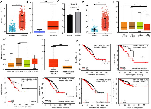
Table 1. Relationships between FAM64A mRNA expression and breast cancer clinicopathological characteristics using the xiantao database.
Table 2. Overall survival analysis in breast cancer patients using the xiantao database.
Table 3. Disease-specific survival analysis of breast cancer patients using the xiantao database.
Clinicopathological and prognostic significance of FAM64A mRNA expression in cervical cancer
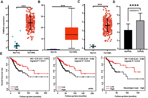
We used xiantao () [95% CI = 3.335: 2.894–3.734)], UALCAN (), TCGA () [95% CI = 2.395: 1.572–3.219)], and GEO () [95% CI = 1.946: 1.269–2.694] databases to perform bioinformatics analysis and observed that FAM64A expression was higher in cervical cancer tissue when compared with normal tissue (p < 0.05). The KM plotter showed that high FAM64A expression was positively correlated with long overall survival rates in all white, neoantigen-high cancer patients (, p < 0.05).
Figure 2. The clinicopathological and prognostic significance of FAM64A mRNA expression in cervical cancer. According to xiantao (A), UALCAN (B), TCGA (C), and GEO (D) databases, FAM64A hyperexpression was detectable in cervical cancer when compared with normal cervical tissue (p < 0.05). In the Kaplan-Meier plotter (E), relationships between FAM64A expression and cervical cancer patient survival data were analysed and stratified by clinicopathological features. Note: N, normal tissue; T, tumour; HR, hazard ratio; ***p < 0.001.
Clinicopathological and prognostic significance of FAM64A mRNA expression in endometrial cancer
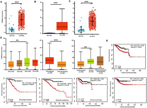
According to xiantao () [95% CI = 2.602: 2.261–2.94)], UALCAN (), and TCGA () [95% CI = 1.835: 1.579–2.097] databases, FAM64A expression was higher in endometrial cancer tissue when compared with normal tissue (p < 0.05). Levels were also higher in stage 3 when compared with stage 1 cancer, in TP53-mutant when compared with non-TP53-mutant patients, and serous adenocarcinoma when compared with endometrioid carcinoma patients (, p < 0.05). As indicated (Supplementary Table 1), FAM64A expression was positively associated with the histological grade and a minimally invasive approach (p < 0.05). From the KM plotter, FAM64A expression was positively correlated with overall survival rates in all, neoantigen-low, mutation-low or mutation-high cancer patients (, p < 0.05). The same results were observed for recurrence-free survival in all cancer patients (, p < 0.05).
Figure 3. The clinicopathological and prognostic significance of FAM64A mRNA expression in endometrial cancer. According to xiantao (A), UALCAN (B), and TCGA (C) databases, FAM64A hyperexpression was detectable in endometrial cancer when compared with normal endometrial tissue (p < 0.05). FAM64A expression was compared with endometrial cancer clinicopathological characteristics in UALCAN (D). In the Kaplan-Meier plotter (E), relationships between FAM64A expression and endometrial cancer patient survival data were analysed and stratified by clinicopathological features. Note: N, normal tissue; T, tumour; HR, hazard ratio; **p < 0.01; ***p < 0.001.
Clinicopathological and prognostic significance of FAM64A mRNA expression in ovarian cancer
We performed a bioinformatics analysis of FAM64A expression in ovarian cancer using xiantao (Supplementary Figure 1A) [95% CI = 2.506: 2.334–2.681] and GEO (Supplementary Figure 1B) [95% CI = 127.431: 91.358–186.589] databases. FAM64A had lower expression in normal ovary tissue when compared with ovarian cancer tissue (p < 0.05). UALCAN data showed higher FAM64A expression at stage 2 when compared with stage 4 ovarian cancer (Supplementary Figure 1C, p < 0.05). KM plotter data showed a positive correlation between FAM64A expression and overall survival rates in White, grade 2, mutation-high and neoantigen-low ovarian cancer patients (Supplementary Figure 1D, p < 0.05).
Relationships between FAM64A mRNA expression and infiltrating immune cells in gynecological cancers
From the xiantao database, FAM64A mRNA expression was positively related to Th2, dendritic (aDC), Treg, Th1, natural killer (NK) CD56dim, B, and Tgd cell infiltration, but negatively related to neutrophil, CD8 T, Tcm, iDC, NK CD56bright, pDC, Th17, eosinophil, NK, and mast cell infiltration in breast cancer (Supplementary Figure 2, p < 0.05).
For cervical cancer, FAM64A mRNA expression was positively related to Th2 infiltration, but negatively related to iDC, Treg, Tgd, Th17, mast, CD8 T, B, T, aDC, macrophage, DC, Th1, cytotoxic, NK CD56dim, and neutrophil cell infiltration (Supplementary Figure 2, p < 0.05).
In endometrial cancer, FAM64A mRNA expression was also positively related to Th2, Tgd, and T helper infiltration, but negatively related to NK CD56dim, B, Treg, follicular helper T (TFH), T, cytotoxic, NK, Th17, mast, iDC, eosinophil, neutrophil, pDC, and NK CD56bright cell infiltration (Supplementary Figure 2, p < 0.05).
For ovarian cancer, FAM64A mRNA expression was positively related to Th2 and T helper cell infiltration, but negatively related to neutrophil, Th1, DC, effector memory T (Tem), T, mast, CD8 T, Tcm, cytotoxic, pDC, NK CD56bright, and Th17 cell infiltration (Supplementary Figure 2, p < 0.05).
FAM64A-related genes and pathways in gynecological cancers
In the xiantao platform, we identified differentially expressed genes between low and high FAM64A expression groups in gynecological cancers. KEGG pathway analyses showed that the top signal pathways related to FAM64A included ligand-receptor interactions and transmembrane transporters, keratinisation, and intermediate filaments in breast cancer; ligand-receptor interactions and signals, extracellular matrix, and digestion in cervical cancer; ligand-receptor interactions, cilium, and peptidases in endometrial cancer; and ligand-receptor interactions, DNA-binding transcription, and synaptic vesicle in ovarian cancer (Supplementary Figure 3A). Additionally, the STRING database was used to identify PPI pairs and Cytoscape used to identify the top ten nodes ranked by connectivity levels (Supplementary Figure 3B). The top hub genes mainly included cyclins and cell cycle-related proteins in breast cancer, mucins and acetylgalactosaminyl transferases in cervical cancer, kinesin family members in endometrial cancer, and synovial sarcoma X and the cancer/testis antigen in ovarian cancer. Genes correlating with FAM64A were analysed in the xiantao database and subjected to KEGG analyses (Supplementary Figure 4). FAM64A-correlated genes involved chromosomal, cell cycle, and DNA replication processes in breast, cervical, endometrial, and ovarian cancers.
Discussion
In breast cancer cells, FAM64A and IκBα competitively interact with the NF-κB REL homology domain to generate sustained NF-κB nuclear accumulation and transcriptional activity by maintaining the NF-κB/IκBα negative feedback loop to promote aggressiveness (Jiang et al. Citation2019). Zhao et al. (Citation2022) reported that FAM64A levels gradually increased from normal to dysplastic to cancerous tissue in a carcinogenic 4-nitroquinoline-1-oxide mouse model, and promoted tumorigenesis in head and neck squamous cell carcinoma (HNSCC) by interacting with forkhead box protein M1 (FOXM1), thereby enhancing its transcriptional activity and regulating its expression via an autoregulation loop. FAM64A expression was up-regulated in neuroblastoma, osteosarcoma, glioma, breast cancer, HNSCC, lung cancer, oesophageal cancer, gastric cancer, colorectal cancer, cholangiocarcinoma, pancreatic cancer, renal clear cell carcinoma (RCC), prostate cancer, bladder cancer, leukaemia, and melanoma (Yao et al. Citation2019, Zhang et al. Citation2019, Jiang et al. Citation2020a, Citation2020b, Jiang et al. Citation2021, Qiu et al. Citation2021, Wang et al. Citation2021a, Citation2021b, Zhu et al. Citation2021, Zhu et al. Citation2022, Fu et al. Citation2023, Yao et al. Citation2023). In our study, higher FAM64A expression was identified in breast, cervical, endometrial, and ovarian cancers when compared with normal tissue. Thus, up-regulated FAM64A mRNA expression was closely linked to carcinogenic processes in gynecological cancers. These processes will be analysed in future studies.
FAM64A knockdown inhibited the proliferation, migration, and epithelial-to-mesenchymal transition (EMT) in breast cancer (Yao et al. Citation2019), prostate cancer (Zhou et al. Citation2021), and RCC (Yao et al. Citation2023) cells, while the opposite was observed for FAM64A overexpression in breast cancer (Jiang et al.Citation2019), glioma cells via the β-catenin pathway (Wang et al. Citation2021b), and colorectal cancer cells via the ERK/MAPK pathway (Zhang et al. Citation2022). Fu et al. (Citation2023) demonstrated that transforming growth factor-β (TGF-β) signalling up-regulated FAM64A, while FAM64A silencing suppressed TGF-β-stimulated EMT processes in glioma. Reportedly, FAM64A expression was positively associated with World Health Organisation grade of glioma (Zhu et al. Citation2022), RCC clinical stages, Tumour-Node-Metastasis stages, histological grades (Zhu et al. Citation2021), breast cancer tumour stemness, clinical grades, metastasis (Jiang et al. Citation2019, Wang et al. Citation2021a), and lung adenocarcinoma clinical stages (Jiang et al. Citation2021). In our study, FAM64A expression was positively correlated with the White race, low T stages, low N stages, low clinical stages, infiltrative ductal carcinoma, and favourable PAM50 classification in breast or ovarian cancer patients, but positively correlated with clinical stages, histological grade and TP53 mutation, and a serous endometrial cancer subtype. Therefore, FAM64A overexpression may be used to indicate aggressive gynecological cancer behaviours.
Reportedly, FAM64A expression was highly associated with decreased survival rates in patients with neuroblastoma, osteosarcoma, lung adenocarcinoma, breast cancer, cholangiocarcinoma, HNSCC, adrenocortical carcinoma, RCC, glioma, mesothelioma, pancreatic adenocarcinoma, prostate cancer, and sarcoma (Jiang et al. Citation2019, Yao et al. Citation2019, Zhang et al. Citation2019, Jiang et al. Citation2020a, Citation2020b, Jiang et al. Citation2021, Qiu et al. Citation2021, Wang et al. Citation2021b, Zhu et al. Citation2021, Zhao et al. Citation2022, Zhu et al. Citation2022, Fu et al. Citation2023, Yao et al. Citation2023). High FAM64A expression, as an independent hazard factor, was absolutely correlated with short overall survival or recurrence-free survival rates in pancreatic cancer (Jiao et al. Citation2019), glioma (Zhu et al. Citation2022), and RCC (Yao et al. Citation2023) patients. In our study, FAM64A expression was negatively associated with overall and/or recurrence-free survival rates in breast and endometrial cancer patients, but positively associated with cervical and ovarian cancer patients. FAM64A appeared to function as an overall and disease-specific survival independent predictor in breast cancer. Thus, FAM64A mRNA expression may be used as a potential prognostic predictor in gynecological cancers.
Zhu et al. (Citation2022) reported that FAM64A expression was mainly classified into mitotic cell cycle, mRNA splicing, DNA repair, Rho GTPase signalling, p53 transcriptional regulation, and translation pathways in glioma. Jiao et al. (Citation2019) found that FAM64A-related pathways included mitotic spindle, Myc target, mTORC1 signalling, G2/M checkpoint, E2F target, DNA repair, glycolysis, and unfolded protein responses in pancreatic cancer. Jiang et al. (Citation2021) observed that FAM64A-related genes were involved in mitotic cell cycle, mRNA splicing, DNA repair, Rho GTPase signalling, TP53 transcriptional regulation, and translation pathways in lung adenocarcinoma. In our study, FAM64A-correlated genes were principally categorised into ligand-receptor interaction, chromosome, cell cycle, and DNA replication processes in breast, cervical, endometrial, and ovarian cancers. The top hub genes primarily included cyclins and related proteins in breast cancer, mucins and acetylgalactosaminyl transferases in cervical cancer, kinesin family members in endometrial cancer, and synovial sarcoma X and the cancer/testis antigen in ovarian cancer. These findings provide novel clues on the roles and molecular mechanisms underlying FAM64A in the tumorigenesis and subsequent progression of gynecological cancers, which should be investigated in future work.
Abnormal FAM64A expression reportedly exerted an impact on immune and interferon signalling pathways in prostate cancer cells (Zhou et al. Citation2021). In glioma, FAM64A was closely related to M1 and M2 macrophage, monocyte, and CD8+ T cell infiltration, and was associated with multiple tumour and immune-related pathways in patient responses to immunotherapy (Zhu et al. Citation2022). In contrast, Jiang et al. (Citation2021) identified negative correlations for FAM64A with T cell, macrophage, B, DC, and CD8+ T cell infiltration in lung adenocarcinoma. Yao et al. (Citation2023) showed that FAM64A in RCC was positively correlated with immune cells, such as Th2 cells, Tregs, Th1 cells, T cells, macrophages, and DCs, while negatively correlated with Th17 cells, mast cells, pDCs, NK cells, and neutrophils. Zhu et al. (Citation2021) reported that FAM64A was co-expressed with genes encoding major histocompatibility complex, immune activation, immunosuppression, chemokine and chemokine receptors, and exerted effects on immune-related pathways, chemokine signalling pathways, RIG-I like receptor signalling pathways, antigen processing and presentation, FC epsilon RI pathways, T cell receptor pathways, and NK cell mediated cytotoxicity. In our study, FAM64A mRNA expression was positively related to Th2 cell infiltration, but negatively with neutrophil and Th17 cell infiltration in breast, cervical, endometrial, and ovarian cancers. Thus, FAM64A may have a role in immune surveillance and therapy and also tumour-associated immune responses in gynecological cancers.
Hussein et al. (Citation2015) used TCGA data to categorise endometrial cancers into four main genetic subtypes: polymerase epsilon (POLE) ultramutated, MSI (microsatellite instability) hypermutated, copy-number (CN) low, and CN high, which helped resolve tumour grade and histotyping limitations, myometrial invasion depth, and cervical and adnexal involvement. Among them, ultramutated (POLE) and hypermutated (MSI-H) patients demonstrated better response to monoclonal antibody therapy against immune checkpoint-associated proteins (Oaknin et al. Citation2022). Additionally, p53-mutant-associated endometrial cancer patients had worse prognoses and high recurrence rates. Endometrial cancer patients carrying CTNNB1 mutations in exon 3 had an increased risk of distant recurrence. For endometrial cancer, the molecular characteristics underpinning worse prognoses were PI3K/Akt mutations, L1CAM (L1 cell adhesion molecule) positivity, and oestrogen and progesterone receptor positivity (Di Donato et al. Citation2023, Golia D’Augè et al. Citation2023). In future studies, we will aim to identify correlations of FAM64A expression with endometrial cancer molecular subtypes, which may in turn guide clinical practice.
In summary, up-regulated FAM64A mRNA expression was closely associated with gynecological carcinogenesis, and may function as an aggressiveness or prognosis biomarker. The prognostic significance of FAM64A mRNA expression depends on the gynecological cancer type.
Our study had some limitations in that we did not validate these bioinformatic data using real-time polymerase chain reactions and laser capture dissection.
Author contributions
All authors approved the final submitted manuscript and were responsible for all aspects of the work. H-CZ: project development, study design, and manuscript writing. Z-MW, D-HR, and C-YZ: manuscript revision, data analysis, and interpreted data. LZ and YC: project development and manuscript revision.
Supplemental Material
Download Zip (3.3 MB)Disclosure statement
The authors claim no competing interests in the publication of this paper. Registration is not appliable. We wrote the article according to Enhancing the Quality and Transparency of Health Research network guidelines and we used the STREGA checklist during our writing (Little et al. Citation2009).
The authors declare that this research was conducted in the absence of any commercial or financial relationships that could be construed as a potential conflict of interest.
Data availability statement
The datasets in this study can be found in online repositories. The names and accession numbers of the repository/repositories can be found in the article/Supplementary Materials.
Additional information
Funding
References
- Aquil, A., et al., 2021. Predictors of mental health disorders in women with breast and gynecological cancer after radical surgery: a cross-sectional study. Annals of Medicine and Surgery, 65, 102278.
- Archangelo, L.F., et al., 2013. The CATS (FAM64A) protein is a substrate of the kinase interacting stathmin (KIS). Biochimica et Biophysica Acta, 1833 (5), 1269–1279.
- Barbutti, I., et al., 2016. CATS(FAM64A) abnormal expression reduces clonogenicity of hematopoietic cells. Oncotarget, 7 (42), 68385–68396.
- Di Donato, V., et al., 2023. Recent advances in endometrial cancer management. Journal of Clinical Medicine, 12 (6), 2241.
- Fu, M., et al., 2023. Cell cycle-related FAM64A could be activated by TGF-β signaling to promote glioma progression. Cellular and Molecular Neurobiology, in press. DOI: 10.1007/s10571-023-01348-2.
- Giannini, A., et al., 2022. Advances on prevention and screening of gynecologic tumors: Are we stepping forward? Healthcare, 10 (9), 1605.
- Golia D’Augè, T., et al., 2023. Novel insights into molecular mechanisms of endometrial diseases. Biomolecules, 13 (3), 499.
- Hussein, Y.R., et al., 2015. Clinicopathological analysis of endometrial carcinomas harboring somatic POLE exonuclease domain mutations. Modern Pathology, 28 (4), 505–514.
- Hashimoto, K., et al., 2017. FAM64A is a novel cell cycle promoter of hypoxic fetal cardiomyocytes in mice. Scientific Reports, 7 (1), 4486.
- Jiang, F., et al., 2021. High expression of PIMREG predicts poor survival outcomes and is correlated with immune infiltrates in lung adenocarcinoma. PeerJ, 9, e11697.
- Jiang, L., et al., 2019. Overexpression of PIMREG promotes breast cancer aggressiveness via constitutive activation of NF-κB signaling. EBioMedicine, 43, 188–200.
- Jiang, Y., et al., 2020a. FAM64A promotes osteosarcoma cell growth and metastasis and is mediated by miR-493. Journal of Oncology, 2020, 2518297.
- Jiao, Y., et al., 2019. Aberrant FAM64A mRNA expression is an independent predictor of poor survival in pancreatic cancer. PLoS One, 14 (1), e0211291.
- Jiang, Z.M., et al., 2020b. PIMREG, a marker of proliferation, facilitates aggressive development of cholangiocarcinoma cells partly through regulating cell cycle-related markers. Technology in Cancer Research & Treatment, 19, 1533033820979681.
- Little, J., et al., 2009. STrengthening the REporting of Genetic Association Studies (STREGA)–an extension of the STROBE statement. Genetic Epidemiology, 33 (7), 581–598.
- Oaknin, A., et al., 2022. Safety and antitumor activity of dostarlimab in patients with advanced or recurrent DNA mismatch repair deficient/microsatellite instability-high (dMMR/MSI-H) or proficient/stable (MMRp/MSS) endometrial cancer: interim results from GARNET-a phase I, single-arm study. Journal for ImmunoTherapy of Cancer, 10 (1), e003777.
- Paepke, D., et al., 2020. Prevalence and predictors for nonuse of complementary medicine among breast and gynecological cancer patients. Breast Care, 15 (4), 380–385.
- Qiu, X., et al., 2021. FAM64A antagonizes tumor suppressive effects of miR-610 in neuroblastoma in vitro. Journal of Neurosurgical Sciences, 2021, 9.
- Segev, Y., et al., 2021. Correlation between an integrative oncology treatment program and survival in patients with advanced gynecological cancer. Supportive Care in Cancer, 29 (7), 4055–4064.
- Seland, M., et al., 2022. Distress, problems and unmet rehabilitation needs after treatment for gynecological cancer. Acta Obstetricia et Gynecologica Scandinavica, 101 (3), 313–322.
- Vargiu, V., et al., 2022. Impact of obesity on sentinel lymph node mapping in patients with apparent early-stage endometrial cancer: the ObeLyX study. Gynecologic Oncology, 165 (2), 215–222.
- Wang, D., et al., 2021a. Tumor-promoting function of PIMREG in glioma by activating the β-catenin pathway. Biotech, 11 (8), 380.
- Wang, L., et al., 2021b. Identification of immune-related therapeutically relevant biomarkers in breast cancer and breast cancer stem cells by transcriptome-wide analysis: a clinical prospective study. Frontiers in Oncology, 10, 554138.
- Xu, Z.S., et al., 2019. FAM64A positively regulates STAT3 activity to promote Th17 differentiation and colitis-associated carcinogenesis. Proceedings of the National Academy of Sciences of the United States of America, 116 (21), 10447–10452.
- Yao, H., et al., 2023. PIMREG is a prognostic biomarker involved in immune microenvironment of clear cell renal cell carcinoma and associated with the transition from G1 phase to S phase. Frontiers in Oncology, 13, 1035321.
- Yao, Z., et al., 2019. Knockdown of FAM64A suppresses proliferation and migration of breast cancer cells. Breast Cancer, 26 (6), 835–845.
- Zhang, J., et al., 2019. Up-regulation of FAM64A promotes epithelial-to-mesenchymal transition and enhances stemness features in breast cancer cells. Biochemical and Biophysical Research Communications, 513 (2), 472–478.
- Zhang, X., et al., 2022. PICALM exerts a role in promoting CRC progression through ERK/MAPK signaling pathway. Cancer Cell International, 22 (1), 178.
- Zhao, X., et al., 2022. FAM64A promotes HNSCC tumorigenesis by mediating transcriptional autoregulation of FOXM1. International Journal of Oral Science, 14 (1), 25.
- Zhao, W.M., et al., 2008. RCS1, a substrate of APC/C, controls the metaphase to anaphase transition. Proceedings of the National Academy of Sciences of the United States of America, 105 (36), 13415–13420.
- Zheng, H.C., et al., 2023. The bioinformatics analysis about the clinicopathological and prognostic significances of oocyte-arresting BTG4 mRNA in gynecological cancers. Journal of Obstetrics and Gynaecology, 43 (1), 2182672.
- Zhou, Y., et al., 2021. FAM64A is an androgen receptor-regulated feedback tumor promoter in prostate cancer. Cell Death & Disease, 12 (7), 668.
- Zhu, H., et al., 2022. Predictive value of PIMREG in the prognosis and response to immune checkpoint blockade of glioma patients. Frontiers in Immunology, 13, 946692.
- Zhu, H., et al., 2021. Pan-cancer analysis of PIMREG as a biomarker for the prognostic and immunological role. Frontiers in Genetics, 12, 687778.