

Abstract
Background
Cervical cancer (CC) is the second most common malignancy in women, and identifying biomarkers of CC is crucial for prognosis prediction. Here, we investigated the expression of AF4/FMR2 Family Member 3 (AFF3) in CC and its association with clinicopathological features and prognosis.
Methods
Tumour and adjacent tissues, along with clinicopathological features and follow-up information, were collected from 78 patients. AFF3 expression was assessed using quantitative real-time polymerase chain reaction and Western blotting. The correlation between AFF3 expression and CC symptoms was using chi-square test. The 5-year overall survival (OS) was analysed using the Kaplan-Meier method. The Univariate analysis of prognostic risk factors was conducted using the COX proportional hazards model, followed by multivariate COX regression analysis including variables with p < 0.01.
Results
AFF3 expression was downregulated in CC, and its levels were correlated with lymph node metastasis (LNM) and International Federation of Gynaecology and Obstetrics (FIGO) stage. Patients with low AFF3 expression had a lower 5-year OS rate (52.78%, 19/36). Postoperative survival was reduced in patients with histological grade 3 (G3), myometrial invasion (depth ≥ 1/2), lymphovascular space invasion, LNM, and advanced FIGO stage. Low expression of AFF3 (HR: 2.848, 95% CI: 1.144–7.090) and histological grade G3 (HR: 4.393, 95% CI: 1.663–11.607) were identified as independent prognostic risk factors in CC patients.
Conclusion
Low expression of AFF3 and histological G3 are independent predictors of poor prognosis in CC patients, suggesting that AFF3 could serve as a potential biomarker for prognostic assessment in CC.
PLAIN LANGUAGE SUMMARY
Cervical cancer is a significant health concern worldwide, responsible for over 300,000 deaths annually and ranking as the fourth most common cancer in women. Existing screening methods have limitations, highlighting the need for innovative therapies. In our research, we identified a specific genetic material that varied significantly among cervical cancer patients with varying survival outcomes, detected in tissue samples obtained post-surgery. Our study demonstrates the considerable potential of this marker for accurately predicting outcomes in our study population. By analysing differences in the expression of this genetic marker, we can forecast the prognosis and progression of cervical cancer. These findings offer valuable insights for advancing cervical cancer treatment strategies, potentially improving outcomes for patients. Early detection and targeted treatment based on this genetic marker could extend patients’ lives and prevent fatalities by enabling timely medical intervention and management.
Keywords:
Introduction
Cervical cancer (CC) ranks as the fourth most prevalent cancer among women globally, with over 500,000 new cases diagnosed globally, resulting in more than 300,000 deaths each year (Cohen et al. Citation2019). Risk factors associated with CC include human papilloma virus infections, parity, smoking, and prolonged use of oral contraceptives (Johnson et al. Citation2019). While surgery remains the primary treatment for CC, including type III open radical hysterectomy with bilateral pelvic lymph node dissection, it can be accompanied by short-term and long-term complications (Poddar and Maheshwari Citation2021). Immunotherapy has emerged as a promising novel treatment for CC, targeting tumour cells more effectively, with pembrolizumab being the only immunotherapy agent currently approved for CC treatment (Ferrall et al. Citation2021). Therefore, there is an ongoing need to identify biomarkers for CC and validate their prognostic and therapeutic potential (Wang et al. Citation2022).
The AF4/FMR2 family (AFF) consists of four members: AFF1, AFF2, AFF3, and AFF4, playing crucial roles in lymphoid cell development, transcription elongation, protein binding, and various cellular processes (Bitoun and Davies Citation2009, Marschalek Citation2010). Among these, AFF3, a transcription factor highly expressed in lymphoid tissues, is implicated in early lymphocyte development (von Bergh et al. Citation2002). Previous research has demonstrated the involvement of AFF3 in the progression of various cancers, including breast cancer (Chen et al. Citation2020), non-small cell lung cancer (Zhang et al. Citation2018), adrenocortical carcinoma (Lefèvre et al. Citation2015), and glioblastoma (Bhargava et al. Citation2017). Dysregulation of AFF3 expression has been associated with poor prognosis and drug resistance in these cancers. For instance, AFF3 may contribute to immune cell infiltration and lymph node metastasis (LNM) in gastric cancer, with high AFF3 expression correlating with poor overall survival (Zeng et al. Citation2022). In breast cancer, overexpression of AFF3 leads to tamoxifen insensitivity and lower overall survival rate rates in patients with primary luminal breast cancers (Shi et al. Citation2018). Despite these findings, the clinical significance of AFF3 expression in CC and its relationship with CC prognosis remain unclear.
In this study, we conducted a prospective cohort study involving patients with CC, collecting cancer tissues and pathological information to investigate the expression of AFF3 in CC tissues. Our primary objective was to examine the association between AFF3 expression and the clinicopathological features and prognosis of CC patients. By exploring new biomarkers that hold the potential for predicting CC prognosis, our study aims to enhance the post-surgery survival rate of patients and contribute to the development of novel cancer treatments.
Methods
Collection of tissues and general data of patients
A prospective cohort study was conducted involving 78 patients diagnosed with CC who underwent surgery at our hospital between January 2015 and January 2018. During surgery, cancer tissue samples and adjacent normal tissues, with a distance from the tumour edge > 2 cm, were collected and prepared into paraffin blocks stored in the pathology department. Subsequently, tissue sections were prepared, deparaffinized, and stained for later analysis. Clinicopathological information, including age, pathological type, histological grade, tumour diameter, myometrial invasion (with or without), lymphovascular space invasion (LVSI) (with or without), lymph node metastasis (with or without), distant metastasis (with or without), and International Federation of Gynaecology and Obstetrics (FIGO) stage, was recorded for all included patients (Bhatla et al. Citation2021), etc.
Inclusion and exclusion criteria
Inclusion criteria were as follows: (1) Meeting the diagnostic criteria of CC (Koh et al. Citation2015); (2) Age over18 years; (3) First-time diagnosis and treatment; (4) Informed consent signed by patients or their relatives; (5) Surgical indications without contraindications to radiotherapy and chemotherapy; (6) Consent to enrol in a long-term follow-up study.
Exclusion criteria were as follows: (1) History of cervical surgery or other cervical diseases; (2) Complications of other malignant tumours, severe organ dysfunction, autoimmune or infectious diseases; (3) Secondary CC; (4) Recurrence of CC.
Treatment and follow-up study
All patients included in the study underwent radical hysterectomy combined with pelvic lymph node dissection. Following surgery, the treatment approach, platinum-based concurrent chemoradiotherapy, was determined based on factors such as age, pathological type, stage, and patient preferences. This treatment regimen commenced between the 2nd and 4th weeks post-surgery. Central pelvic radiotherapy was administered at a total dose of 45 Gy, with a fraction size of 1.8 Gy/fraction, 1 fraction/day, and 5 fractions/week. Chemotherapy was performed once a week.
All patients were followed up for 5 years post-surgery, primarily through regular outpatient reviews and follow-up via Wechat or telephone. Outpatient reviews included gynecological examinations, ThinPrep cytologic tests, pelvic ultrasound, pelvic magnetic resonance imaging, among other examinations. Follow-up visits were scheduled every 3 months during the first and second years post-surgery, and every 6 months thereafter. Overall survival (OS) was calculated as the time (in months) from surgery to death.
Quantitative real-time polymerase chain reaction (qRT-PCR)
CC tissues and adjacent tissues were homogenised post-grinding and centrifuged at 4 °C and 450 × g for 10 minutes. Total RNA extraction from tissues was performed using the TRIzol method (Thermo Fisher, Waltham, USA), followed by reverse-transcription into complementary DNA (cDNA) using PrimeScriptTM RT MasterMix (TaKaRa, Tokyo, Japan). qRT-PCR was performed on a Mastercycler®nexus X2 (Eppendorf, Hamburg, Germany) with cDNA as the template. Data were analysed using the 2-ΔΔCt method (Livak and Schmittgen Citation2001), and the relative expression of AFF3 mRNA was calculated using glyceraldehyde-3-phosphate dehydrogenase (GAPDH) as the internal reference gene. Primer sequences are provided in Supplementary Table S1.
Western blot (WB) assay
Tissue samples were collected and lysed using radio immunoprecipitation assay (RIPA) lysis buffer, followed by vibration and centrifugation to extract total protein. The total protein content was quantified using the bicinchoninic acid (BCA) method (Pierce, Rockford, IL, USA). Equal amounts of proteins were separated by sodium dodecyl sulfate-polyacrylamide gel electrophoresis (SDS-PAGE) and transferred onto polyvinylidene fluoride membranes (Millipore, Billerica, MA, USA). The membranes were incubated overnight at 4 °C with primary antibodies against AFF3 (ab106231, 1:500, Abcam, Cambridge, MA, USA) and β-actin (ab213262, 1: 1000, Abcam). Following washing, horseradish peroxidase-conjugated anti-rabbit Immunoglobulin G (IgG) secondary antibody (ab205718, 1:2000, Abcam) was added and incubated for 2 hours at room temperature. The enhanced chemiluminescence system (CW Biotech, Beijing, China) was used for colour development, and bands were obtained by scanning with Amersham Imager 680.
Bioinformatics
The expression of AFF3 mRNA in CC tissues was investigated using UALCAN (http://ualcan.path.uab.edu/) (Chandrashekar et al. Citation2017) and GEPIA (http://gepia.cancer-pku.cn/) (Tang et al. Citation2017).
Statistical analysis
Descriptive analysis of qualitative data frequency (N) and constituent ratio (%) was conducted using SPSS 22.0 software. Quantitative data were expressed as mean ± standard deviation (mean ± SD). The Chi-square test was utilised to detect differences in clinicopathological features between low and high AFF3 expression groups. Survival curves for the low and high AFF3 expression groups and patients with different clinicopathological features were plotted using the Kaplan-Meier method, with differences between the curves analysed using the log-rank test. The COX proportional hazards model was employed to analyse the prognostic risk factors based on clinical information in CC patients, and hazard ratios (HR) and 95% confidence interval (CI) were calculated. A significance level of p < 0.05 was considered statistically significant.
Results
Clinical information of patients with CC
A total of 78 patients were enrolled in the study, with a median age of 45 (range: 28–77) years (). Following postoperative pathological examination, statistical analysis was conducted on the patients’ pathological classification, histological grade, tumour size, LNM, and other relevant information. Detailed data are presented in .
Figure 1. Study process.
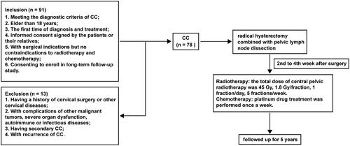
Table 1. Clinical information of CC patients.
AFF3 expression in CC tissues
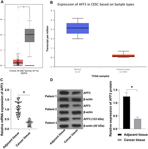
To confirm the differential expression of AFF3 in normal and tumour tissues, we utilised the GEPIA and UALCAN websites. The analysis revealed a reduction in AFF3 expression in CC tissues compared to normal tissues (). Furthermore, mRNA and protein levels of AFF3 were evaluated in tissue specimens and paired adjacent normal tissues from 78 patients with CC. Our results demonstrated a significant decrease in AFF3 expression in CC tissues compared to adjacent tissues (p < 0.05, ).
Figure 2. AFF3 expression in CC tissues. (A, B): AFF3 expression was assessed using GEPIA and UALCAN databases; cancer tissue samples and adjacent tissues were collected from CC patients; (C): Relative expression of AFF3 mRNA in the tissues was measured by qRT-PCR; (D): Relative expression of AFF3 protein was detected by WB, with representative bands shown. Data were expressed as mean ± standard deviation. n = 78 for (C, D). Data (C, D) were analysed using the paired t test. *p < 0.05.
Association of AFF3 expression with clinicopathological features
To explore the relationship between AFF3 expression and clinicopathological features of CC patients, patients were divided into low AFF3 expression group (AFF3 ≤ 0.30, n = 36) and high AFF3 expression group (AFF3 > 0.30, n = 42) based on the average relative mRNA expression of AFF3. Correlation analysis revealed significant associations between AFF3 expression and LNM as well as FIGO stage (p < 0.05). Patients with LNM exhibited higher odds of low AFF3 expression, and those with advanced FIGO stage showed higher odds of low AFF3 expression in cancer tissues. However, no clear relationship was observed between mRNA expression of AFF3 and patient’s age, pathological type, histological grade, tumour diameter, depth of myometrial invasion, lymphovascular space invasion (LVSI), and distant metastasis (p > 0.05). Additional details are provided in .
Table 2. AFF3 expression is associated with the clinicopathological features of CC patients.
Clinicopathological features and AFF3 expression associated with 5-year OS
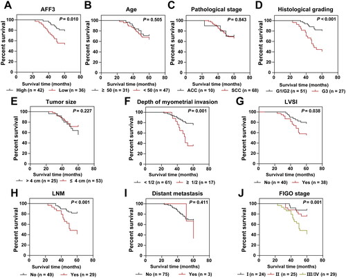
Previous studies have highlighted the significance of AFF3 in cancer progression and prognosis in various cancers, including breast cancer (Chen et al. Citation2020), non-small cell lung cancer (Zhang et al. Citation2018), and gastric cancer (Zeng et al. Citation2022). We investigated the relationship between AFF3 expression in CC and patient prognosis over a 5-year follow-up period. Among the enrolled population, 26 patients died within the 5-year follow-up period, resulting in a 5-year survival rate of 66.67%. Kaplan-Meier curve analysis and log-rank test revealed a lower survival rate among CC patients with low AFF3 expression (52.78%, 19/36) compared to those with high AFF3 expression (78.57%, 33/42) (p < 0.05, ). Additionally, patients with histological G3, depth of myometrial invasion ≥ 1/2, lymphovascular space invasion (LVSI), lymph node metastasis (LNM), and advanced FIGO stage exhibited a lower postoperative survival rate (all p < 0.05, )). However, no significant difference in postoperative survival rate was observed among patients of different ages, pathological types, tumour diameters, and distant metastasis (all p > 0.05, ).
Figure 3. Association between clinicopathological features, AFF3 expression, and 5-year OS of CC patients. (A–J): OS analysis based on AFF3 expression and clinicopathological features using Kaplan-Meier curves. Log-rank test was performed.
AFF3 as an independent prognostic factor for CC patients
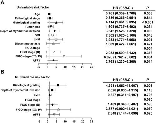
Kaplan-Meier analysis revealed the association between AFF3 expression level, clinicopathological factors, and prognosis. COX univariate analysis indicated that AFF3 expression, FIGO stage, LNM, LVSI, depth of myometrial invasion, and histological grade were related to patient prognosis (all p < 0.05, ). Multivariate COX regression analysis, including variables with p < 0.1 in univariate analysis, identified low expression of AFF3 as an independent risk factor for overall survival (OS) in CC patients after surgery. Patients with low expression of AFF3 had a 2.848 times higher risk of death compared to those with high AFF3 expression (HR: 2.848, 95%CI: 1.144–7.090, p = 0.025). Moreover, histological grade 3 (G3) was also identified as an independent risk factor for the prognosis of CC patients (HR: 4.393, 95%CI: 1.663–11.607, p = 0.003) (all p < 0.05, ).
Figure 4. AFF3 as an independent prognostic factor for CC patients. (A): COX univariate analysis illustrating the relationship between AFF3 expression, clinicopathological factors, and prognosis. (B): COX multivariate analysis identifying independent prognostic factors for CC. Line segments represent 95%CI.
Discussion
Recent years have witnessed extensive exploration into prognostic markers for cervical cancer (CC), including CD177 (Liao et al. Citation2023), interleukin 6 receptor (Luan et al. Citation2018), and Prostaglandin E receptor type 3 (Heidegger et al. Citation2017). Molecular-targeted therapies based on these biomarkers have been developed to specifically target cancer-related pathways, thereby enhancing treatment efficacy while minimising side effects (Tsuda et al. Citation2016). For instance, downregulation of MCM5, a marker highly expressed in CC, has been shown to reduce CC cell proliferation, with correlations to clinical stage, lymph node metastasis (LNM), distant metastasis, and histological grade. (Wang et al. Citation2018). Similarly, CYP4Z1 expression, along with histological stage, tumour invasion, and LNM, has been significantly linked to overall survival (OS) in CC (Al-Saraireh et al. Citation2021). Our study contributes to this body of research by identifying low expression of AFF3 in CC tissues. Patients with low AFF3 expression were more likely to exhibit LNM and advanced FIGO stage. Furthermore, AFF3 expression, histological grade 3 (G3), depth of myometrial invasion ≥ 1/2, lymphovascular space invasion (LVSI), LNM, and late FIGO stage were associated with shorter postoperative survival time. Importantly, low AFF3 expression and histological G3 emerged as independent prognostic factors for CC patients.
The AFF family members have been implicated in a spectrum of diseases, spanning leukaemia (Tirtakusuma et al. Citation2022), epilepsy (Zou et al. Citation2022), and keloids (Gao et al. Citation2022). Particularly noteworthy is their involvement in cancer progression across various malignancies. For example, AFF1 inhibits neuropeptide neurotensin transcription, enhancing the prognosis of lung cancer, where high expression of AFF1 correlates with improved overall survival (Yue et al. Citation2021). Conversely, AFF4 is notably downregulated in colorectal cancer (CRC), and its deletion amplifies the metastatic capacity of CRC cells in vivo (Fang et al. Citation2022). AFF3, also known as LAF4, a member of the AFF family, exerts a pivotal role in cancer progression with varying expression patterns across different malignancies. In adrenocortical cancer, AFF3 activates oncogenic Wnt/β-catenin signalling pathways, promoting cancer progression (Lefèvre et al. Citation2015), while in breast cancer, inhibition of AFF3 degradation augments ER signalling (Liang et al. Citation2022). Conversely, overexpression of AFF3 has been noted in B cells and endothelial cells in lung adenocarcinoma (Song et al. Citation2023) and pancreatic cancer cells (Zang et al. Citation2023). However, a positive correlation between AFF3 expression and disease-free survival has been observed in prostate cancer (Fan et al. Citation2022). Importantly, AFF3 is down-regulated in the white race population with CC (Chandra et al. Citation2022). Our study corroborates these findings, revealing decreased AFF3 levels in CC tissues. While no correlation was found between AFF3 expression and patient age, pathological type, histological grade, tumour diameter, depth of myometrial invasion, LVSI, and distant metastasis, low AFF3 expression was associated with LNM and advanced FIGO stage. This aligns with prior research indicating a potential association between AFF3 mRNA expression and LNM in small-cell lung cancer (Kong et al. Citation2023), as well as a significant correlation between high AFF3 levels and LNM in advanced gastric cancer (Zeng et al. Citation2022). Further investigations are warranted to elucidate the divergent roles of AFF3 in different cancers. We speculate that the differential expression of AFF3 between lung adenocarcinoma or pancreatic cancer and CC concerning LNM staging may be linked to its distinct regulatory roles in immune-related functions.
Previous research has highlighted the prognostic significance of AFF in various cancers. For example, AFF3 has been positively correlated with recurrence-free survival in prostate cancer (Fan et al. Citation2022), while its overexpression in lung cancer has been linked to gefitinib resistance, impacting the prognosis of lung cancer patients (Song et al. Citation2023). Consistent with these findings, our 5-year follow-up study revealed a substantial difference in the overall survival (OS) rate between patients with low AFF3 expression (52.78%, 19/36) and those with high AFF3 expression (78.57%, 9/42). This observation underscores the importance of AFF3 as a prognostic marker in cervical cancer (CC). Moreover, our study corroborates previous research indicating that lymph node metastasis (LNM) and advanced FIGO stage contribute to poor prognosis in CC patients (Merz et al. Citation2020, Zhang et al. Citation2020). Our COX univariate analysis further identified AFF3 expression, FIGO stage, LNM, lymphovascular space invasion (LVSI), depth of myometrial invasion, and histological grade as factors influencing the prognosis of CC patients. Subsequent multivariate COX regression analysis confirmed low AFF3 expression and histological grade 3 (G3) as independent risk factors for OS in CC patients after surgery.
However, our study is not without limitations. Firstly, the relatively limited number of patients included in this study may introduce biases in the clinical data, and the patient’s pathological conditions and AFF3 expression may be influenced by other incidental factors. Secondly, the non-uniform distribution of some prognostic factors among patients could affect the reliability of our results. Thirdly, the mechanistic role of AFF3 in CC remains unexplored. Additionally, all patients in our study received standardised treatment comprising cisplatin and radiotherapy. Nonetheless, inherent resistance to cisplatin or radiotherapy in certain patients may have led to treatment failure, potentially influencing the experimental outcomes. Moreover, we did not dynamically track changes in AFF3 expression with CC progression. In future studies, we intend to address these limitations by expanding the sample size, ensuring a sufficient number of cases to mitigate biases, and investigating the mechanistic role of AFF3 in CC. We plan to explore upstream and downstream mechanisms of AFF3 and conduct animal and cellular-level experiments to elucidate its effects on the growth, invasion, migration, and other malignant behaviours of CC cells, thereby contributing new insights into CC treatment.
Conclusions
In conclusion, our study revealed that AFF3 expression is significantly lower in CC tissues, and patients with low AFF3 expression are more likely to exhibit lymph node metastasis (LNM) and advanced FIGO stage. Moreover, the survival rate of CC patients with low AFF3 expression is notably lower compared to those with high AFF3 expression. These findings underscore the potential of AFF3 as a crucial biomarker for predicting the prognosis of CC. Moving forward, our future research will prioritise animal and cellular experiments to further elucidate the impact of AFF3 on the development of CC cells. Additionally, we aim to expand the sample size to enhance the robustness of our findings and validate the utility of AFF3 as a prognostic indicator in CC.
Ethical approval
This study received approval from the Ethics Committee of YanBian University Hospital (2023207).
Informed consent
Informed consent was obtained from all study participants.
Supplemental Material
Download MS Word (15.4 KB)Acknowledgements
We express our gratitude to all the participants who contributed to this study.
Data availability statement
The data that support the findings of this study are available from the corresponding author upon reasonable request.
Additional information
Funding
References
- Al-Saraireh, Y. M., et al., 2021. Cytochrome 4Z1 expression is correlated with poor prognosis in patients with cervical cancer. Current Oncology (Toronto, Ont.), 28 (5), 1–9.
- Bhargava, S., et al., 2017. Elucidation of the genetic and epigenetic landscape alterations in RNA binding proteins in glioblastoma. Oncotarget, 8 (10), 16650–16668.
- Bhatla, N., et al., 2021. Cancer of the cervix uteri: 2021 update. International Journal of Gynaecology and Obstetrics: The Official Organ of the International Federation of Gynaecology and Obstetrics, 155 Suppl 1Suppl 1 (Suppl 1), 28–44.
- Bitoun, E. and Davies, K. E., 2009. The robotic mouse: understanding the role of AF4, a cofactor of transcriptional elongation and chromatin remodelling, in purkinje cell function. Cerebellum (London, England), 8 (3), 175–183.
- Chandra, S., et al., 2022. Molecular heterogeneity of cervical cancer among different ethnic/racial populations. Journal of Racial and Ethnic Health Disparities, 9 (6), 2441–2450.
- Chandrashekar, D. S., et al., 2017. UALCAN: a portal for facilitating tumor subgroup gene expression and survival analyses. Neoplasia (New York, N.Y.), 19 (8), 649–658.
- Chen, F., et al., 2020. RNA-seq analysis identified hormone-related genes associated with prognosis of triple negative breast cancer. Journal of Biomedical Research, 34 (2), 129–138.
- Cohen, P. A., et al., 2019. Cervical cancer. Lancet (London, England), 393 (10167), 169–182.
- Fan, A., et al., 2022. A novel prognostic model for prostate cancer based on androgen biosynthetic and catabolic pathways. Frontiers in Oncology, 12, 950094.
- Fang, Y., et al., 2022. AFF4 predicts the prognosis of colorectal cancer patients and suppresses colorectal cancer metastasis via promoting CDH1 expression. Frontiers in Oncology, 12, 797392.
- Ferrall, L., et al., 2021. Cervical cancer immunotherapy: facts and hopes. Clinical Cancer Research: An Official Journal of the American Association for Cancer Research, 27 (18), 4953–4973.
- Gao, H., et al., 2022. Circular RNA hsa_circ_0057452 facilitates keloid progression by targeting the microRNA-1225-3p/AF4/FMR2 family member 4 axis. Bioengineered, 13 (5), 13815–13828.
- Heidegger, H., et al., 2017. The prostaglandin EP3 receptor is an independent negative prognostic factor for cervical cancer patients. International Journal of Molecular Sciences, 18 (7), 1571.
- Johnson, C. A., et al., 2019. Cervical cancer: an overview of pathophysiology and management. Seminars in Oncology Nursing, 35 (2), 166–174.
- Koh, W. J., et al., 2015. Cervical cancer, version 2.2015. Journal of the National Comprehensive Cancer Network: JNCCN, 13 (4), 395–404; quiz 404.
- Kong, K., et al., 2023. Integrative genomic profiling reveals characteristics of lymph node metastasis in small cell lung cancer. Translational Lung Cancer Research, 12 (2), 295–311.
- Lefèvre, L., et al., 2015. Combined transcriptome studies identify AFF3 as a mediator of the oncogenic effects of beta-catenin in adrenocortical carcinoma. Oncogenesis, 4 (7), e161–e161.
- Liang, X., et al., 2022. DLGAP1-AS2 promotes estrogen receptor signalling and confers tamoxifen resistance in breast cancer. Molecular Biology Reports, 49 (5), 3939–3947.
- Liao, W., et al., 2023. Diagnostic, prognostic, and immunological roles of CD177 in cervical cancer. Journal of Cancer Research and Clinical Oncology, 149 (1), 173–189.
- Livak, K. J. and Schmittgen, T. D., 2001. Analysis of relative gene expression data using real-time quantitative PCR and the 2(-Delta Delta C(T)) Method. Methods (San Diego, Calif.), 25 (4), 402–408.
- Luan, S., et al., 2018. Interleukin 6 receptor (IL-6R) was an independent prognostic factor in cervical cancer. Histology and Histopathology., 33 (3), 269–276.
- Marschalek, R., 2010. Mixed lineage leukemia: roles in human malignancies and potential therapy. The FEBS Journal, 277 (8), 1822–1831.
- Merz, J., et al., 2020. Revised FIGO staging for cervical cancer - a new role for MRI. Rofo: Fortschritte Auf Dem Gebiete Der Rontgenstrahlen Und Der Nuklearmedizin, 192 (10), 937–944.
- Poddar, P. and Maheshwari, A., 2021. Surgery for cervical cancer: consensus & controversies. The Indian Journal of Medical Research, 154 (2), 284–292.
- Shi, Y., et al., 2018. AFF3 upregulation mediates tamoxifen resistance in breast cancers. Journal of Experimental & Clinical Cancer Research: Cr, 37 (1), 254.
- Song, P., et al., 2023. The underlying mechanism involved in gefitinib resistance and corresponding experiment validation in lung cancer. Mediators of Inflammation, 2023, 9658912–9658927.
- Tang, Z., et al., 2017. GEPIA: a web server for cancer and normal gene expression profiling and interactive analyses. Nucleic Acids Research, 45 (W1), W98–W102.
- Tirtakusuma, R., et al., 2022. Epigenetic regulator genes direct lineage switching in MLL/AF4 leukemia. Blood, 140 (17), 1875–1890.
- Tsuda, N., et al., 2016. Chemotherapy and molecular targeting therapy for recurrent cervical cancer. Chinese Journal of Cancer Research = Chung-Kuo Yen Cheng Yen Chiu, 28 (2), 241–253.
- von Bergh, A. R., et al., 2002. LAF4, an AF4-related gene, is fused to MLL in infant acute lymphoblastic leukemia. Genes, Chromosomes & Cancer, 35 (1), 92–96.
- Wang, D., et al., 2018. The role of MCM5 expression in cervical cancer: Correlation with progression and prognosis. Biomedicine & Pharmacotherapy = Biomedecine & Pharmacotherapie, 98, 165–172.
- Wang, R., et al., 2022. PD-L1: can it be a biomarker for the prognosis or a promising therapeutic target in cervical cancer? International Immunopharmacology, 103, 108484.
- Yue, J., et al., 2021. Suppression of the NTS-CPS1 regulatory axis by AFF1 in lung adenocarcinoma cells. The Journal of Biological Chemistry, 296, 100319.
- Zang, L., et al., 2023. Machine learning algorithm integrates bulk and single-cell transcriptome sequencing to reveal immune-related personalized therapy prediction features for pancreatic cancer. Aging,), 15 (23), 14109–14140.
- Zeng, Y., et al., 2022. AFF3 is a novel prognostic biomarker and a potential target for immunotherapy in gastric cancer. Journal of Clinical Laboratory Analysis., 36 (6), e24437.
- Zhang, C., et al., 2020. FABP5 promotes lymph node metastasis in cervical cancer by reprogramming fatty acid metabolism. Theranostics, 10 (15), 6561–6580.
- Zhang, D. L., et al., 2018. Genome-wide identification of transcription factors that are critical to non-small cell lung cancer. Cancer Letters, 434, 132–143.
- Zou, D., et al., 2022. AFF2 is associated with X-Linked partial (focal) epilepsy with antecedent febrile seizures. Frontiers in Molecular Neuroscience, 15, 795840.