

Abstract
Background
Primary amenorrhoea (PA) refers to an ailment when adolescent girls do not attain menarche naturally. It is one of the most common gynaecological disorders specified. Chromosomal abnormalities play a pivotal role in PA. Cytogenetic analysis is an indispensable diagnostic tool to determine the abnormality of the chromosome. In an emerging country like India, cytogenetic analysis is at a nascent stage. There are very few studies on Cytogenetics present in eastern India, including West Bengal. In rural and suburban areas PA sufferers often experience late diagnosis and struggle to access suitable curative management. The aim of the study is to evaluate the various types of chromosomal abnormalities in patients suffering from PA for accurate, better management of the same and further counselling.
Methods
A total of 40 PA cases were referred by obstetricians and gynaecologists to the Department of Genetics of Nirnayan Health Care, Kolkata. To screen the chromosomal abnormalities, human leukocyte culture was accomplished with their peripheral venous blood followed by G-banding and then karyotyping was executed according to ISCN-2020.
Result
Out of 40 patients, 29 were normal among which 46,XX was found in 70% cases (n = 28) and 46,XX,9qh + in 2.5% (n = 1). The remaining 11 showed different types of abnormalities. 45,X was found in 10% (n = 4), 46,X,i(X)(q10) in 2.5% (n = 1), 46,X,del(X)(p11.2) in 2.5% (n = 1), 46,X,del(X)(p22.1) in 2.5% (n = 1), 46,X,del(X)(q24) in 2.5% (n = 1), 46,XY in 2.5% (n = 1), mos 45,X[22]/46,Xi(X)(q.10)[8] in 2.5% (n = 1) and mos 45,X[16]/46,XY[14] (2.5%) in 2.5% (n = 1).
Conclusion
This study indicates the importance of chromosomal study which must be included in early diagnosis of PA. Karyotyping at the appropriate phase of life will not only help in the judicial management of this disorder but will also give young girls a better lifestyle.
PLAIN LANGUAGE SUMMARY
Primary amenorrhoea is a common gynecological disorder reported in adolescent girls, often linked to chromosomal abnormalities. In Eastern India, including West Bengal, where cytogenetic analysis is still in its nascent stage, late diagnosis and limited access to curative management are prevalent issues. A study conducted from January 2021 to May 2023 at Nirnayan Healthcare, Kolkata aimed to evaluate chromosomal abnormalities in 40 PA cases. Out of these, 28 exhibited normal karyotypes (46,XX); one patient was reported with 46,XX,9qh + which is considered a normal karyotype, while the remaining 11 revealed diverse abnormalities, including 45,X; sex reversal & several structural variations. The study underscores the significance of cytogenetic analysis in the early diagnosis of Primary Amenorrhoea. Early karyotyping not only facilitates judicious management but also ensures a better lifestyle for affected girls.
Introduction
Menstruation is an important parameter of healthy adolescence in girls. Generally, the onset of menstruation occurs at the age of 11–15 years (Menarche) (Patavegar et al. Citation2015). Primary Amenorrhoea (PA) refers to a phenomenon when a girl does not attain menarche spontaneously at this age. This condition may or may not be associated with the delayed development of secondary sexual characters. It is the most common gynaecological disorder in pubertal girls (Mujumdar et al. Citation2015). The main clinical findings of this disorder are hypoplastic uterus, imperforated hymen, bilateral undescended testes, gonadal dysgenesis, Mullerian agenesis etc.
The cause of PA may be improper functioning of the hypothalamus or pituitary, defect in the anatomy and functioning of ovaries and uterus or genetic abnormalities at the chromosome or gene level (Merin et al. Citation2012, Kanaan et al. Citation2022, 2023). Some studies have shown that the frequency of chromosomal abnormality in these patients varies from 15.9% to 63.3% (Pal et al. Citation2019). In PA, there may be structural or numerical abnormalities of the sex chromosome. Numerical abnormalities include Turner Syndrome (45,X) or Triple X syndrome (47,XXX) (Birmingham Citation2008) and structural abnormalities include deletion or inversion of some genes of X chromosome (Safaei et al. Citation2010).
Due to the advancement of cytogenetics, the detection of chromosomal abnormality is much easier and also a standard economical method than microarray and gene sequencing. Unlike Western countries, cytogenetics study is at a nascent stage in a developing country like India. There are very few laboratories that provide the facility for chromosome study in eastern India including West Bengal. In this study, we will emphasise the different chromosomal abnormalities in PA individuals with the help of karyotyping. PA is a very crucial health parameter to be taken care of. In India, women’s health issues are much neglected due to a lack of awareness, difficulties in reaching proper healthcare facilities and social taboos.
The study aimed to evaluate the chromosomal abnormalities in patients with PA and accurate diagnosis for better management and further counselling and to investigate the prevalence and types of chromosomal abnormalities associated with PA. The purpose of this study was also to create a database for further studies.
Methods
A study was conducted on the patients with symptoms of PA from urban, suburban and rural areas of West Bengal and other parts of eastern India, referred by Gynaecologists and Obstetricians to the Department of Genetics of Nirnayan Health Care Private Limited, Kolkata. In this observational study comprehensive case history and informed written consent of 40 patients were collected. The detailed study design is documented in the STROBE flowchart (). Peripheral venous blood samples (PVB) from the patients were collected aseptically in a Na-heparinized vial (green vial) and human leukocyte culture was performed by Moorhead et al. Citation1960. The cultures were processed according to the standard laboratory technique protocol with minor modifications (Safaei et al. 2010). Rendering to the procedure 600 μl PVB samples were aseptically transferred into sterile falcon tubes with 4 ml of RPMI-1640 medium each (Gibco, Life Technologies, supplemented with L-glutamine), 1 ml Foetal Bovine Serum (Gibco, Life Technologies), 200 μl Phytohaemagglutinin (Gibco, Life Technologies), 20 μl Benzylpenicillin-streptomycin-Gentamycin cocktail solution and incubated at 37 °C for 69 hours in 5% CO2 incubator (with humidity). 50 μl of colchicine (Gibco, Life Technologies) (Potency: 10 μg/ml) was added to the culture at the 69th hour and again kept for one hour to arrest the cells at metaphase state. Harvesting was done in the conventional method using 0.075 M hypotonic KCl and Fixative [3:1; Methanol: Glacial acetic acid] solution. The slides were prepared, hand dried and aged at 90 °C for 15 minutes. GTG-banding was done using KARYOMAX® Giemsa stain improved R66 solution “Gurr” [Gibco by life technologies™ REF: 10092-013]. The slides were dipped in Trypsin solution (0.026 gm trypsin in 50 ml PBS) for 10 sec, then kept in chilled PBS for few seconds followed by Giemsa solution for 15 min, which was prepared using 5 ml of phosphate buffer and 50 ml of distilled water, 3 ml was decreased from the solution and 3 ml stain was added. Well-spread G-banded 20 metaphases plates in each case were observed to determine the abnormalities with the help of DSS-Cytovision Microscope (Olympus) attached with ASI- GenASIs™ software (Version: GeneASIs 7.2.5.16308). Karyotypes were prepared following ISCN 2020: An International System for Human Cytogenomic Nomenclature (2020) guideline.
Nirnayan Healthcare is registered under Health & Family Welfare Department of West Bengal, licenced under clinical establishment act, 2017 (Licence no. 33735154). The study was ethically cleared.
Results
A total of 40 cases came up with a history of PA [median age of 40 cases:17 years; age range:14-36 years], who were focused in this study. Most of the patients were reported with the absence of menarche, some had a hypoplastic uterus, in a few cases ovaries were absent, some had short stature and in one patient imperforated hymen was present. Out of 40 cases, 28 (70%) [median age was 16.5 years; age range:14-36 years] patients were with normal karyotypes, one patient (2.5%) was reported with 46,XX,9qh + which is considered as normal karyotype as it has no known clinical significance. The remaining 11 cases (27.5%) [median age of 12 cases with chromosomal abnormalities:17.5 years; age range:15-25 years] showed different types of chromosomal abnormalities which include sex chromosomal structural and numerical changes. The most common chromosomal abnormality was 45,X which was present in 4 (10%) patients. One patient (2.5%) was found with 46,XY, i.e., genotypically male karyotype. Two (5%) different mosaicisms were found in the patients where 30 metaphases were analysed to find the results, those are mos 45,X[22]/46,Xi(X)(q.10)[8] and mos 45,X[16]/46,XY[14]. The other results were 46,XX, 9qh + in 2.5%, 46,X,del(X)(p11.2) in 2.5%, 46,X,i(X)(q10) in 2.5%, 46,X,del(X)(q24) in 2.5%, and 46,X,del(X)(p22.1) in 2.5% cases. The pictorial demonstration of all the chromosomal abnormalities of this study is documented in .
Discussion
Primary amenorrhoea is a condition in which a female has not experienced her first menstruation by the age of 11–15 years (Patavegar et al. Citation2015). Women with normal menstruation are considered potentially fertile individuals. A normal karyotype is very essential for normal development of a person. Abnormality in any chromosome indicates abnormal developments or disorders. Over the years, researchers have investigated the genetic basis of menstrual disorders and many studies reported the relationship between PA and abnormal karyotypes involving sex chromosomes (Kara et al. Citation2012, Vijayalakshmi et al. Citation2010). Our study on the genetic basis of PA revealed 27.5% (n = 11) abnormalities among the patients ().
Table 1. The comparative result of the present study on the primary amenorrhoea patients with their clinical complications present at the time of presentation, Karyotype and a number of cases with the corresponding percentage
Normal female development requires two X chromosomes. However, in some PA cases with a normal karyotype of 46,XX are hard to be explained. There may be some mutations or abnormal epigenetic changes in the genome. Environmental pollution, hormonal disorders, and mutagens may play a crucial role in these cases (Anitha et al. Citation2015).
In our study, PA was mainly due to numerical changes of chromosome i.e., 45,X (Turner Syndrome). As per previous studies, monosomy X is the leading cause of PA (Vijayalakshmi et al. Citation2010). There was the occurrence of sex reversal (i.e., 46,XY in a phenotypic female), isochromosome, deletion and mosaicism. During gametogenesis, anomalies in the segregation phase of chromosomes in meiosis can result in abnormal gametes. Subsequently, fertilisation of the abnormal gametes may develop an embryo with an aneuploid karyotype (Khamees and Al-Ouqaili Citation2022). On the other hand, postzygotic abnormal mitosis results in mosaicism of chromosomes (Pal et al. Citation2019). In the case of sex reversal of the phenotypical female individual, female genitalia develops due to the absence of Mullerian inhibiting factor and testosterone (El-Dahtory Citation2012, Taha et al. Citation2023). A phenotypic female with 46,XY chromosomes is termed Swyer Syndrome (Pal et al. Citation2019). There may be a mutation or deletion of the SRY gene present on the Y chromosome (John et al. Citation2014, Al-Qaisi et al. Citation2020, Al-Ouqaili et al. Citation2022). Autosomal genes also play an important role in gonad development and PA (Kalantaridou and Chrousos Citation2002). As mentioned earlier, cytogenetic analysis is an important tool for the early diagnosis of root cause of PA. Presence of Isochromosome and mosaicism can only be detected through karyotyping, not by microarray. Moreover, isochromosome cases can only be clarified by Cytogenetics, as in microarray studies the imbalance could also have other explanations like a ‘rec’ chromosome, but microarray can be more confirmative in some other amenorrhoea cases followed by cytogenetic study.
In our study, only 19 patients seek medical advice before the age of 16 years and the rest 21 patients approached after the age of 16 years. As currently there is no treatment for genetic disorders, prompt referral and early diagnosis of PA cases can help with the management of this condition. This includes genetic testing and hormonal treatment to promote the development of secondary sexual characters, avoid oestrogen deficiency and unnecessary hormonal supplementation (Chung et al. Citation2013). PA patients may face some psychological disorders like anxiety, depression and sometimes suicidal tendencies. It is necessary to provide psychological support during counselling (Butnariu et al. Citation2011).
Strength and limitation
Karyotyping plays a crucial role in investigating primary amenorrhoea, offering insights into genetic abnormalities. Identifying numerical or structural chromosomal anomalies aids in understanding the root causes. However, limitations arise as not all cases stem from chromosomal issues, necessitating a comprehensive approach. Some cases may be better understood with microarray in associated with karyotyping. Despite these limitations, chromosomal studies remain a valuable tool in the complex genetic causes of primary amenorrhoea, contributing to improved clinical management and personalised care.
Conclusion
The study highlights the significance of chromosomal abnormalities in the aetiology of PA. The findings underscore the importance of karyotyping in the accurate diagnosis of chromosomal abnormalities. Currently, there is no curative treatment for genetic disorders or diseases. By understanding the underlying causes of this condition, effective treatments and genetic counselling can be done. The patients with an abnormal karyotype were counselled about their lifestyle, emphasising that being a woman extends beyond motherhood as sole purpose. Further research is necessary to understand the complex mechanisms involved in the development of PA, it’s long-term health implications, treatment outcomes and psychological impacts.
Figure 1. STROBE flow chart.
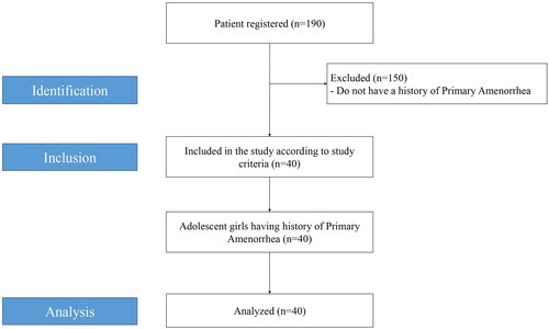
Figure 2. Karyotype of 11 primary amenorrhoea patients, where 45,X was found in 10% (n = 4); 46,X,i(X)(q10) in 2.5% (n = 1); 46,X,del(X)(p11.2) in 2.5% (n = 1); 46,X,del(X)(p22.1) in 2.5% (n = 1); 46,X,del(X)(q24) in 2.5% (n = 1); 46,XY in 2.5% (n = 1); mos 45,X[22]/46,Xi(X)(q.10)[8] in 2.5% (n = 1) and mos 45,X[16]/46,XY[14] in 2.5% (n = 1). The photographs of each karyotype are arranged in the figure sequentially.
![Figure 2. Karyotype of 11 primary amenorrhoea patients, where 45,X was found in 10% (n = 4); 46,X,i(X)(q10) in 2.5% (n = 1); 46,X,del(X)(p11.2) in 2.5% (n = 1); 46,X,del(X)(p22.1) in 2.5% (n = 1); 46,X,del(X)(q24) in 2.5% (n = 1); 46,XY in 2.5% (n = 1); mos 45,X[22]/46,Xi(X)(q.10)[8] in 2.5% (n = 1) and mos 45,X[16]/46,XY[14] in 2.5% (n = 1). The photographs of each karyotype are arranged in the figure sequentially.](/cms/asset/b75316bb-20d3-4f73-a4b0-d54e149496bf/ijog_a_2348085_f0002_b.jpg)
Acknowledgement
We would like to express our sincere gratitude to Nirnayan Healthcare Private Limited, Kolkata for providing the necessary infrastructure for this study. We acknowledge the invaluable contribution of the institute to the successful completion of the article.
Disclosure statement
No potential conflict of interest was reported by the author(s).
Data availability statement
The data that support the findings of this study are available on request from the corresponding author. The data are not publicly available as it can compromise the privacy of research participants.
Additional information
Funding
References
- Al-Ouqaili, M.T., et al., 2022. Detection of partial and/or complete Y chromosome microdeletions of azoospermia factor a (AZFa) sub-region in infertile Iraqi patients with azoospermia and severe oligozoospermia. Journal of Clinical Laboratory Analysis, 36 (3), 1.
- Al-Qaisi, M.N., Al-Ouqaili, M. and Al-Hadithi, D.T., 2020. Molecular analysis for azoospermia factor microdeletions in the Y chromosome for Azoospermic and severe oligospermic infertile Iraqi patients. Systematic Reviews in Pharmacy, 11 (8), 562–6.
- Anitha, G.S., Tejeswini, K.K. and Shivamurthy, G., 2015. A clinical study of primary amenorrhea. Journal of South Asian Federation of Obstetrics and Gynaecology, 7 (3), 158–166.
- Birmingham, A., 2008. Current evaluation of amenorrhea. Fertility and Sterility, 90, 219–225.
- Butnariu, L., et al., 2011. Clinical and cytogenetic correlation in primary and secondary amenorrhea: Retrospective study on 531 patients. Revista româna˘ de medicina˘ de laborator., 19, 149–160.
- Chung, S.H., et al., 2013. Primary amenorrhea of 16 year old girl that no chromosomal abnormalities. World Journal of Pharmaceutical and Medical Research, 1, 4–5.
- El-Dahtory, F., 2012. Chromosomal abnormalities and hormonal disorders of primary amenorrhea patients in Egypt. Indian Journal of Human Genetics, 18 (2), 183–186.
- John, A.M., et al., 2014. A case of primary amenorrhea. The Journal of the Association of Physicians of India, 62 (8), 753–755.
- Kalantaridou, S.N. and Chrousos, G.P., 2002. Clinical review 148: monogenic disorders of puberty. The Journal of Clinical Endocrinology and Metabolism, 87 (6), 2481–2494.
- Kanaan, B.A., Mts, A.-O. and Murshed, R.M., 2022. In terms of the PCRRFLP technique, genetic screening of Ala575Val inactivating mutation in patients with amenorrhea. Journal of Emergency Medicine, Trauma & Acute Care, 6, 8.
- Kanaan, B.A., Al-Ouqaili, M.T.S. and Murshid, R.M., 2023. Cytogenetic screening of chromosomal abnormalities and genetic analysis of FSH receptor Ala307Thr and Ser680Asn genes in amenorrheic patients. PeerJ., 11, e15267.
- Kara, N., et al., 2012. Cytogenetics finding of patients with ameorrhea in Turkish population: a retrospective study. International Journal of Human Genetics, 12 (2), 87–92.
- Khamees, D.A. and Al-Ouqaili, M.T.S., 2022. Cross-sectional study of chromosomal aberrations and immunologic factors in Iraqi couples with recurrent pregnancy loss. PeerJ., 10, e12801.
- Malla, T.M., et al., 2016. Frequency and pattern of cytogenetic alterations in primary amenorrhea cases of Kashmir, North India. Egyptian Journal of Medical Human Genetics., 17 (1), 25–31.
- Merin, T., et al., 2012. Amenorrhea: cytogenetic studies and beyond. The American Journal of Molecular Cell Biology, 1, 25–32.
- Moorhead, P.S., et al., 1960. Chromosome preparations of leucocytes cultured from human peripheral blood. Experimental Cell Research, 20 (3), 613–616.
- Mujumdar, P., Ghosh, S. and Day, S.K., 2015. Association between primary amenorrhea and early maternal age: a population study. Indian Journal of Science and Technology., 8, 1–6.
- Pal, A.K., et al., 2019. A study on chromosomal analysis of patients with primary amenorrhea. Journal of Human Reproductive Sciences, 12 (1), 29–34.
- Patavegar, B.N., et al., 2015. Menstrual pattern and menstrual disorders among school going adolescent girls in Delhi. International Journal of Applied and Basic Medical Research, 11, 241–246.
- Safaei, A., Vasei, M. and Ayatollahi, H., 2010. Cytogenetic analysis of patients with primary amenorrhea in Southwest of Iran. Iranian Journal of Pathology, 5, 121–125.
- Safai, A., et al., 2012. Chromosomal abnormality in patients with secondary amenorrhea. Archives of Iranian Medicine, 15 (4), 232–234.
- Taha, R.F., Al-Ouqaili, M.T.S. and Abdullah, S.A., 2023. The association of anti-mullerian hormone and infertility hormonal imbalance with polycystic ovarian syndrome among Iraqi patients. Pakistan Journal of Biological Sciences, 26 (5), 241–248.
- Vijayalakshmi, J., et al., 2010. Cytogenetic analysis of patients with primary amenorrhea. International Journal of Human Genetics, 10 (1-3), 71–76.