
Abstract
Synapses are the functional units of the nervous system, and their number and protein composition undergo changes over a wide time scale. These synaptic changes manifest into differential behavioural outputs and, in turn, changes in the external conditions to the individual may elicit changes in synapses. We review here publications appeared during the last 10 years in which advances on molecular and cellular mechanisms for synapse changes have been reported. We focus on synaptic changes occurring in the time range of minutes to hours, mainly.
Introduction
In our High School years, neural endowment was considered as a treasure to be maintained against the constant wearing of daily life. Thus, we were told that drinking a whiskey, among other improper actions, will cost us many thousands of synapses which will never come back. This conservationist attitude toward the nervous system experienced a substantial change over the past three decades. Synapses in particular are now considered highly dynamic structures in terms of number, molecular constituents and functional properties. The time frame in which synaptic changes can take place spans from the nanosecond, during the phosphorylation of proteins, all the way to years, during normal aging. We review here the current state of opinion on several molecular mechanisms that mediate changes at the synapse for which progress have been achieved during the past 10 years. To keep published materials within a manageable amount, we focus on synaptic changes that occur in the time range of minutes and days. This time range implies that we will focus on molecular, as opposed to cellular, changes mainly. Although it has been customary to discriminate between the studies carried out in vertebrates from those done in invertebrates, the conservation of mechanisms and the continuum among all living forms render unjustified to maintain this separation. Data will be duly referred to the organism in which they were obtained but, beyond the diversification and specialization of protein family members, the synapse should be viewed as a conserved device for fast communication between neural cells in all animals; in essence, a form of Ca2+-dependent localized exocytosis which constitutes the functional unit of all nervous systems. Mechanisms involved in neurotransmitter release and vesicle fusion operate in the millisecond time range and have been reviewed recently; thus, will not be considered here (Körber & Kuner, Citation2016).
It is quite appropriate that this review is dedicated to prof. Harold L. Atwood on the occasion of his academic retirement. Over two hundred original publications on the biology of the synapse is a compelling evidence to sustain his significant contribution to the field. He is a colleague and a friend with whom some articles emerged from our past collaboration.
Building a synapse, a two sides task
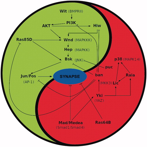
Although signalling pathways are usually considered as strings of molecular interactions, the in vivo context must be closer to a fluctuating matrix operating in a largely unknown landscape of compartmentalized cellular domains. Thus, one encounters signalling molecules in synaptogenesis which are also known to participate in other signalling processes; hence the need to refer to the cellular context when assigning a function to a given protein. For example, the phosphoinositide 3 kinase class I (PI3K), widely known for its role in growth control, is also involved in synaptogenesis in Drosophila and vertebrates (Cuesto et al., Citation2011; Jordan-Alvarez, Fouquet, Sigrist, & Acebes, Citation2012; Jordan-Alvarez, Santana, Casas-Tintó, Acebes, & Ferrús, Citation2017; Martín-Peña et al., Citation2006). The recently discovered PI3K pathway for synaptogenesis includes the Wit receptor, the ligand Gbb, and the MAPkinases cascade (. Further down in the hierarchy, Bsk/JNK undergoes regulation by Puc and Ras85D which results in a narrow range of activity of Bsk/JNK to determine normalcy of synapse number. The transcriptional output of this synaptogenesis pathway involves the Fos/Jun complex and the repressor Cic. Interestingly, this pro-synaptogenesis pathway has an anti-synaptogenesis counterpart which is similarly composed of small GTPases and MAPkinases, such as Ras64B, Ras-like-a, p38a and Licorne, along with Mad and Medea as transcriptional effectors (Jordan-Alvarez et al., Citation2017). The antagonistic cross-regulation between components of both pathways yields a balanced output that seems to determine the number of synapses in a neuron. This type of equilibrium-based mechanisms can respond very fast to changes in the physiological status of the cell, and provide remarkable precision to the trait to be regulated, synapse number in this case (Marucci et al., Citation2009).
Figure 1. Summary of signalling interactions between pro- and anti-synaptogenesis pathways in Drosophila. Activating and repressing interactions are marked by ↑ and т symbols, respectively. Cross interactions between members of both pathways sustain the balanced equilibrium of signalling, hence the use of the yin-yang symbol. The actual number of synapses established by a neuron over its target should result from the signalling output determined by the functional status of both cells. Note the similar nature of signalling members in both pathways; for example: small GTPases (Ras85D and 64B), or MAPKs (Wnd, Hep and p38, Lic), etc. The acronyms for the corresponding vertebrate homologues are shown in parenthesis. Original data are from Ferrús’s lab.
Context-dependent functions are illustrated by the ubiquitin ligase Hiw, another signal included in the pro-synaptogenesis pathway described above for fly motor neurons, which, in the giant fibre interneuron, promotes axon pruning of neurites instead. In addition to these cell-autonomous roles, Hiw also acts on the morphology and activity of the giant fibre interneuron through signalling from the midline glia (Borgen et al., Citation2017). Another ubiquitin ligase, Smurf, regulates the ubiquitination of Mad and is required in glia to mediate clearance of synaptic debris and pruning of the fly’s neuromuscular junction (Chen, Yin, Cao, & Ho, Citation2017). We will review glia–neuron interactions below.
Pre- and postsynaptic partners exhibit more or less conspicuous electron-dense specializations under the transmission electron microscope (). Most, but not all, synapses can be found in the distal portion of neurites which, in motor neurons, often show the form of varicosities (a.k.a. boutons). Neurites, boutons and synapses are morphologically different features in a neuron and, consequently, the signalling for their genesis is also different (reviewed in Acebes & Ferrus, Citation2000; Gallo, Citation2011). Genetic manipulation of synaptogenisis signals demonstrate that each of these three neural features are modified independently. For example, increasing MAPkinases Wnd or Hep elicit neurite outgrowth but only Wnd can increase synapses as well. Likewise, down-expression of Hiw increases neurites but not synapses (Jordan-Alvarez et al., Citation2017). In a similar context, LIMK1 binds the synaptogenic receptor Wit to stabilize synapses, but the binding is dispensable for synapse growth and function (Eaton & Davis, Citation2005). Finally, although fly motorneuron boutons usually contain one or more synapses it is not unfrequent to observe empty boutons and synapses outside boutons (e.g. See Supplementary Figure S3 in Jordan-Alvarez et al., Citation2017).
Mechanistic differences between neuritogenesis and synaptogenesis are also evident in vertebrates. The hepatocyte growth factor (HFG) induces phosphorylation of the MET receptor triggering two different signalling pathways mediated by the kinases ERK1/2, and Akt. These events are accompanied by two major morphological changes: increase in total dendritic growth and increase of synapse density. Selective inhibition of each signalling pathway, ERK1/2 or Akt, alters only one of the two distinct events. The ERK pathway inhibition significantly reduces the HGF-induced increase in dendritic length, but had no effect on synapse density. In contrast, inhibition of the PI3K/Akt pathway reduces the HGF-induced increments in synapse density, with no effect on dendritic length (Eagleson et al., Citation2016).
Pre- and postsynaptic specializations compose a full synapse. Since both components need to be properly localized in register for normal function, coordinated signalling to control the dynamics of their proteins assembly must be at work. Genetic driving of PI3K in fly motor neurons, even late in development, elicits the clustering of postsynaptic densities and GluRII receptors. Overexpressing PI3K selectively in the muscle target cell, however, does not elicit an equivalent reaction by the genetically normal motor neuron. This is yet another demonstration that the presynaptic cell has the capacity to induce a response of the postsynaptic cell to complete a full synapse (Jordan-Alvarez et al., Citation2012).
Signalling for synapse dynamics is likely to depend on the developmental stage and dosage. In flies, the genetic down-regulation of GSK3β increases the number of synapses, while its upregulation leads to synapse loss. Likewise, in three weeks cultured rat hippocampal neurons, the pharmacological inhibition of GSK3β increases synapse density and Synapsin expression. However, experiments on younger cultures (12 days) yield an opposite effect, a reduction of synapse density. This result may unveil an age- and dosage-dependent differential response of mammalian neurons to the stimulation/inhibition of GSK3β, a feature that must be considered in the context of human adult neurogenesis and pharmacological treatments for Alzheimer’s disease based on GSK3β antagonists (Cuesto et al., Citation2015).
Once the signalling for synaptogenesis has resolved the site where to build a synapse, the sequential assembly of active zone components proceeds. The sequence is now well established in Drosophila (). Liprinα precedes by several hours the arrival of the ERC/CAST family protein Bruchpilot (BRP). The latter contributes to the effective clustering of Ca2+ channels. Coincident in time with BRP arrival at the active zone, the postsynaptic GluR receptors become clustered as well (Fouquet et al., Citation2009). Proper alignment between pre- and postsynaptic sides is achieved by Syndecan (Syd-1) (). This transynaptic protein helps to localize, but it acts independent from, presynaptic Liprinα and it binds to BRP (Owald et al., Citation2010). Syd-1 also interacts with Neurexin (Nrx-1) to control synapse formation. Syd-1 and Nrx-1 form a complex in vivo, to cluster and immobilize Nrx-1. On the postsynaptic side, clustering of Neuroligin1, Nlg-1, is altered in Syd-1 mutants, as it is GluR incorporation in Syd-1, Nrx-1 and Nlg-1 fly mutants. Thus, the three proteins cooperatively help the early assembly processes between pre- and postsynaptic membranes (Owald et al., Citation2012). The fine contour of the presynaptic active zone seems defined by the positioning of Spinophilin, another scaffold protein that binds the C-terminus of Nrx limiting the extent of the Nrx-1/Nlg-1 signalling by antagonizing Syd-1 (Muhammad et al., Citation2015). An additional group of adhesion molecules with a role in synaptogenesis is represented by the Calsyntenins (CST). It has been proposed that a CST-3 may contribute from the presynaptic side to the formation of inhibitory synapses through an indirect interaction with Nrx (Um et al., Citation2014).
The Ras-related in brain 3 (Rab3) interacting molecules (RIM) are key elements for proper location of Ca2+ channels in the active zone of the synapse. RIMs tether N- and P/Q-type Ca2+ channels to presynaptic active zones via a direct PDZ-domain-mediated interaction (Kaeser et al., Citation2011). In the inner hair cells of the mouse auditory system, three RIMs, 2alpha, 2beta and 3gamma contribute to the clustering of Cav1.3 Ca2+ channels and their depletion cause hearing impairment (Jung et al., Citation2015). The RIM-binding proteins (RBPs) at the murine retina synapses between the rod bipolar and the amacrine type 2 cells, tether the L-type Ca2+ channels to the active zones allowing to synchronize vesicle exocytosis and, thus, effective neurotransmitter release (Luo, Liu, Sudhof, & Acuna, Citation2017). A RIM-related protein from Drosophila, Fife, positions the clusters of Ca2+ channels and vesicle release sites within nanometre distance of each other (Bruckner et al., Citation2017).
The assembly of clusters of postsynaptic proteins also requires anchoring proteins to stabilize them. The trans-synaptic organizing protein ephrin-B3 controls the localization and stability of postsynaptic density-95 (PSD-95). A MAPK-dependent phosphorylation site on ephrin-B3, Ser332, mediates the activity dependent changes in PSD95 clustering (Hruska, Henderson, Xia, Le Marchand, & Dalva, Citation2015). Simultaneous time-lapse imaging of PSD-95 and Ca2+/calmodulin-dependent protein kinase II (CaMKII) at the synapse, demonstrate the high turnover of these synapse components. Both proteins move into and out of the same synapse independently, so that synapses cycle rapidly between states in which they are enriched for none, one or both proteins. Phospho-CaMKII and PSD-95 are present more often at stable than labile contacts, and both of them can change localization over a time-scale of hours (Taft & Turrigiano, Citation2014).
It is well established that activity-induced shrinkage and retraction of dendritic spines depend on activation of the NMDA-type glutamate receptor (NMDAR), which leads to influx of extracellular Ca2+ ions and activation of Ca2+-dependent phosphatases that modify the spine cytoskeleton, suggesting that influx of extracellular Ca2+ drives spine shrinkage. However, non-ionotropic function of the NMDAR also regulates synaptic strength (Nabavi et al., Citation2013). Additional support to this phenomenon has been obtained by two-photon glutamate uncaging and time-lapse imaging of rat hippocampal CA1 neurons, in the presence of the NMDAR d-serine/glycine binding site antagonist 7-chlorokynurenic acid (7CK), which blocks the Ca2+ influx (Stein, Gray, & Zito, Citation2015).
Work with primary cultured cortical neurons show that Arc/Arg3.1 mRNA and Arc/Arg3.1 protein dynamically increase within 24 hr after glutamate treatment. This increase could be prevented by the NMDAR inhibitor DL-AP5, but not the AMPAR inhibitor NBQX, or by blocking the activation of ERK or CREB. That is, the glutamate-induced increase of Arc/Arg3.1 relies on a NMDAR mechanism that is independent of Ca2+ influx (Chen, et al., Citation2017).
Fine tuning of synapses
Activity-dependent changes at the synapse can be detected remarkably fast. High-resolution light microscopy on the Drosophila larval NMJ revealed an increase in the amount of the active zone protein Bruchpilot and an enlargement of the presynaptic cytomatrix structure which regulates the number of release-ready vesicles within minutes of presynaptic strengthening (Weyhersmüller, Hallermann, Wagner, & Eilers, Citation2011).
In general, neurons with large number of synapses exhibit a low probability of neurotransmitter release per synapse and vice versa. This antagonistic correlation was considered until recently as an epiphenomenon resulting from neuron homeostasis. However, mutants in the Drosophila Ca2+-binding protein Frequenin (Frq) offer a possible mechanism to explain the opposite co-regulation. Loss-of-function Frq mutants increase the number of synapses while reducing the probability of release per synapse. In turn, excess-of-function Frq mutants exhibit the reverse phenotypes (Dason, Romero-Pozuelo, Atwood, & Ferrús, Citation2012; Romero-Pozuelo, Dason, Atwood, & Ferrús, Citation2007). The Frq mechanism includes the functional interaction with the α1 subunit of Ca2+ channels (Dason et al., Citation2009). Drosophila and Danio exhibit two Frq genes, Frq1 and Frq2, whose protein sequences are 95% identical. Fly Frq2, but not Frq1, bind the guanyl-exchange factor Ric8a, as it does the single vertebrate homologue, NCS-1 (Romero-Pozuelo et al., Citation2014). Consistent with the GEF nature of Ric8a, specific members of the Gα protein family are involved, either in synaptogenesis (Gs) or in synapse release (Gq) (see below). Thus, the antagonistic co-regulation seems to result from a common path, Frq2/NCS-1 binding to Ric8a, which diverges later on toward specific Gα interactions to regulate synapse number or release. Identifying the specific partners of Frq1 or alternative binding sites in NCS-1 would help to complete this co-regulatory process.
Optimal function results from a bidirectional modulation between the stoichiometry of specific proteins and the activity regime of the synapse. Relevant protein species include neurotransmitter receptors, voltage gated Ca2+ channels and Ca2+-binding proteins among others. Recent progress in this scenario incorporates the axonal transport system as an additional level of regulation. This has been illustrated in the Caenorhabditis elegans homolog of CAMKII, UNC43, which targets the kinesin light chain to mediate the transport, plasma membrane insertion and removal of the AMPA receptor (Hoerndli et al., Citation2015). CAMKIIα also binds the metabotropic receptor 1 (mGluR1) which is coupled to Gα(q) (see section on G-protein coupled receptors below). The binding is augmented by Ca2+, it implies the phosphorylation of the receptor at the T871 site and constitutes a feedback loop that facilitates desensitization of mGluR1 (Jin et al., Citation2013).
In addition, AMPA receptor transport requires Ca2+ release from internal deposits. Upon Ca2+ release from the endoplasmic reticulum (ER) via the IP3 and ryanodine receptors, CaMKII becomes activated and enters a PICK1-containing complex that interacts with the GluA2 C-terminal domain of AMPA and stimulates its exit from the ER and cell surface trafficking (Lu et al., 2014). An equivalent transport system for pore-forming Ca2+ cannel subunits of the P/Q and T types seems to depend on the actin-binding protein Kelch-like 1 (KLHL1), albeit the mechanism is still poorly known (Perissinotti et al., Citation2014). Another case of biologically significant equilibrium activity is represented by the α isoform of the phosphatase calcineurin (CNAα) and the kinase cyclin-dependent kinase 5 (CDK5). Acute or chronic loss of these enzymatic activities results in a sevenfold increase on single action potential-driven exocytosis. This control is achieved through the Cav2.2 (N-type) channel (Kim & Ryan, Citation2013). The balanced equilibrium between these enzymatic activities seems responsible of maintaining certain synapses unresponsive to sparse action potentials.
Structural changes at the postsynaptic spine result from synaptic activity and, thus, Ca2+ transients. Microtubules enter spines from specific sites at the base of spines as part of the process of spine activation. The Ca2+ dynamics promotes the transient and local polymerization of F-actin. Further, F-actin fibres interact with microtubules through a mechanism mediated by Debrin (Merriam et al., Citation2013). Ca2+ channel associated factors can exert profound effects on the channel permeability and, thus, on synapse activity. A remarkable case is the family of glycosylphosphatidylinositol (GPI)-anchored subunits represented by the several α2δ. Three of them, α2δ-1, α2δ-2 and α2δ-3, are involved in pain perception. They contribute to Ca2+-channel driven exocytosis at the synapse through an extracellular metal ion-dependent adhesion site (MIDAS) which is located within the Willebrand A domain of these α2δ. Interestingly, the MIDAS-dependent function is independent from an additional role of α2δ protecting exocytosis from blockade by intracellular Ca2+ chelators (Hoppa, Lana, Margas, Dolphin, & Ryan, Citation2012). The dual role of the channel associated factors justifies their pharmacological relevance, as the neuropathic analgesics gabapentin and pregabalin target α2δ-1, α2δ-2 and α2δ-3, respectively. Another GPI-anchored peptide is quiver (qvr) (a.k.a sleepless/sss) whose mutants exhibit strong motor effects and synapse excitability alterations (Ruan et al., Citation2017). In this case, qvr peptide seems to have functional interactions with K+ channels (Shaker), nicotinic Ach receptor (Wu, Liu, & Joiner, Citation2016), GABA transaminase (Chen et al., Citation2015), among other targets.
Maintenance of synapse morphology is dependent on activity. Ca2+ influx through the L-type Cav1.3a is required for proper size of ribbon synapses in sensory hair cells of zebra fish (Sheets, Kindt, & Nicolson, Citation2012). The proper number of ion channels is also regulated by scaffolding proteins coupled to the ubiquitin degradation pathway. This is the case of Harmonin and the Cav1.3 (Gregory et al., Citation2011) among others. Fine tuning of synapse activity is also achieved by sets of counteracting ion channels. For example, the Ca2+ influx that promotes neurotransmitter release may also trigger a Cl− flux that shortens action potential duration, dampens excitatory synaptic potentials, impede temporal summation and raises the threshold for action potential generation in hippocampal neurons. Further, out of the two reported types of Ca2+-activated chloride channels, TMEM16A and TMEM16B, only the latter seems effective in the hippocampus (Huang et al., Citation2012). The widespread regulatory effects of the Ca2+-binding protein Calmodulin (CAM) are further enriched by the identification of functions for the Ca2+-free form (apoCAM). That form, on its own, can bind and favour the opening of Ca2+ channels. Actually, splice variants of these channels exhibit different affinities for apoCAM (Adams, Ben-Johny, Dick, Inoue, & Yue, Citation2014).
Synapses must work under environmental conditions external to the organism and under the metabolic rate of the neuron. Temperature is a functional constrain for synapse activity, in particular when high frequency firing is required. Studies on Ca2+ dynamics in Drosophila have revealed that the thermal limit of synaptic transmission may be directly linked to the stability of ATP-dependent mechanisms that regulate intracellular ion concentrations (Klose, Atwood, & Robertson, Citation2008; Klose, Boulianne, Robertson, & Atwood, Citation2009). In addition to synapse strengthening, de novo synaptogenesis is required for long term memory (LTM). In a α-calcium/calmodulin kinase II autophosphorylation-deficient (T286A) mutant that impairs synapse strengthening, contextual LTM formation is linked to PSD95 up-regulation followed by the generation of multi-innervated spines and activation of mTOR signalling (Radwanska et al., Citation2011). Either excess or deficit in the number of synapses can lead to pathology. In the case of the Fragile X-syndrome (FXS), neurons of patients or fmr1 mice or fly mutants show postsynaptic spines at increased density and with long neck morphology which has been interpreted as defects in synapse maturation or pruning (Antar, Li, Zhang, Carroll, & Bassell, Citation2006; Gatto & Broadie, Citation2008; Pfeiffer & Huber, Citation2007). As in humans, homozygous and heterozygous fmr1 mutants in Drosophila are deficient in associative learning and memory (Kanellopoulos, Semelidou, Kotini, Anezaki, & Skoulakis, Citation2012). Interfering with the Frq2/NCS1-Ric8a binding described above in fly fmr1 mutants restores the synapse number and the learning abilities to normal values (Mansilla et al., Citation2017).
Synapse elimination toward the end of early development, the so-called “pruning” effect, is clearly relevant for normal biology. However, the mechanisms that lead to building an apparent excess of synapses to be followed by their pruning remain largely unknown. Mice deficient in the complement protein C1q or the downstream C3, fail to prune synapses during refinement of retinogeniculate connections. Neuronal C1q is normally downregulated in the adult CNS; however, in a mouse model of glaucoma, C1q becomes upregulated and synaptically relocalised in the adult retina, indicating that dispensable synapses are tagged by complement and this tagging is abnormally reactivated in the disease (Stevens et al., Citation2007). Synapse pruning is also evident in Drosophila’s embryonic motor neuron growth cones where low-frequency Ca2+ oscillations are required for synaptic refinement. Ca2+ transients mature over several hours into regular low-frequency (0.03 Hz) oscillations. The subsequent signalling cascade involves the phosphatases Calcineurin and protein phosphatase-1, as well the serine/threonine kinases CaMKII and PKA. This cascade modulates the neuron’s response to the muscle’s Sema2a chemorepellant, critical for the removal of off-target contacts (Vonhoff & Keshishian, Citation2017).
G-protein coupled receptors at the synapse
Receptors coupled to G proteins (GCPR) can modulate synapse activity. One of them is the metabotropic glutamate receptor (mGluR). The three component types of the G protein complex, α, β and γ, play a wide repertoire of regulatory activities. In cone photoreceptors of salamander retinas, activation of mGluR reduces Ca2+ influx and neurotransmission. However, both effects are mechanistically independent because the βγ dimer interacts with SNAP-25 to modulate synaptic vesicle fusion without altering significantly the Ca2+ influx (Van Hook et al., Citation2017). Other GPCRs, however, use the Gβγ dimer to elicit an increase of the Ca2+ influx and promote spinogenesis, as illustrated by Frizzled9 (FZD9) receptor upon binding to its ligand the growth factor Wnt-5a in hippocampal neurons (Ramirez, Ramos-Fernandez, Henriquez, Lorenzo, & Inestrosa, Citation2016). The mammalian Wnt is represented in Drosophila, where it was first identified, and named Wingless (wg). In both sets of species, this family of growth factors is involved in multiple functions including, segment polarity, cell differentiation, synapse maintenance and, when defective, may result in cancer or neurodegeneration (rev. Arnés & Casas-Tintó, Citation2017).
Classical GPCRs such as α2, GABA(B) and CB1 use the βγ dimer to interact with Ca2+ channels, K+ channels or other synaptic proteins that mediate neurotransmitter release. In certain contexts, however, it is the Gα monomer that inhibits presynaptic Ca2+ channels via a cAMP cascade which includes the 4 phosphodiesterase (PDE4). This feature occurs in retinal rod bipolar synapses and is remarkably similar to the Gβγ–cGMP–guanylate cyclase/PDE6 strategy that occurs in phototransduction (Dong, Guo, Ye, & Hare, Citation2014). This is another example of two similar, albeit distinct, mechanisms to attain the same biological effect, down regulation of Ca2+ influx, operating in two different cellular contexts, synapse transmission and phototransduction, in the retina. A family of GPCRs exhibits an extracellular N-terminal region with multiple adhesion domains connected to the core GPCR moieties by G-protein proteolysis sites. Humans have 33 of them but their functions remain largely unknown. At least one case is involved in spino- and synaptogenesis. The brain-specific angiogenesis inhibitor 1 (BAI1) receptor recruits proteins to synapses through its interaction with the polarity protein Par3/Tiam1 (Duman et al., Citation2013). Tiam1 is a Rac1-guanine nucleotide exchange factor that becomes restricted to spines, which implies a spatial control of Rac1 activation. This allows Par3 to promote synapse formation (Um et al., Citation2014).
A structurally related family of GPCRs is the latrophilin (LPHN) characterized by their high affinity binding to Teneurins, a ubiquitous group of multifunctional transmembrane proteins represented in vertebrates by four members. All Teneurin genes encode in their terminal exons small peptides that can bind to Teneurin C termini, TCAP1–4. TCAP-1 interacts with LPHN, through the association with β-dystroglycan, to induce a tissue-dependent signal cascade that modulates cytoskeletal dynamics. TCAP-1 reduces stress-induced behaviours associated with anxiety, addiction and depression in a variety of models, in part, by regulating synaptic plasticity (Woelfle, D'Aquila, Pavlovic, Husic, & Lovejoy, Citation2015).
Some GPCRs play specific roles only at juvenile stages of development. This is the case of dopamine 2 receptors (D2R) for spine morphogenesis. Activation of D2R reduces spine number via GluN2B- and cAMP-dependent mechanisms in mice. If D2R are overactivated, due to mutations in the schizophrenia-related gene Dtnbp1, spines are reduced in number, the connectivity in the entorhinal–hippocampal circuit is altered and the spatial working memory is impaired. These effects can be prevented to a large extent if young mice are fed with D2R blockers (Jia, Zhao, Hu, Lindberg, & Li, Citation2013). A peculiar case of synaptic modulation by GPCRs is illustrated by Fmrf receptor (FR) and Drosomyosupressin Receptor-2 (DmsR-2). Both receptors share the same ligand, the peptide DPKQDFMRFamide. The coincident detection by both receptors modulates synapse activity through the Ca2+-induced Ca2+ release from cell internal deposits (Klose, Dason, Atwood, Boulianne, & Mercier, Citation2010).
Many proteins exhibit a structural motif known as PDZ (postsynaptic density protein of 95 kDa, disc large, zona occludens-1) which facilitates interactions among PDZ containing partners, many of which are synaptic. This extensive field has been recently reviewed (Dunn & Ferguson, Citation2015) and, consequently, will not be covered here. An exhaustive account of GPCR interactions relevant for spine morphogenesis and structural reorganizations in general, can be found in another recent review (Leung & Wong, Citation2017).
Circadian changes of synapses
That synapses change during the light/dark (L/D) cycle has been known for a long time and documented in several species. However, the current scenario shows a wide diversity of context-dependent changes (rev. Frank & Cantera, Citation2014) (see below). The molecular components of the circadian clock known to date include the heterodimer CLOCK/CYCLE (CLK/CYC) which binds to the regulatory regions of timeless (tim) and period (per) genes. Both, tim and per genes are transcribed during the day, but by early night, TIM and PER protein levels rise, dimerize and translocate to the nucleus, where they bind to CLK/CYC and block their own transcription (rev in Jarabo & Martin, Citation2017). In this way, the system generates a transcriptional feedback loop that constitutes the foundation of circadian periodicity. CRYPTOCHROME (CRY) is a cell-autonomous blue light photoreceptor that mediates the entrainment of the core clock by light, through degradation of TIM upon light stimulation (rev in Ozkaya & Rosato, Citation2012). Concerning the architecture of the neuronal system, in adult Drosophila the circadian pacemaker network includes several groups of neurons comprising about 150 cells (rev in Hardin, Citation2011).
In addition to its circadian activities, Drosophila CRY plays a role in magnetoreception, phototransduction and synapse activity. Based on a yeast-two hybrid assay, CRY physically interacts with the presynaptic protein Bruchpilot (BRP). The CRY-BRP complex is located in visual synapses mainly. Light activated CRY seems to decrease BRP levels in the terminals of the visual lamina neurons by targeting it to degradation (Damulewicz et al., Citation2017). In the same line, fly lamina neurons (L2 monopolar interneurons) exhibit structural and synapse number rhythmic changes that depend on the normal activity of the per gene, and involve the cyclic expression of the synaptogenic signalling by TOR, PI3K and AKT, as well as that of the autophagy genes Atg5 and Atg7 (Kijak & Pyza, Citation2017).
Circadian changes can be detected in the structure of clock neurons themselves. The Drosophila s-LNv neurons change their amount of axonal material in line with the fasciculation/defasciculation cycles. These features are mediated by rhythmic Rho1 activity that retracts axonal termini by increasing myosin phosphorylation concomitant with pre-synaptic and dendritic markers. Rhythmic Rho1 activity is regulated by the cyclic transcription of Puratrophin-1-like (Pura), a Rho1 GEF (Petsakou, Sapsis, & Blau, Citation2015). The sLNv neurons also undergo changes in connectivity partners. Synapses are dismantled or built between late night and the following morning but, based on the sensitive GRASP technique these neurons seem to contact different partners at different times along the day which implies different paths of information processing (Gorostiza, Depetris-Chauvin, Frenkel, Pirez, & Ceriani, Citation2014).
Circadian changes in synapse number have been documented also in living zebrafish larvae by means of time-lapse two-photon imaging of the presynaptic marker Synaptophysin in hypocretin/orexin (HCRT) neurons. The mechanism sustaining these changes includes the rhythmic expression of NPTX2, a protein that helps to cluster AMPA receptors. The NPTX2 encoding gene is mostly expressed in hypothalamic and pineal gland cells and its overexpression increases synapse number and abolishes rhythmicity in HCRT axons. These cyclic changes are also affected by sleep deprivation because NPTX2 overexpressing fish are resistant to sleep-promoting effects of melatonin (Appelbaum et al., Citation2010). Also in this fish, the depletion of per2 activity reduces the retina ribbon synapses, causes abnormal ON response in a visual motor assay, and reduces transcriptional expression of opsins, as well as that of other clock genes such as cry1ba and bmal1b (Huang et al., Citation2018).
The suprachiasmatic nucleus (SCN) of the hypothalamus is a central pacemaker for light-dark cycle related behaviours. Its synapses contain relatively high levels of C1QL3 protein that seems to be involved in their maintenance. Knockout mice for this gene show reduced density of excitatory synapses and weak activity during the active portions of the day (Chew, Fernandez, Hattar, Sudhof, & Martinelli, Citation2017). The balance between excitatory/inhibitory synapses shows circadian changes through the oscillation of transcription of Neurexin genes, in particular in SCN cells. The Neurexin genes (NRXN1/2/3) encode two families (α, β) of polymorphic proteins. Neural activity and memory formation impinge upon NRXN1/2/3α expression and alternative splicing at exons sites 3 and 4. Diurnal rhythms in NRXN1α and NRNXN2α, but not NRXN3α, transcription, exon 3/4 selective splicing, and protein levels of Neurexin-2α, PSD-95 and gephyrin have been documented in the SCN (Shapiro-Reznik, Jilg, Lerner, Earnest, & Zisapel, Citation2012).
Downstream from the intrinsic SCN synapse changes caused by circadian rhythmicity, SCN efferents expressing either vasopressin (AVP) or vasoactive intestinal peptide (VIP) are differentially involved in day/night SCN neuroglial changes. Synaptic inputs received by the VIP neurons, which are major integrators of photic signals in the retinal SCN subregion, increase during the day while those received by the AVP neurons remain unchanged (Girardet, Becquet, Blanchard, Francois-Bellan, & Bosler, Citation2010). Glial changes include morphological coverage of dendritic versus somatic regions of VIP expressing neurons, coincident with quantitative changes in the expression of glial fibrillary acidic protein (GFAP) (Becquet, Girardet, Guillaumond, Francois-Bellan, & Bosler, Citation2008).
The pineal gland hormone melatonin peaks during the night in diurnal mammals. Its actions may be channelled through its intrinsic antioxidant properties or through its binding to two GPCRs, MT1 and MT2. A set of interactome data for both receptors shows 378 putative partners, some of which are selective for MT1 and are residents in the pre-synapse. The MT1 receptor is found in the hypothalamus, striatum and cortex and it physically interacts with Cav2.2 channels to inhibit Ca2+ entry in an agonist-independent manner (Benleulmi-Chaachoua et al., Citation2016). Circadian changes in the synapse have been monitored through the metabotropic glutamate receptor5 (mGluR5) in mice along the day. mGluR5 levels increase during the light-on (07:00 to 15:00 hr), or sleep phase for rodents, by approximately 10% (Elmenhorst et al., Citation2016). Noticeably, altered density of mGluR5 seems to occur in psychiatric disorders and, as indicated by these monitoring data, mGluR5 changes may also be involved in human mood changes. Similar to GluR5, quantitatively minor oscillations of certain protein levels during the day may have noticeable biological effects. This is the case of synaptic Shank3 in hippocampal and striatal brain neurons. Shank3 changes in these regions correlate also with serum melatonin levels and, as expected, with motor activity. The experimental increase of motor exercise leads to a rapid increase of Shank3α expression in thalamus, and cortex, but to a decrease in striatum (Sarowar et al., Citation2016). This illustrates the hierarchical superposition of brain regions concerning molecular cyclic changes toward coherence of the behavioural output of the organism.
Circadian rhythmicity in synapse number is documented throughout species but not all synapse types follow the circadian changes in the same manner. In mice kept under LD conditions, the total density of synapses and the density of excitatory synapses are higher during the light period (rest phase). In contrast, the density of inhibitory synapses increases during the dark period (activity phase). Under DD conditions, the upregulation of the inhibitory synapses is retained, but that of excitatory synapses is not. Thus, the circadian plasticity concerns only synapses located on spines, as opposed to dendritic shafts, and excitatory and inhibitory synapses are differently regulated during the 24 hr cycle: the excitatory ones are modified by light, whilst the inhibitory synapses are driven by the endogenous circadian clock (Jasinska et al., Citation2015). Sometimes the observed circadian changes in synapse number seem counterintuitive. The flight motor neuron MN5 of Drosophila under LD condition shows more synapses at midnight that at midday, suggesting that the rest period is used to form new synapses which are disassembled during flight activity at light time (Ruiz et al., Citation2013). The vertebrate’s somatosensory cortex also shows cyclic changes in synapse number (Jasinska et al., Citation2014).
Some synapses may exhibit infra-circadian cyclic changes. For example, in the Drosophila lamina neurons of the optic lobe, the number of the so-called tetrad synapse increases twice a day, once in the morning and again in the evening (Gorska-Andrzejak et al., Citation2013). The same neurons exhibit other type of synapse, the so-called feedback synapse. One scaffold component of most fly synapses, the Bruchpilot (BRP) protein, exists in two isoforms, 190 and 170 kD. The quantification of tetrads and feedback synapses in isoform specific mutants suggests that each BRP isoform contributes differently to each synapse type and time of the day (Woznicka et al., Citation2015). The heterogeneity of circadian changes expands also to regions of the same nervous system and protein components of the synapse. In the Drosophila optic lamina, the levels of the postsynaptic density Disc Large (DLG) oscillates both, under LD and DD conditions. However, in contrast to the lamina, the transcription of dlg gene in the central brain oscillates under the LD condition only. Other synaptic genes, as those encoding BRP or SYN, do not cycle their transcription in the central brain under LD neither DD conditions (Krzeptowski et al., Citation2014).
Electrical synapses are also subject to circadian changes. Rod photoreceptors in the mice kept under circadian conditions show a median junctional conductance of 98 pS during the subjective day and 493 pS during the subjective night. Conductance in dark adapted animals is about 140 pS regardless of the time of the day, while adaptation to bright light reduces it to near 0 pS, at all times during the circadian cycle (Jin & Ribelayga, Citation2016).
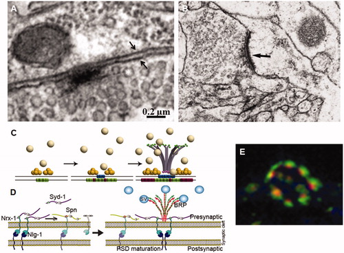
Figure 2. Assembly of synapse components. (A) Synapse in a larval neuromuscular junction. Note the electron dense T bar shaped presynaptic specialization around which synaptic vesicles (asterisk) accumulate. Two small arrows indicate the pre- and postsynaptic cell membranes. (B) Human cortex synapse. Note the pre- and post-synaptic electron-dense specializations. Bar in A = 0.2 and 0.6 µm in B. (C) Sequential assembly of synapse components. Liprin-α (yellow spheres) cluster on the presynaptic membrane followed by Ca2+ channels (blue barrels). Finally, the BRP protein forms a bouquet centred in the active zone attracting the synaptic vesicles (SV, grey spheres) through its Nc82 epitope (green dots). Meanwhile, the postsynaptic membrane accumulates GluRIIA (green barrels) and, later, GluRIIB (red barrels) receptors. (D) The register between the pre- and postsynaptic components is established by the interaction between the presynaptic Neurexin-1 (Nrx-1) and the postsynaptic Neuroliguin1 (Nrl1). Syndecan-1 (Syd-1) is thought to hold the BRP bouquet in place while the peripheral limits of the synapse are determined by the Syd-1 antagonist Spinophilin (Spn). (E) Motor neuron bouton showing several synapses immunolabelled for BRP (red) and GluRIIC (green) showing the perfect register between the pre- and postsynaptic components. Original data are from Ferrus’s lab (A and E); De Felipe’s lab (B); Sigrist’s lab modified from Fouquet et al. (Citation2009) (C) and Muhammad et al. (Citation2015) (D).
Glial cells also undergo circadian changes in activity and molecular components as illustrated by the astrocyte brain fatty acid binding protein Fabp7 which shows a diurnal regulation of gene and protein expression throughout the mouse brain. The mechanism that mediates this phenomenon includes the binding of the 3′UTR region of the Fabp7 mRNA to the cytoplasmic polyadenylation element-binding protein 1 (CPEB1), that effectively regulates trafficking and translation of the mRNA (Gerstner et al., Citation2012). Microglia also exhibits a circadian clock. One of the effects is the cyclic secretion of Cathepsin S (CatS). Mice deficient in that gene lose the diurnal variation of evoked synaptic response of cortical neurons, and behave abnormally during social interactions (Takayama, Zhang, Hayashi, Wu, & Nakanishi, Citation2017). Circadian rhythm includes oscillations of glucocorticoids. Peak concentrations of the later promote synaptogenesis and facilitate learning. In turn, sustained or excessive glucocorticoid levels, as those attained during stress, lead to synapse elimination and disrupt previously acquired memories. The synaptogenic effect can be achieved through the LIM-kinase-Cofilin pathway while the synapse elimination effect requires transcriptional changes and mineralocorticoid receptor activation (Liston et al., Citation2013).
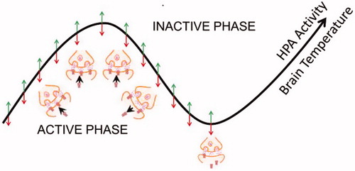
The body of data available so far clearly shows that changes in synapse number, protein composition and neurite morphology are specific to the neuronal system and activity regime of the circuit. As an attempt to provide a frame where to accommodate current data, a model has been proposed: the “State-clock model of synaptic plasticity” (, Frank & Cantera, Citation2014). In essence, the model aims to reconcile the activity cycle present in all organisms and cells with the experience-dependent changes that all synapses undergo. The direction and phase of the cycle where synaptic changes may occur, however, are diverse. Beyond the obvious differences that diurnal versus nocturnal physiologies represent in all animals, experimental conditions in the laboratory are an additional matter of concern. While laboratory studies are carried out under 12 hr/12 hr of L/D conditions, natural environment include the key phases of light transitions: sunset and sunrise. The first lab study to incorporate these transitions already showed significant changes in the activity pattern along the 24 hr period. In particular, the Drosophila resting period in the middle of the day (“siesta” time) and the morning anticipation do not appear under natural-like conditions (Vanin et al., Citation2012). The raising debate on the significance of these effects to draw conclusions applicable to humans, whose current living habits are also quite different to those of our ancestors, ensures unexpected findings to come (Jarabo & Martin, Citation2017).
Figure 3. State-Clock model of synaptic changes. As proposed (Frank and Cantera, Citation2014), the biological clocks would drive the 24 hr rhythms in synaptic plasticity. The experience dependent changes in synapses would occur during the activity phase and be consolidated during the inactive phase, but the direction of these changes would not be fixed. In mammals, oscillations in the hypothalamic-pituitary axis (HPA) activity would increase global cortical synaptic activity.
Glia, the third element
Early in the last decade, it became evident that glial cells play essential roles in neuron biology. Work in C. elegans showed that glial ablated animals exhibit profound sensory deficits, abnormal neuron morphology and behaviour. One of the glia enriched genes first identified, fig-1, encodes a labile protein with a conserved thrombospondin 1 (TSP1) domain (Bacaj, Tevlin, Lu, & Shaham, Citation2008). Transcriptomic databases of astrocytes at the times of synapse formation and elimination show that this type of glial cells are particularly enriched in metabolic, lipidogenic and phagocytic pathways such as those mediated by draper/Megf10 and Mertk/integrin alpha(v)beta5. Further, the transcriptional profile of astrocytes reveals it as very different from that of oligodendrocytes consistent with the mounting evidences on glial cell heterogeneity (Cahoy et al., Citation2008). The repertoire of glial types and their putative homologies across species have been reviewed recently (Losada-Pérez, Citation2018).
Certain types of glia play specific roles in synapse biology. For example, reactive astrocytes enwrap, engulf and may digest dystrophic neurites in the hippocampus of amyloid precursor protein/presenilin-1 (APP/PS1) mice and Alzheimer disease patients. However, microglia, the brain phagocytic population, is apparently not engaged in this clearance (Gomez-Arboledas et al., Citation2018). However, a different report with data from AD patients suggests a role for microglia promoting amyloid clearance accompanied by synapse loss. The DNA-RNA binding protein TDP-43 mediates this process since mutant mice for this gene exhibit reduced amyloid load in an AD model and, at the same time, display a drastic synapse loss, even in the absence of amyloid (Paolicelli et al., Citation2017).
Microglia also controls inhibitory synapses, albeit of the glycinergic, rather than the GABAergic, type. This is achieved via modulation of the diffusion dynamics and synaptic trapping of glycine (GlyR), but not GABAA, receptors in the spinal cord. The microglia-synapse cross talk requires production of prostaglandin E2 by microglia which activates neuronal EP2 receptors and cyclic adenosine monophosphate-dependent protein kinase (Cantaut-Belarif et al., Citation2017).
In addition to microglia, astrocytes also contribute to synapse elimination during development and in the adult. This process requires the MEGF10 and MERTK phagocytic pathways, and is strongly dependent on neuronal activity. Developing mice deficient in both astrocyte pathways fail to refine their retino-geniculate connections and retain excess of functional synapses. In the adult mouse brain, astrocytes continuously engulf both excitatory and inhibitory synapses (Chung et al., Citation2013). Among the several mechanisms that mediate astrocyte-initiated changes in synapse number under stress conditions are those represented by the CRH family of peptides and their Urocortin (UCNs) partners. The activation of the CRH receptor 1 (CRHR1) attenuates synaptogenesis in the hippocampus. UCN2 and 3 are agonists selective for a different, but structurally similar, receptor CRHR2. However, UCN2, but not UCN3, treated slice cultures increase SynapsinI and PSD95 levels. Consistently, the CRHR2 antagonist Astressin 2B reverses this effect. If isolated neuronal cultures are used instead of slices, UCN2 decreases the number of synapses via CRHR2, pointing toward a key role for the missing cells, the glia. Actually, treatment of hippocampal neurons with media from UCN2-treated astrocytes yields the expected increase of SynapsinI and PSD95-labelled terminals, and this effect requires the induction and secretion of NGF from astrocytes. As a corollary, UCN2 and UNC3 must play differential roles when signalling through CRHR2 (Zheng, Zhang, & Ni, Citation2016). NGF secretion also mediates the expression of GluN2A and GluN2B, two subunits of NMDAR which protect synapses from Aβ toxicity (Li et al., Citation2016).
Astrocytes may suffer deleterious effects by long exposure to amyloid β40 peptide, and these effects can be transmitted to naïve hippocampal neurons when co-cultured. The reflected neural dysfunctions include decrements of EPSCs and mEPSC frequency, along with reduced number of excitatory synapses. Interestingly, release probability per synapse is increased under this co-culture condition (Kawano et al., Citation2017). Likely, the increase of release probability per synapse results from the antagonistic co-regulation with synapse number described above in the context of Frq2/NCS1 versus Ric8a mechanism.
Bidirectional interactions between astrocytes and neurons have been studied under several types of abnormal conditions. Under brain ischemia, neurons release urokinase-type plasminogen activator (uPA) and astrocytes recruit the uPA receptor (uPAR) to their plasma membrane. Binding of neuronal uPA to astrocytic uPAR activates them, and activated astrocytes promote synaptic recovery in neurons. The neuronal recovery mechanism does not require Plasmin, but instead is mediated by ERK1/2-regulated STAT3 phosphorylation, astrocytic thrombospondin-1 (TSP1) and synaptic low-density lipoprotein receptor-related protein-1 (LRP1) (Diaz et al., Citation2017). Work on eye-globe pressure models in macroglia/neuron cultures indicate that pressure activated macroglia alters presynaptic proteins of retinal neurons, and macroglia-derived thrombospondin 2 may modulate these changes via binding to its neuronal receptor α2δ-1 (Wang et al., Citation2017).
Astrocytic response to axonal insult may occur at long distance from the injury site. Experimental transection of mice extracranial facial nerve causes a remote reaction in astrocytes through the signal transducer and activator of transcription-3 (STAT3) protein. STAT3 leads to the re-expression of a synaptogenic molecule, thrombospondin-1 (TSP-1), independently from supporting neuronal integrity (Tyzack et al., Citation2014). In Drosophila, the range of astrocytic signalling for the modulation of synaptic transmission via glutamate transport (Eaat1) may occur within 1 µm of the synapse site in the absence of glial wrapping of neuron terminal (MacNamee et al., Citation2016).
Changes in synapse number can be elicited by the same type of molecule acting from neuron or from glia. The major histocompatibility complex class I (MHCI), although mainly expressed in neurons, it is upregulated in astrocytes following systemic immune activation. Mice injected with polyinosinic-polycytidylic acid or interferon (IFNγ), show this upregulation in medial prefrontal cortex (mPFC) astrocytes. In addition, these molecules over-activate microglial cells, reduce parvalbumin-positive cell number and reduce dendritic spine density in mPFC. These cellular and molecular changes result in social and cognitive deficits (Sobue et al., Citation2018). Similarly, type I IFN stimulated microglia can engulf neuronal and synaptic materials in the auto-immune disease systemic lupus erythematosus (Bialas et al., Citation2017). Glial-to-neuron communication may involve extracellular vesicles enriched in miRNAs that regulate the expression of key synaptic proteins. The miR-146a-5p, a microglia-specific miRNA not present in hippocampal neurons, controls the expression of presynaptic synaptotagmin1 (Syt1) and postsynaptic neuroligin1 (Nlg1), an adhesion protein required in spine formation and excitatory synaptic stability (Prada et al., Citation2018).
In addition to neurons, glial cells contribute to memory-related functional changes such as long-term potentiation (LTP). Ca2+-dependent release of D-serine from a single astrocyte controls NMDAR-dependent plasticity in many thousands of excitatory synapses nearby (Henneberger, Papouin, Oliet, & Rusakov, Citation2010). Astrocytes contribute to LTP of CA3-CA1 hyppocampal synapses using ephrin-A3, a ligand of Eph4 which resides in postsynaptic CA1 neurons. Ephrin-A3 modulates glutamate transporter currents in astrocytes (Filosa et al., Citation2009). Astrocytic ephrin-B1 also contributes to injury-induced synapse remodelling through the activation of STAT3 signalling (Nikolakopoulou et al., Citation2016). Another mechanism by which astrocytes contribute to memory formation is the recycling of gliotransmitters. Astrocytes recycle BDNF previously secreted by neurons as pro-neurotrophin during LTP induction. Upon recycling, temporal and localized TrkB phosphorylation on adjacent neurons occurs, thus expanding BDNF action (Vignoli et al., Citation2016).
The role of glia in memory formation is also illustrated in Drosophila by the adhesion molecule Klingon (Klg). It localizes to the neuron/glia interface and its expression in both cells is required for long term memory (LTM). Also, klg mutants under-express the glia-specific transcription factor Repo. Actually, glial expression of Repo is necessary and sufficient to restore LTM in klg mutants (Matsuno et al., Citation2015). Further, one of the Repo targets identified by chromatin immunoprecipitation is Wingless, a well-known growth factor for glutamatergic synapses in Drosophila. Thus, glial cells help the assembly of glutamate receptors at the postsynaptic site of neurons (Kerr et al., Citation2014). Whether glial Wg acts via secretion or by selective neuronal capture from the glial membrane is still controversial.
Many neural dysfunctions that manifest in the adult result from altered synaptogenesis during development. Immune challenges during second postnatal week in the mouse promote excitatory synapse formation that enhances seizure susceptibility. This process is mediated by the activation of Toll-like receptor 4 (TLR4) in astrocytes and the subsequent increase of Erk1/2 and phospho-Erk1/2 levels (Shen et al., Citation2016). Complement activation and microglia-mediated synaptic pruning are drivers, as opposed to consequences, of some neurodegenerative processes. In the deficiency in frontotemporal dementia (FTD) caused by the deletion of the mouse progranulin gene (Grn), there is an age-dependent, progressive upregulation of lysosomal and innate immunity genes, increased complement production, and enhanced synaptic pruning by microglia. These glial cells infiltrate and eliminate preferentially inhibitory synapses in the ventral thalamus which results in hyperexcitability of thalomocortical circuits and, thus, obsessive-compulsive disorder (OCD)-like grooming behaviours. Deleting the complement-signalling C1qa gene significantly attenuates all these features in the Grn mutant mice (Lui et al., Citation2016).
Based on human astrocyte/rat hippocampal neuron co-cultures, it has been reported that Down’s syndrome (DS) astrocytes are involved in spine formation and reduced synaptic density through the astrocyte secreted protein Thrombospondin 1 (TSP-1) which is depleted in the disease. Mutant mice for TSP-1 reproduce similar structural deficits as DS astrocytes (Garcia, Torres, Helguera, Coskun, & Busciglio, Citation2010). In addition, DS astrocytes exhibit higher levels of reactive oxygen species and lower levels of synaptogenic molecules. DS astrocyte conditioned medium is toxic to neurons and fails to promote neurogenesis of endogenous neural stem cells in vivo. It is reported that the antibiotic minocycline partially corrects these pathological effects of DS astrocytes by modulating the expression of S100B, GFAP, iNOS and thrombospondins 1 and 2 in DS astrocytes (Chen et al., Citation2014). The same antibiotic seems effective to prevent the microglia-originated inflammatory effects in the oculoleptomeningeal amyloidosis where TNFα and IL-6 eventually cause synapse loss and extensive neuronal apoptosis (Azevedo et al., Citation2013).
Neuroinflammation is a common feature of many neuropathologies, and microglia and complement are usually involved. Experimental inhibition of C1q, the trigger of the classical complement cascade, C3, or the microglial complement receptor CR3 reduces the number of phagocytic microglia, as well as the extent of synapse loss in an Alzheimer’s disease mouse model (Hong et al., Citation2016). In response to inflammation, GABA receptor subunits are downregulated via TNFα signalling. TNFα enhances the association of protein phosphatase 1 to GABAAR β3 subunit dephosphorylating a site in β3 known to regulate phosphor-dependent interactions with the endocytic machinery (Pribiag & Stellwagen, Citation2013). Microglia secreted IL-1β causes axonal demyelinization and synapse loss, through the activation of p38-MAPK signalling (Han et al., Citation2017).
Lipid metabolism is another aspect of synapse function in which glial cells play a relevant role. Hippocampal expression of the sterol regulatory element-binding protein (SREBP) and its target gene fatty acid synthase (Fasn) occurs in astrocytes but not in neurons. Mutant mice in which astrocyte SREBP activity is attenuated due to a mutation in the SREBP cleavage-activating protein (SCAP) yield decreased cholesterol and phospholipid secretion by astrocytes. These SCAP mutant mice show more immature synapses, lower presynaptic SNAP-25 levels as well as fewer synaptic vesicles, indicating impaired development of the presynaptic terminal (Van Deijk et al., Citation2017). Astrocyte derived fatty acid binding protein (FAB7) influences synapse activity. Excitatory synapse number is decreased in the medial prefrontal cortex (mPFC) of Fabp7 KO mice and in neurons co-cultured with Fabp7 KO astrocytes. Accordingly, whole-cell voltage-clamp recording in brain slices from pyramidal cells in the mPFC show decreased amplitude and frequency of mEPSCs in Fabp7 KO mice. Since these mutant mice show behaviours reminiscent of human schizophrenia, it seems that lipid homeostasis may be relevant in certain neuropsychiatric disorders (Ebrahimi et al., Citation2016).
Selective excitatory, as opposed to inhibitory, synaptogenesis is elicited by astrocytic ALK5/TGF-β pathway which, if triggered by extravasated albumin after brain injury, may precede the appearance of seizures. A treatment with SJN2511, an inhibitor of the ALK5/TGFβ pathway is reported to prevent synaptogenesis and epilepsy (Weissberg et al., Citation2015). Postnatal synapse pruning involves several signals originated in glial cells. In murine neuromuscular junctions synapse elimination requires the glial isoform of neurofascin (Nfasc155) from the myelinating glia and the neurofilament light protein (NF-L) from the neuron. Mice lacking NF-L recapitulate the delayed synapse elimination phenotype observed in mice lacking Nfasc155, suggesting that glial cells regulate synapse elimination, at least in part, through modulation of the axonal cytoskeleton (Roche et al., Citation2014).
Synapse loss under glioma conditions results from a shift of signalling mechanisms between neurons and glia. Gliomas overtake neuronal glutamate signalling for their own growth advantage. Reactive oxygen species (ROS) activate transient receptor potential (TRP) channels and, thereby, TRP channels can potentiate glutamate release. Excessive glutamate released via the glutamate/cysteine antiporter xCT (system xc-, SLC7a11) renders cancer cells resistant to chemotherapeutics and exacerbate the tumour microenvironment toxic for neurons (Lee et al., Citation2011; Savaskan, Fan, Broggini, Buchfelder, & Eyüpoglu, Citation2015). Neuronal activity also promotes glioma growth by the secretion of Neuroligin-3 (NLGN3) from the neuron and the subsequent signalling through the PI3K-mTOR pathway and feed forward expression of NLGN3 in glioma cells (Venkatesh et al., Citation2015). In general, genes up-regulated in cancer are often down-regulated in neurodegenerative disorders and vice versa (Ibáñez, Boullosa, Tabarés-Seisdedos, Baudot, & Valencia, Citation2014).
Concluding remarks
Synapses have revealed as rapidly changing structures which sustain neural physiology and, hence, behaviour. A synapse can be fully built or dismantled within hours, substitute protein constituents in seconds/minutes, and modify the conformation of some of its components in milliseconds. Whether a given synapse will undergo rapid changes or, by contrast, remain stable for a long period of time is still largely unknown. In general, rates of activity either above or below uncharacterized thresholds may elicit increase or decrease of synapse number, respectively. Unravelling the mechanisms that control these thresholds is particularly relevant because, if we accept that synapses are the true functional units of the nervous system, their dynamic nature defies understanding memory formation and fidelity of its retrieval. In essence, a hypothetical neural code could not be sustained by unstable units.
Disclosure statement
No potential conflict of interest was reported by the authors.
Additional information
Funding
References
- Acebes, A., & Ferrus, A. (2000). Cellular and molecular features of axon collaterals and dendrites. Trends in Neuroscience, 23, 557–565. doi:10.1016/S0166-2236(00)01646-5
- Adams, P.J., Ben-Johny, M., Dick, I.E., Inoue, T., & Yue, D.T. (2014). Apocalmodulin itself promotes ion channel opening and Ca(2+) regulation. Cell, 159, 608–622. doi:10.1016/j.cell.2014.09.047
- Antar, L.N., Li, C., Zhang, H., Carroll, R.C., & Bassell, G.J. (2006). Local functions for FMRP in axon growth cone motility and activity-dependent regulation of filopodia and spine synapses. Molecular and Cellular Neuroscience, 32, 37–48. doi:10.1016/j.mcn.2006.02.001
- Appelbaum, L., Wang, G., Yokogawa, T., Skariah, G.M., Smith, S.J., Mourrain, P., & Mignot, E. (2010). Circadian and homeostatic regulation of structural synaptic plasticity in hypocretin neurons. Neuron, 68, 87–98. doi:10.1016/j.neuron.2010.09.006
- Arnés, M., & Casas-Tintó, S. (2017). Aberrant Wnt signaling: A special focus in CNS diseases. Journal of Neurogenetics, 31, 216–222. doi:10.1080/01677063.2017.1338696
- Azevedo, E.P., Ledo, J.H., Barbosa, G., Sobrinho, M., Diniz, L., Fonseca, A.C., … Foguel, D. (2013). Activated microglia mediate synapse loss and short-term memory deficits in a mouse model of transthyretin-related oculoleptomeningeal amyloidosis. Cell Death & Disease, 4, e789. doi:10.1038/cddis.2013.325
- Bacaj, T., Tevlin, M., Lu, Y., & Shaham, S. (2008). Glia are essential for sensory organ function in C. elegans. Science, 322, 744–747. doi:10.1126/science.1163074
- Becquet, D., Girardet, C., Guillaumond, F., Francois-Bellan, A.M., & Bosler, O. (2008). Ultrastructural plasticity in the rat suprachiasmatic nucleus. Possible involvement in clock entrainment. Glia, 56, 294–305. doi:10.1002/glia.20613
- Benleulmi-Chaachoua, A., Chen, L., Sokolina, K., Wong, V., Jurisica, I., Emerit, M.B., … Jockers, R. (2016). Protein interactome mining defines melatonin MT1 receptors as integral component of presynaptic protein complexes of neurons. Journal of Pineal Research, 60, 95–108. doi:10.1111/jpi.12294
- Bialas, A.R., Presumey, J., Das, A.V.D., Poel, C.E., Lapchak, P.H., Mesin, L., … Carroll, M.C. (2017). Microglia-dependent synapse loss in type I interferon-mediated lupus. Nature, 546, 539–543. doi:10.1038/nature22821
- Borgen, M., Rowland, K., Boerner, J., Lloyd, B., Khan, A., & Murphey, R. (2017). Axon termination, pruning, and synaptogenesis in the giant fiber system of Drosophila melanogaster is promoted by highwire. Genetics, 205, 1229–1245. doi:10.1534/genetics.116.197343
- Bruckner, J.J., Zhan, H., Gratz, S.J., Rao, M., Ukken, F., Zilberg, G., & O’Connor-Giles, K.M. (2017). Fife organizes synaptic vesicles and calcium channels for high-probability neurotransmitter release. Journal of Cell Biology, 216, 231–246. doi:10.1083/jcb.201601098
- Cahoy, J.D., Emery, B., Kaushal, A., Foo, L.C., Zamanian, J.L., Christopherson, K.S., … Barres, B.A. (2008). A transcriptome database for astrocytes, neurons, and oligodendrocytes: A new resource for understanding brain development and function. Journal of Neuroscience, 28, 264–278. doi:10.1523/JNEUROSCI.4178-07.2008
- Cantaut-Belarif, Y., Antri, M., Pizzarelli, R., Colasse, S., Vaccari, I., Soares, S., … Bessis, A. (2017). Microglia control the glycinergic but not the GABAergic synapses via prostaglandin E2 in the spinal cord. Journal of Cell Biology, 216, 2979–2989. doi:10.1083/jcb.201607048
- Chen, C., Jiang, P., Xue, H., Peterson, S.E., Tran, H.T., McCann, A.E., … Deng, W. (2015). Role of astroglia in Down's syndrome revealed by patient-derived human-induced pluripotent stem cells. Nature Communications, 5, 4430. doi:10.1038/ncomms5430
- Chen, W.F., Maguire, S., Sowcik, M., Luo, W., Koh, K., & Sehgal, A. (2015). A neuron-glia interaction involving GABA transaminase contributes to sleep loss in sleepless mutants. Molecular Psychiatry, 20, 240–251. doi:10.1038/mp.2014.11.
- Chen, C., Yin, S., Cao, W., & Ho, M.S. (2017). Drosophila ubiquitin E3 ligase dSmurf is required for synapse remodeling and axon pruning by glia. The Journal of Genetics and Genomics, 44, 67–70. doi:10.1016/j.jgg.2016.10.007
- Chen, Y., Wang, B., Liu, D., Li, J.J., Xue, Y., Sakata, K., … Liao, F.F. (2014). Hsp90 chaperone inhibitor 17-AAG attenuates Abeta-induced synaptic toxicity and memory impairment. Journal of Neuroscience, 34, 2464–2470. doi:10.1523/JNEUROSCI.0151-13.2014
- Chen, T., Zhu, J., Yang, L.K., Feng, Y., Lin, W., & Wang, Y.H. (2017). Glutamate-induced rapid induction of Arc/Arg3.1 requires NMDA receptor-mediated phosphorylation of ERK and CREB. Neuroscience Letters, 661, 23–28. doi:10.1016/j.neulet.2017.09.024
- Chew, K.S., Fernandez, D.C., Hattar, S., Sudhof, T.C., & Martinelli, D.C. (2017). Anatomical and behavioral investigation of c1ql3 in the mouse suprachiasmatic nucleus. Journal of Biological Rhythms, 32, 222–236. doi:10.1177/0748730417704766
- Chung, W.S., Clarke, L.E., Wang, G.X., Stafford, B.K., Sher, A., Chakraborty, C., … Barres, B.A. (2013). Astrocytes mediate synapse elimination through MEGF10 and MERTK pathways. Nature, 504, 394–400. doi:10.1038/nature12776
- Cuesto, G., Enriquez-Barreto, L., Caramés, C., Cantarero, M., Gasull, X., Sandi, C., … Morales, M. (2011). PI3K activation controls synaptogenesis and spinogenesis in hippocampal neurons. Journal of Neuroscience, 31, 2721–2733. doi:10.1523/JNEUROSCI.4477-10.2011
- Cuesto, G., Jordan-Alvarez, S., Enriquez-Barreto, L., Ferrús, A., Morales, M., & Acebes, Á. (2015). GSK3β inhibition promotes synaptogenesis in Drosophila and mammalian neurons. PLoS One, 10, e0118475. doi:10.1371/journal.pone.0118475
- Damulewicz, M., Mazzotta, G.M., Sartori, E., Rosato, E., Costa, R., & Pyza, E.M. (2017). Cryptochrome is a regulator of synaptic plasticity in the visual system of Drosophila melanogaster. Frontiers in Molecular Neuroscience, 10, 165. doi:10.3389/fnmol.2017.00165
- Dason, J.S., Romero-Pozuelo, J., Atwood, H.L., & Ferrús, A. (2012). Multiple roles for frequenin/ncs-1 in synaptic function and development. Molecular Neurobiology, 45, 388–402. doi:10.1007/s12035-012-8250-4
- Dason, J.S., Romero-Pozuelo, J., Marin, L., Iyengar, B.G., Klose, M.K., Ferrus, A., & Atwood, H.L. (2009). Frequenin/NCS-1 and the Ca2+ channel α1-subunit co-regulate synaptic transmission and nerve terminal growth. Journal of Cell Science, 122, 4109–4121. doi:10.1242/jcs.055095
- Diaz, A., Merino, P., Manrique, L.G., Ospina, J.P., Cheng, L., Wu, F., … Yepes, M. (2017). A cross talk between neuronal urokinase-type plasminogen activator (uPA) and astrocytic uPA receptor (uPAR) promotes astrocytic activation and synaptic recovery in the ischemic brain. Journal of Neuroscience, 37, 10310–10322. doi:10.1523/JNEUROSCI.1630-17.2017
- Dong, C.J., Guo, Y., Ye, Y., & Hare, W.A. (2014). Presynaptic inhibition by α2 receptor/adenylate cyclase/PDE4 complex at retinal rod bipolar synapse. Journal of Neuroscience, 34, 9432–9440. doi:10.1523/JNEUROSCI.0766-14.2014
- Duman, J.G., Tzeng, C.P., Tu, Y.K., Munjal, T., Schwechter, B., Ho, T.S., & Tolias, K.F. (2013). The adhesion-GPCR BAI1 regulates synaptogenesis by controlling the recruitment of the Par3/Tiam1 polarity complex to synaptic sites. Journal of Neuroscience, 33, 6964–6978. doi:10.1523/JNEUROSCI.3978-12.2013
- Dunn, H.A., & Ferguson, S.S. (2015). PDZ protein regulation of g protein-coupled receptor trafficking and signaling pathways. Molecular Pharmacology, 88, 624–639. doi:10.1124/mol.115.098509
- Eagleson, K.L., Lane, C.J., McFadyen-Ketchum, L., Solak, S., Wu, H.H., & Levitt, P. (2016). Distinct intracellular signaling mediates C-MET regulation of dendritic growth and synaptogenesis. Developmental Neurobiology, 76, 1160–1181. doi:10.1002/dneu.22382
- Eaton, B.A., & Davis, G.W. (2005). LIM Kinase1 controls synaptic stability downstream of the type II BMP receptor. Neuron, 47, 695–708. doi:10.1016/j.neuron.2005.08.010
- Ebrahimi, M., Yamamoto, Y., Sharifi, K., Kida, H., Kagawa, Y., Yasumoto, Y., … Owada, Y. (2016). Astrocyte-expressed FABP7 regulates dendritic morphology and excitatory synaptic function of cortical neurons. Glia, 64, 48–62. doi:10.1002/glia.22902
- Elmenhorst, D., Mertens, K., Kroll, T., Oskamp, A., Ermert, J., Elmenhorst, E.M., … Bauer, A. (2016). Circadian variation of metabotropic glutamate receptor 5 availability in the rat brain. Journal of Sleep Research, 25, 754–761. doi:10.1111/jsr.12432
- Filosa, A., Paixao, S., Honsek, S.D., Carmona, M.A., Becker, L., Feddersen, B., … Klein, R. (2009). Neuron-glia communication via EphA4/ephrin-A3 modulates LTP through glial glutamate transport. Nature Neuroscience, 12, 1285–1292. doi:10.1038/nn.2394
- Fouquet, W., Owald, D., Wichmann, C., Mertel, S., Depner, H., Dyba, M., … Sigrist, S.J. (2009). Maturation of active zone assembly by Drosophila Bruchpilot. Journal of Cell Biology, 186, 129–145. doi:10.1083/jcb.200812150
- Frank, M.G., & Cantera, R. (2014). Sleep, clocks, and synaptic plasticity. Trends in Neuroscience, 37, 491–501. doi:10.1016/j.tins.2014.06.005
- Gallo, G. (2011). The cytoskeletal and signaling mechanisms of axon collateral branching. Developmental Neurobiology, 71, 201–220. doi:10.1002/dneu.20852
- Garcia, O., Torres, M., Helguera, P., Coskun, P., & Busciglio, J. (2010). A role for thrombospondin-1 deficits in astrocyte-mediated spine and synaptic pathology in Down's syndrome. PLoS One, 5, e14200. doi:10.1371/journal.pone.0014200
- Gatto, C.L., & Broadie, K. (2008). Temporal requirements of the fragile X mental retardation protein in the regulation of synaptic structure. Development, 135, 2637–2648. doi:10.1242/dev.022244
- Gerstner, J.R., Vanderheyden, W.M., LaVaute, T., Westmark, C.J., Rouhana, L., Pack, A.I., … Landry, C.F. (2012). Time of day regulates subcellular trafficking, tripartite synaptic localization, and polyadenylation of the astrocytic Fabp7 mRNA. Journal of Neuroscience, 32, 1383–1394. doi:10.1523/JNEUROSCI.3228-11.2012
- Girardet, C., Becquet, D., Blanchard, M.P., Francois-Bellan, A.M., & Bosler, O. (2010). Neuroglial and synaptic rearrangements associated with photic entrainment of the circadian clock in the suprachiasmatic nucleus. European Journal of Neuroscience, 32, 2133–2142. doi:10.1111/j.1460-9568.2010.07520.x
- Gomez-Arboledas, A., Davila, J.C., Sanchez-Mejias, E., Navarro, V., Nunez-Diaz, C., Sanchez-Varo, R., … Gutierrez, A. (2018). Phagocytic clearance of presynaptic dystrophies by reactive astrocytes in Alzheimer's disease. Glia, 66, 637–653. doi:10.1002/glia.23270
- Gorostiza, E.A., Depetris-Chauvin, A., Frenkel, L., Pirez, N., & Ceriani, M.F. (2014). Circadian pacemaker neurons change synaptic contacts across the day. Current Biology, 24, 2161–2167. doi:10.1016/j.cub.2014.07.063
- Gorska-Andrzejak, J., Makuch, R., Stefan, J., Gorlich, A., Semik, D., & Pyza, E. (2013). Circadian expression of the presynaptic active zone protein Bruchpilot in the lamina of Drosophila melanogaster. Developmental Neurobiology, 73, 14–26. doi:10.1002/dneu.22032
- Gregory, F.D., Bryan, K.E., Pangrsic, T., Calin-Jageman, I.E., Moser, T., & Lee, A. (2011). Harmonin inhibits presynaptic Cav1.3 Ca²+ channels in mouse inner hair cells. Nature Neuroscience, 14, 1109–1111. doi:10.1038/nn.2895
- Han, Q., Lin, Q., Huang, P., Chen, M., Hu, X., Fu, H., … Deng, Y. (2017). Microglia-derived IL-1β contributes to axon development disorders and synaptic deficit through p38-MAPK signal pathway in septic neonatal rats. Journal of Neuroinflammation, 14, 52. doi:10.1186/s12974-017-0805-x
- Hardin, P.E. (2011). Molecular genetic analysis of circadian timekeeping in Drosophila. Advances in Genetics, 74, 141–173. doi:110.1016/B978-0-12-387690-4.00005-2
- Henneberger, C., Papouin, T., Oliet, S.H., & Rusakov, D.A. (2010). Long-term potentiation depends on release of D-serine from astrocytes. Nature, 463, 232–236. doi:10.1038/nature08673
- Hoerndli, F.J., Wang, R., Mellem, J.E., Kallarackal, A., Brockie, P.J., Thacker, C., … Maricq, A.V. (2015). Neuronal activity and CaMKII regulate kinesin-mediated transport of synaptic AMPARs. Neuron, 86, 457–474. doi:10.1016/j.neuron.2015.03.011
- Hong, S., Beja-Glasser, V.F., Nfonoyim, B.M., Frouin, A., Li, S., Ramakrishnan, S., … Stevens, B. (2016). Complement and microglia mediate early synapse loss in Alzheimer mouse models. Science, 352, 712–716. doi:10.1126/science.aad8373
- Hoppa, M.B., Lana, B., Margas, W., Dolphin, A.C., & Ryan, T.A. (2012). alpha2delta expression sets presynaptic calcium channel abundance and release probability. Nature, 486, 122–125. doi:10.1038/nature11033
- Hruska, M., Henderson, N.T., Xia, N.L., Le Marchand, S.J., & Dalva, M.B. (2015). Anchoring and synaptic stability of PSD-95 is driven by ephrin-B3. Nature Neuroscience, 18, 1594–1605. doi:10.1038/nn.4140
- Huang, D.F., Wang, M.Y., Yin, W., Ma, Y.Q., Wang, H., Xue, T., … Hu, B. (2018). Zebrafish lacking circadian gene per2 exhibit visual function deficiency. Frontiers in Molecular Neuroscience, 12, 53. doi:10.3389/fnbeh.2018.00053
- Huang, W.C., Xiao, S., Huang, F., Harfe, B.D., Jan, Y.N., & Jan, L.Y. (2012). Calcium-activated chloride channels (CaCCs) regulate action potential and synaptic response in hippocampal neurons. Neuron, 74, 179–192. doi:10.1016/j.neuron.2012.01.033
- Ibáñez, K., Boullosa, C., Tabarés-Seisdedos, R., Baudot, A., & Valencia, A. (2014). Molecular evidence for the inverse comorbidity between central nervous system disorders and cancers detected by transcriptomic meta-analyses. PLoS Genetics, 10, e1004173. doi:10.1371/journal.pgen.1004173
- Jarabo, P., & Martin, F.A. (2017). Neurogenetics of Drosophila circadian clock: Expect the unexpected. Journal of Neurogenetics, 31, 250–265. doi:10.1080/01677063.2017.1370466
- Jasinska, M., Grzegorczyk, A., Jasek, E., Litwin, J.A., Kossut, M., Barbacka-Surowiak, G., & Pyza, E. (2014). Daily rhythm of synapse turnover in mouse somatosensory cortex. Acta Neurobiologiae Experimentalis, 74, 104–110.
- Jasinska, M., Grzegorczyk, A., Woznicka, O., Jasek, E., Kossut, M., Barbacka-Surowiak, G., … Pyza, E. (2015). Circadian rhythmicity of synapses in mouse somatosensory cortex. European Journal of Neuroscience, 42, 2585–2594. doi:10.1111/ejn.13045
- Jia, J.M., Zhao, J., Hu, Z., Lindberg, D., & Li, Z. (2013). Age-dependent regulation of synaptic connections by dopamine D2 receptors. Nature Neuroscience, 16, 1627–1636. doi:10.1038/nn.3542
- Jin, D.Z., Guo, M.L., Xue, B., Fibuch, E.E., Choe, E.S., Mao, L.M., & Wang, J.Q. (2013). Phosphorylation and feedback regulation of metabotropic glutamate receptor 1 by calcium/calmodulin-dependent protein kinase II. Journal of Neuroscience, 33, 3402–3412. doi:10.1523/JNEUROSCI.3192-12.2013
- Jin, N.G., & Ribelayga, C.P. (2016). Direct evidence for daily plasticity of electrical coupling between rod photoreceptors in the mammalian retina. Journal of Neuroscience, 36, 178–184. doi:10.1523/JNEUROSCI.3301-15.2016
- Jordan-Alvarez, S., Fouquet, W., Sigrist, S.J., & Acebes, A. (2012). Presynaptic PI3K activity triggers the formation of glutamate receptors at neuromuscular terminals of Drosophila. Journal of Cell Science, 125, 3621–3629. doi:10.1242/jcs.102806
- Jordan-Alvarez, S., Santana, E., Casas-Tintó, S., Acebes, Á., & Ferrús, A. (2017). The equilibrium between antagonistic signaling pathways determines the number of synapses in Drosophila. PLoS One, 12, e0184238. doi:10.1371/journal.pone.0184238
- Jung, S., Oshima-Takago, T., Chakrabarti, R., Wong, A.B., Jing, Z., Yamanbaeva, G., … Moser, T. (2015). Rab3-interacting molecules 2alpha and 2beta promote the abundance of voltage-gated CaV1.3 Ca2+ channels at hair cell active zones. Proceedings of the National Academy of Sciences of the United States of America, 112, E3141. doi:10.1073/pnas.1417207112
- Kaeser, P.S., Deng, L., Wang, Y., Dulubova, I., Liu, X., Rizo, J., & Südhof, T.C. (2011). RIM proteins tether Ca2+ channels to presynaptic active zones via a direct PDZ-domain interaction. Cell, 144, 282–295. doi:10.1016/j.cell.2010.12.029
- Kanellopoulos, A.K., Semelidou, O., Kotini, A.G., Anezaki, M., & Skoulakis, E.M. (2012). Learning and memory deficits consequent to reduction of the fragile X mental retardation protein result from metabotropic glutamate receptor-mediated inhibition of cAMP signaling in Drosophila. Journal of Neuroscience, 32, 13111–13124. doi:10.1523/JNEUROSCI.1347-12.2012
- Kawano, H., Oyabu, K., Yamamoto, H., Eto, K., Adaniya, Y., Kubota, K., … Iwasaki, K. (2017). Astrocytes with previous chronic exposure to amyloid beta-peptide fragment 1-40 suppress excitatory synaptic transmission. Journal of Neurochemistry, 143, 624–634. doi:10.1111/jnc.14247
- Kerr, K.S., Fuentes-Medel, Y., Brewer, C., Barria, R., Ashley, J., Abruzzi, K.C., … Budnik, V. (2014). Glial wingless/Wnt regulates glutamate receptor clustering and synaptic physiology at the Drosophila neuromuscular junction. Journal of Neuroscience, 34, 2910–2920. doi:10.1523/JNEUROSCI.3714-13.2014
- Kijak, E., & Pyza, E. (2017). TOR signaling pathway and autophagy are involved in the regulation of circadian rhythms in behavior and plasticity of L2 interneurons in the brain of Drosophila melanogaster. PLoS One, 12, e0171848. doi:10.1371/journal.pone.0171848
- Kim, S.H., & Ryan, T.A. (2013). Balance of calcineurin Aα and CDK5 activities sets release probability at nerve terminals. Journal of Neuroscience, 33, 8937–8950. doi:10.1523/JNEUROSCI.4288-12.2013
- Klose, M.K., Atwood, H.L., & Robertson, R.M. (2008). Hyperthermic preconditioning of presynaptic calcium regulation in Drosophila. Journal of Neurophysiology, 99, 2420–2430. doi:10.1152/jn.01251.2007
- Klose, M.K., Boulianne, G.L., Robertson, R.M., & Atwood, H.L. (2009). Role of ATP-dependent calcium regulation in modulation of Drosophila synaptic thermotolerance. Journal of Neurophysiology, 102, 901–913. doi:10.1152/jn.91209.2008
- Klose, M.K., Dason, J.S., Atwood, H.L., Boulianne, G.L., & Mercier, A.J. (2010). Peptide-induced modulation of synaptic transmission and escape response in Drosophila requires two G-protein-coupled receptors. Journal of Neuroscience, 30, 14724–14734. doi:10.1523/JNEUROSCI.3612-10.2010
- Körber, C., & Kuner, T. (2016). Molecular machines regulating the release probability of synaptic vesicles at the active zone. Frontiers in Synaptic Neuroscience, 8, 5. doi:10.3389/fnsyn.2016.00005
- Krzeptowski, W., Gorska-Andrzejak, J., Kijak, E., Garlich, A., Guzik, E.B., Moore, G., & Pyza, E.M. (2014). External and circadian inputs modulate synaptic protein expression in the visual system of Drosophila melanogaster. Frontiers in Physiology, 5, 102. doi:10.3389/fphys.2014.00102
- Lee, S.G., Kim, K., Kegelman, T.P., Dash, R., Das, S.K., Choi, J.K., … Fisher, P.B. (2011). Oncogene AEG-1 promotes glioma-induced neurodegeneration by increasing glutamate excitotoxicity. Cancer Research, 71, 6514–6523. doi:10.1158/0008-5472.CAN-11-0782
- Leung, C.C.Y., & Wong, Y.H. (2017). Role of G protein-coupled receptors in the regulation of structural plasticity and cognitive function. Molecules, 22, 1239. doi:10.3390/molecules22071239
- Li, Y., Chang, L., Song, Y., Gao, X., Roselli, F., Liu, J., … Wu, Y. (2016). Astrocytic GluN2A and GluN2B oppose the synaptotoxic effects of amyloid-β1-40 in hippocampal cells. Journal of Alzheimers Disease, 54, 135–148. doi:10.3233/JAD-160297
- Liston, C., Cichon, J.M., Jeanneteau, F., Jia, Z., Chao, M.V., & Gan, W.B. (2013). Circadian glucocorticoid oscillations promote learning-dependent synapse formation and maintenance. Nature Neuroscience, 16, 698–705. doi:10.1038/nn.3387
- Losada-Pérez, M. (2018). Glia: From 'just glue' to essential players in complex nervous systems: A comparative view from flies to mammals. Journal of Neurogenetics, 32, 78–91. doi:10.1080/01677063.2018.1464568
- Lu, W., Khatri, L., & Ziff, E.B. (2014). Trafficking of alpha-amino-3-hydroxy-5-methyl-4-isoxazolepropionic acid receptor (AMPA) receptor subunit GluA2 from the endoplasmic reticulum is stimulated by a complex containing Ca2+/calmodulin-activated kinase II (CaMKII) and PICK1 protein and by release of Ca2+ from internal stores. Journal of Biological Chemistry, 289, 19218–19230. doi:10.1074/jbc.M113.511246
- Lui, H., Zhang, J., Makinson, S.R., Cahill, M.K., Kelley, K.W., Huang, H.Y., … Huang, E.J. (2016). Progranulin deficiency promotes circuit-specific synaptic pruning by microglia via complement activation. Cell, 165, 921–935. doi:10.1016/j.cell.2016.04.001
- Luo, F., Liu, X., Sudhof, T.C., & Acuna, C. (2017). Efficient stimulus-secretion coupling at ribbon synapses requires RIM-binding protein tethering of L-type Ca(2+) channels. Proceedings of the National Academy of Sciences of the United States of America, 114, E8081–E8090. doi:10.1073/pnas.1702991114
- MacNamee, S.E., Liu, K.E., Gerhard, S., Tran, C.T., Fetter, R.D., Cardona, A., … Oland, L.A. (2016). Astrocytic glutamate transport regulates a Drosophila CNS synapse that lacks astrocyte ensheathment. Journal of Comparative Neurology, 524, 1998. doi:10.1002/cne.24016
- Mansilla, A., Chaves-Sanjuan, A., Campillo, N.E., Semelidou, O., Martinez-González, L., Infantes, L., … Sánchez-Barrena, M.J. (2017). Interference of the complex between NCS-1 and Ric8a with phenothiazines regulates synaptic function and is an approach for fragile X syndrome. Proceedings of the National Academy of Sciences of the United States of America, 114, E999–E1008. doi:10.1073/pnas.1611089114
- Martín-Peña, A., Acebes, A., Rodríguez, J.R., Sorribes, A., de Polavieja, G.G., Fernández-Fúnez, P., & Ferrús, A. (2006). Age-independent synaptogenesis by phosphoinositide 3 kinase. Journal of Neuroscience, 26, 10199–10208. doi:10.1523/JNEUROSCI.1223-06.2006
- Marucci, L., Barton, D.A.W., Cantone, I., Ricci, M.A., Cosma, M.P., Santini, S., … di Bernardo, M. (2009). How to turn a genetic circuit into a synthetic tunable oscillator, or a bistable switch. PloS One, 4, e8083. doi:10.1371/journal.pone.0008083
- Matsuno, M., Horiuchi, J., Yuasa, Y., Ofusa, K., Miyashita, T., Masuda, T., & Saitoe, M. (2015). Long-term memory formation in Drosophila requires training-dependent glial transcription. Journal of Neuroscience, 35, 5557–5565. doi:10.1523/JNEUROSCI.3865-14.2015
- Merriam, E.B., Millette, M., Lumbard, D.C., Saengsawang, W., Fothergill, T., Hu, X., … Dent, E.W. (2013). Synaptic regulation of microtubule dynamics in dendritic spines by calcium, F-actin, and drebrin. Journal of Neuroscience, 33, 16471–16482. doi:10.1523/JNEUROSCI.0661-13.2013
- Muhammad, K., Reddy-Alla, S., Driller, J.H., Schreiner, D., Rey, U., …., Sigrist, S.J. (2015). Presynaptic spinophilin tunes neurexin signalling to control active zone architecture and function. Nature Communications, 6, Article number: 8362. doi:10.1038/ncomms9362
- Nabavi, S., Kessels, H.W., Alfonso, S., Aow, J., Fox, R., & Malinow, R. (2013). Metabotropic NMDA receptor function is required for NMDA receptor-dependent long-term depression. Proceedings of the National Academy of Sciences of the United States of America, 110, 4027–4032. doi:10.1073/pnas.1219454110
- Nikolakopoulou, A.M., Koeppen, J., Garcia, M., Leish, J., Obenaus, A., & Ethell, I.M. (2016). Astrocytic ephrin-b1 regulates synapse remodeling following traumatic brain injury. ASN Neuro, 8, 175909141663022. doi:10.1177/1759091416630220
- Owald, D., Fouquet, W., Schmidt, M., Wichmann, C., Mertel, S., Depner, H., … Sigrist, S.J. (2010). A Syd-1 homologue regulates pre- and postsynaptic maturation in Drosophila. Journal of Cell Biology, 188, 565–579. doi:10.1083/jcb.200908055
- Owald, D., Khorramshahi, O., Gupta, V.K., Banovic, D., Depner, H., Fouquet, W., … Sigrist, S.J. (2012). Cooperation of Syd-1 with Neurexin synchronizes pre- with postsynaptic assembly. Nature Neuroscience, 15, 1219–1226. doi:10.1038/nn.3183
- Ozkaya, O., & Rosato, E. (2012). The circadian clock of the fly: A neurogenetics journey through time. Advances in Genetics, 77, 79–123.
- Paolicelli, R.C., Jawaid, A., Henstridge, C.M., Valeri, A., Merlini, M., Robinson, J.L., … Rajendran, L. (2017). TDP-43 depletion in microglia promotes amyloid clearance but also induces synapse loss. Neuron, 95, 297–308. doi:10.1016/j.neuron.2017.05.037
- Perissinotti, P.P., Ethington, E.G., Cribbs, L., Koob, M.D., Martin, J., & Piedras-Renteria, E.S. (2014). Down-regulation of endogenous KLHL1 decreases voltage-gated calcium current density. Cell Calcium, 55, 269–280. doi:10.1016/j.ceca.2014.03.002
- Petsakou, A., Sapsis, T.P., & Blau, J. (2015). Circadian rhythms in rho1 activity regulate neuronal plasticity and network hierarchy. Cell, 162, 823–835. doi:10.1016/j.cell.2015.07.010
- Pfeiffer, B.E., & Huber, K.M. (2007). Fragile X mental retardation protein induces synapse loss through acute postsynaptic translational regulation. Journal of Neuroscience, 27, 3120–3130. doi:10.1523/JNEUROSCI.0054-07.2007
- Prada, I., Gabrielli, M., Turola, E., Iorio, A., D’Arrigo, G., Parolisi, R., … Verderio, C. (2018). Glia-to-neuron transfer of miRNAs via extracellular vesicles: A new mechanism underlying inflammation-induced synaptic alterations. Acta Neuropathology, 135, 529–550. doi:10.1007/s00401-017-1803-x
- Pribiag, H., & Stellwagen, D. (2013). TNF-α downregulates inhibitory neurotransmission through protein phosphatase 1-dependent trafficking of GABA(A) receptors. Journal of Neuroscience, 33, 15879–15893. doi:10.1523/JNEUROSCI.0530-13.2013
- Radwanska, K., Medvedev, N.I., Pereira, G.S., Engmann, O., Thiede, N., Moraes, M.F.D., … Giese, K.P. (2011). Mechanism for long-term memory formation when synaptic strengthening is impaired. Proceedings of the National Academy of Sciences of the United States of America, 108, 18471–18475. doi:10.1073/pnas.1109680108
- Ramirez, V.T., Ramos-Fernandez, E., Henriquez, J.P., Lorenzo, A., & Inestrosa, N.C. (2016). Wnt-5a/Frizzled9 receptor signaling through the galphao-gbetagamma complex regulates dendritic spine formation. Journal of Biological Chemistry, 291, 19092–19107. doi:10.1074/jbc.M116.722132
- Roche, S.L., Sherman, D.L., Dissanayake, K., Soucy, G., Desmazieres, A., Lamont, D.J., … Gillingwater, T.H. (2014). Loss of glial neurofascin155 delays developmental synapse elimination at the neuromuscular junction. Journal of Neuroscience, 34, 12904–12918. doi:10.1523/JNEUROSCI.1725-14.2014
- Romero-Pozuelo, J., Dason, J.S., Atwood, H.L., & Ferrús, A. (2007). Chronic and acute alterations in the functional levels of Frequenins 1 and 2 reveal their roles in synaptic transmission and axon terminal morphology. European Journal of Neuroscience, 26, 2428–2443. doi:10.1111/j.1460-9568.2007.05877.x
- Romero-Pozuelo, J., Dason, J.S., Mansilla, A., Baños-Mateos, S., Sardina, J.L., Chaves-Sanjuán, A., … Ferrús, A. (2014). The guanine-exchange factor Ric8a binds the calcium sensor NCS-1 to regulate synapse number and probability of release. Journal of Cell Science, 127, 4246–4259. doi:10.1242/jcs.152603
- Ruan, H., Ueda, A., Xing, X., Wan, X., Strub, B., Mukai, S., … Wu, C.-F. (2017). Generation and characterization of new alleles of quiver (qvr) that encodes an extracellular modulator of the Shaker potassium channel. Journal of Neurogenetics, 31, 325–336. doi:10.1080/01677063.2017.1393076
- Ruiz, S., Ferreiro, M.J., Menhert, K.I., Casanova, G., Olivera, A., & Cantera, R. (2013). Rhythmic changes in synapse numbers in Drosophila melanogaster motor terminals. PLoS One, 8, e67161. doi:10.1371/journal.pone.0067161
- Sarowar, T., Chhabra, R., Vilella, A., Boeckers, T.M., Zoli, M., & Grabrucker, A.M. (2016). Activity and circadian rhythm influence synaptic Shank3 protein levels in mice. Journal of Neurochemistry, 138, 887–895. doi:10.1111/jnc.13709
- Savaskan, N.E., Fan, Z., Broggini, T., Buchfelder, M., & Eyüpoglu, I.Y. (2015). Neurodegeneration and the brain tumor microenvironment. Current Neuropharmacology, 13, 258–265. doi:10.2174/1570159X13666150122224158
- Shapiro-Reznik, M., Jilg, A., Lerner, H., Earnest, D.J., & Zisapel, N. (2012). Diurnal rhythms in neurexins transcripts and inhibitory/excitatory synapse scaffold proteins in the biological clock. PLoS One, 7, e37894. doi:10.1371/journal.pone.0037894
- Sheets, L., Kindt, K.S., & Nicolson, T. (2012). Presynaptic CaV1.3 channels regulate synaptic ribbon size and are required for synaptic maintenance in sensory hair cells. Journal of Neuroscience, 32, 17273–17286. doi:10.1523/JNEUROSCI.3005-12.2012
- Shen, Y., Qin, H., Chen, J., Mou, L., He, Y., Yan, Y., … Zhou, Y.D. (2016). Postnatal activation of TLR4 in astrocytes promotes excitatory synaptogenesis in hippocampal neurons. Journal of Cell Biology, 215, 719–734. doi:10.1083/jcb.201605046
- Sobue, A., Ito, N., Nagai, T., Shan, W., Hada, K., Nakajima, A., … Yamada, K. (2018). Astroglial major histocompatibility complex class I following immune activation leads to behavioral and neuropathological changes. Glia, 66, 1034–1052. doi:10.1002/glia.23299
- Stein, I.S., Gray, J.A., & Zito, K. (2015). Non-ionotropic nmda receptor signaling drives activity-induced dendritic spine shrinkage. Journal of Neuroscience, 35, 12303–12308. doi:10.1523/JNEUROSCI.4289-14.2015
- Stevens, B., Allen, N.J., Vazquez, L.E., Howell, G.R., Christopherson, K.S., Nouri, N., … Barres, B.A. (2007). The classical complement cascade mediates CNS synapse elimination. Cell, 131, 1164–1178. doi:10.1016/j.cell.2007.10.036
- Taft, C.E., & Turrigiano, G.G. (2014). PSD-95 promotes the stabilization of young synaptic contacts. Philosophical Transactions of the Royal Society of London. Series B, Biological Sciences, 369, 20130134. doi:10.1098/rstb.2013.0134
- Takayama, F., Zhang, X., Hayashi, Y., Wu, Z., & Nakanishi, H. (2017). Dysfunction in diurnal synaptic responses and social behavior abnormalities in cathepsin S-deficient mice. Biochemical and Biophysical Research Communications, 490, 447–452. doi:10.1016/j.bbrc.2017.06.061
- Tyzack, G.E., Sitnikov, S., Barson, D., Adams-Carr, K.L., Lau, N.K., Kwok, J.C., … Lakatos, A. (2014). Astrocyte response to motor neuron injury promotes structural synaptic plasticity via STAT3-regulated TSP-1 expression. Nature Communications, 5, 4294. doi:10.1038/ncomms5294
- Um, K., Niu, S., Duman, J.G., Cheng, J.X., Tu, Y.K., Schwechter, B., … Tolias, K.F. (2014). Dynamic control of excitatory synapse development by a Rac1 GEF/GAP regulatory complex. Developmental Cell, 29, 701–715. doi:10.1016/j.devcel.2014.05.011
- Um, J.W., Pramanik, G., Ko, J.S., Song, M.Y., Lee, D., Kim, H., … Ko, J. (2014). Calsyntenins function as synaptogenic adhesion molecules in concert with neurexins. Cell Report, 6, 1096–1109. doi:10.1016/j.celrep.2014.02.010
- Van Deijk, A.F., Camargo, N., Timmerman, J., Heistek, T., Brouwers, J.F., Mogavero, F., … Verheijen, M.H. (2017). Astrocyte lipid metabolism is critical for synapse development and function in vivo. Glia, 65, 670–682. doi:10.1002/glia.23120
- Van Hook, M.J., Babai, N., Zurawski, Z., Yim, Y.Y., Hamm, H.E., & Thoreson, W.B. (2017). A Presynaptic group iii mglur recruits gbetagamma/snare interactions to inhibit synaptic transmission by cone photoreceptors in the vertebrate retina. Journal of Neuroscience, 37, 4618–4634. doi:10.1523/JNEUROSCI.2948-16.2017
- Vanin, S., Bhutani, S., Montelli, S., Menegazzi, P., Green, E.W., Pegoraro, M., … Kyriacou, C.P. (2012). Unexpected features of Drosophila circadian behavioural rhythms under natural conditions. Nature, 484, 371–375. doi:10.1038/nature10991
- Venkatesh, H.S., Johung, T.B., Caretti, V., Noll, A., Tang, Y., Nagaraja, S., … Monje, M. (2015). Neuronal activity promotes glioma growth through neuroligin-3 secretion. Cell, 161, 803–816. doi:10.1016/j.cell.2015.04.012
- Vignoli, B., Battistini, G., Melani, R., Blum, R., Santi, S., Berardi, N., & Canossa, M. (2016). Peri-synaptic glia recycles brain-derived neurotrophic factor for ltp stabilization and memory retention. Neuron, 92, 873–887. doi:10.1016/j.neuron.2016.09.031
- Vonhoff, F., & Keshishian, H. (2017). In vivo calcium signaling during synaptic refinement at the drosophila neuromuscular junction. Journal of Neuroscience, 37, 5511–5526. doi:10.1523/JNEUROSCI.2922-16.2017
- Wang, S., Hu, T., Wang, Z., Li, N., Zhou, L., Liao, L., … Chen, D. (2017). Macroglia-derived thrombospondin 2 regulates alterations of presynaptic proteins of retinal neurons following elevated hydrostatic pressure. PLoS One, 12, e0185388. doi:10.1371/journal.pone.0185388
- Weissberg, I., Wood, L., Kamintsky, L., Vazquez, O., Milikovsky, D.Z., Alexander, A., … Kaufer, D. (2015). Albumin induces excitatory synaptogenesis through astrocytic TGF-beta/ALK5 signaling in a model of acquired epilepsy following blood-brain barrier dysfunction. Neurobiology of Disease, 78, 115–125. doi:10.1016/j.nbd.2015.02.029
- Weyhersmüller, A., Hallermann, S., Wagner, N., & Eilers, J. (2011). Rapid active zone remodeling during synaptic plasticity. Journal of Neuroscience, 31, 6041–6052. doi:10.1523/JNEUROSCI.6698-10.2011
- Woelfle, R., D'Aquila, A.L., Pavlovic, T., Husic, M., & Lovejoy, D.A. (2015). Ancient interaction between the teneurin C-terminal associated peptides (TCAP) and latrophilin ligand-receptor coupling: A role in behavior. Frontiers in Neuroscience, 9, 146. doi:10.3389/fnins.2015.00146
- Woznicka, O., Görlich, A., Sigrist, S., & Pyza, E. (2015). BRP-170 and BRP190 isoforms of Bruchpilot protein differentially contribute to the frequency of synapses and synaptic circadian plasticity in the visual system of Drosophila. Frontiers in Cell Neuroscience, 9, 238. doi:10.3389/fncel.2015.00238
- Wu, M., Liu, C.Z., & Joiner, W.J. (2016). Structural analysis and deletion mutagenesis define regions of Quiver/sleepless that are responsible for interactions with Shaker-type potassium channels and nicotinic acetylcholine receptors. PLoS One, 11, e0148215. doi: 10.1371/journal.pone.0148215
- Zheng, Y., Zhang, Y.M., & Ni, X. (2016). Urocortin 2 but not urocortin 3 promotes the synaptic formation in hippocampal neurons via induction of ngf production by astrocytes. Endocrinology, 157, 1200–1210. doi:10.1210/en.2015-1812