
Abstract
Several large or mid-scale collections of Drosophila enhancer traps have been recently created to allow for genetic swapping of GAL4 coding sequences to versatile transcription activators or suppressors such as LexA, QF, split-GAL4 (GAL4-AD and GAL4-DBD), GAL80 and QS. Yet a systematic analysis of the feasibility and reproducibility of these tools is lacking. Here we focused on InSITE GAL4 drivers that specifically label different subpopulations of olfactory neurons, particularly local interneurons (LNs), and genetically swapped the GAL4 domain for LexA, GAL80 or QF at the same locus. We found that the major utility-limiting factor for these genetic swaps is that many do not fully reproduce the original GAL4 expression patterns. Different donors exhibit distinct efficacies for reproducing original GAL4 expression patterns. The successfully swapped lines reported here will serve as valuable reagents and expand the genetic toolkits of Drosophila olfactory circuit research.
Introduction
Powerful fly genetic toolkits have flourished at the forefront of genetically modified animal systems since the GAL4-UAS system was established (Brand & Perrimon, Citation1993). As part of the effort to utilize GAL4 in meaningful ways, large-scale collections of GAL4 lines have been generated either through enhancer traps – including the GT1 (Bellen et al., Citation2004; Lukacsovich et al., Citation2001) and NP (Hayashi et al., Citation2002) collections – or through transgenic methods, wherein enhancer fragments were systematically fused to GAL4 coding sequences – such as the Janelia GAL4 (Jenett et al., Citation2012) and VT-GAL4 (Kvon et al., Citation2014) collections. One important feature of these collections is that the insertion sites and GAL4 expression patterns of individual lines have been analyzed in multiple tissues (NP/GETDB, Janelia GAL4/FlyLight and VT collection).
Based on a need to further restrict GAL4 expression to a small subset of cells or to simultaneously manipulate different sets of cells in a given tissue through two binary systems, split-GAL4 (Luan, Peabody, Vinson, & White, Citation2006), GAL80 (Lee & Luo, Citation1999) and related constructs or variants (Bohm et al., Citation2010; Gordon & Scott, Citation2009; McGuire, Le, & Davis, Citation2001), the LexA-LexOP system (Lai & Lee, Citation2006) and the QF-QUAS system (Potter, Tasic, Russler, Liang, & Luo, Citation2010) were later established to complement the GAL4-UAS system. However, the numbers of available lines for split-GAL4, GAL80, LexA and QF are far lower than the original GAL4 enhancer collections. To tackle this issue, a new GAL4 enhancer trap collection, known as the InSITE system, was established to allow genetic swapping of GAL4 to another component, i.e. LexA, GAL80, QF, GAL4-AD and GAL4-DBD (Gohl et al., Citation2011). In this system, a ϕC31 integrase attP site and a Cre recombinase loxP site flank the GAL4 coding region in the recipient enhancer trap line (Supplementary Figure 1(A)). The donor sequences are flanked by two Flp recombinase FRT sites. Transient FLP expression will induce recombination between these two FRT sites and produce a donor plasmid in cells. Once the attB site in the donor plasmid recognizes the attP site flanking the recipient GAL4 line, the donor sequence will integrate into the recipient enhancer trap genome through ϕC31 integrase-mediated site-specific integration. The GAL4 coding region on the recipient chromosome will be further excised via Cre-mediated recombination, producing a swap line with a donor element inserted in the original GAL4 insertion site (Supplementary Figure 1(B)) (Gohl et al., Citation2011).
Similarly, a large-scale gene-trap collection, MiMIC (Venken et al., Citation2011), was established to allow genetic swapping of different components into the attP sites. In this system, ϕC31 integrase attP sites were introduced into the genome through Minos-mediated random insertions. Donors carrying different elements, such as GAL4, are then introduced to the attP sites through the site-specific integrase, ϕC31. Both InSITE and MiMIC systems are completely independent of the existing GAL4 enhancer trap collections; however, a few methods have been recently developed to swap the GAL4 coding region in the existing GAL4 enhancer trap lines to versatile components. These methods include the HACK technique (Lin & Potter, Citation2016), or the introduction of exchangeable exon cassettes into MiMIC lines (Diao et al., Citation2015). In addition, new collections of LexA lines, QF lines and split-GAL4 lines have been generated through different methods (Dionne, Hibbard, Cavallaro, Kao, & Rubin, Citation2018; Simpson, Citation2016; Stowers, Citation2011).
Understanding the feasibility, reproducibility and limitations of these different genetic swap methods is a key step toward the widespread adoption of the techniques by fly researchers. However, a systematic analysis of the feasibility and reproducibility of the swapped strains is lacking. Here we focus on a set of InSITE lines that express GAL4 in different subsets of neurons in the Drosophila olfactory system. We swapped the GAL4 domains to LexA, GAL80 or QF and found that the genetic swap efficacy varied, depending on the LexA, GAL80 and QF donor. Although we generated some swaps that we considered successful, the major hurdle for a successful genetic swap was fully recapitulating the original GAL4 expression patterns. We hope our sharing of these efforts and experiences will be helpful to the research community.
Methods
Fly strains
Flies were raised at 25 °C with 12 h light–dark cycles, unless otherwise noted for temperature shift experiments. Except for the original InSITE GAL4 expression experiment that used only female brains (), male and female brains were not separately analyzed in other experiments. The InSITE GAL4 flies and donor lines, LexA.L19.2L (BDSC-33235), LexA.L44.2 (BDSC-33237), GAL80.E6.1 (BDSC-33222), GAL80.E22.1 (BDSC-33225) and QF.Q10B (BDSC-33231) were on an Oregon-R isogenic background (Gohl et al., Citation2011). All flies used for swap experiments and resultant flies were also on the Oregon-R isogenic background. Other flies used were UAS-nuclacZ, UAS-mCD8GFP (Lee & Luo, Citation1999), UAS-mCD8GFP (II) (Oregon-R background (Gohl et al., Citation2011)), 13xLexAop2-mCD8::GFP (attP2) (BDSC-32203) (Pfeiffer et al., Citation2010), LexAop-rCD2::GFP (5–2) (Lai & Lee, Citation2006), QUAS-mtdTomato-3xHA, UAS-mCD8GFP (X) (Potter et al., Citation2010), QUAS-mtdTomato-3xHA (24) (Potter et al., Citation2010), QUAS-mtdTomato-3xHA (26) (Potter et al., Citation2010), krasavietz-GAL4 (Dubnau et al., Citation2003), LCCH3-GAL4 (Chou et al., Citation2010) and NP3056-GAL4 (Chou et al., Citation2010). The full genotypes of flies used in individual figures can be found in Supplementary Table 1.
Genetic swap of GAL4
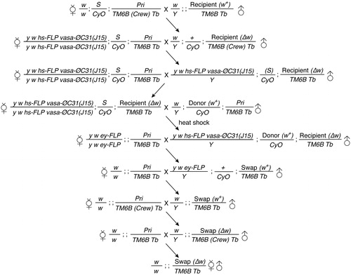
To conduct genetic swaps that replace the GAL4 domain with donor elements, the White marker of recipient GAL4 lines was first removed by introducing ubiquitously expressed Cre (, Supplementary Figure 2). Germ-cell specific ϕC31 Integrase and hs-Flipase were introduced to the w- recipient line. After introducing the w+ donor to the recipient cells, larvae were subject to one-hour 37 °C heat shock each day for five consecutive days. The male progenies with mosaic eyes were selected to build independent swap lines. Such males carried both donor and recipient elements and were crossed to females carrying ey-FLP. Potential true swap candidates have red eyes. Each red-eyed individual served as a founder to establish independent lines that allow removal of the original GAL4 through Cre. The progenies of each independent line were subjected to genotyping to verify true integration. Three to eight Cre/loxP crosses were established for each founder.
Figure 1. Crossing scheme for conducting genetic swaps. An example of the crossing strategy to genetically swap the original InSITE Gal4 inserted on the third chromosome with a donor inserted on the second chromosome.
Genotyping
A single fly genomic DNA preparation protocol was adapted from (Gloor et al., Citation1993). Briefly, a fly was anesthetized with CO2 and quickly squeezed in 50 μl squishing buffer (SB, 10 mM Tris–Cl pH 8.2, 1 mM EDTA, 25 mM NaCl and 200 μg/ml Proteinase K (P8102S, New England BioLabs, Ipswich, MA)) with a pipette tip and in a 1.5 ml tube. Proteinase K was freshly diluted before the experiment. The sample was incubated at 37 °C for 30 min, followed by 95 °C for 5 min to inactivate Proteinase K. Recombinant Taq DNA polymerase (11615010, Invitrogen, Carlsbad, CA) or Phusion® High-Fidelity DNA Polymerase (M0530S, New England BioLabs, Ipswich, MA) were used for polymerase chain reaction (PCR). The primer sets used to verify insertions were: set #1, GAL4-ML and GAL4-MR; set #2, LexAF and LexAR; set#3, LexAF and PBac3'R4, set #4, GAL80-5p and GAL80-3p; set #5, GAL80-5p and PBac3'R4; set #6, QF-F1 and QF-R1; set #7, QF3'F1 and PBac3'R4. The sequences of these primers are as follows:
GAL4-ML: CAGTGGAGGCAGAAAACAAATAC
GAL4-MR: TTTGTAAAAACTTTGGTGCCTGT
LexAF: CCAGGCAACAAGAGGTGTTT
LexAR: TCAGGCGCTTAACGGTAACT
GAL80-5p: GCAATCAAGACACATTACCCCGCCATACTG
GAL80-3p: ACCAACCGCCATTTCCAGCAATCTCG
QF-F1: GCCTAAACGCAAGACACTCA
QF-R1: CCGAGAAAGTCAAGTGAGGC
QF3'F1: GATCCGCAGTTCATGACGAA
PBac3'R4: CGCATGTGTTTTATCGGTCTGT
Immunohistochemistry
Larval or adult brains were dissected in 4% paraformaldehyde (15713-S, Electron Microscopy Sciences, Hatfield, PA) in PBS (UR-PBS001-1L, UniRegion Bio-Tech, Taiwan) and fixed at room temperature (RT) for 20 min. The brains were washed with PBST (0.3% Triton X-100 (X100, Sigma-Aldrich, St. Louis, MO) in PBS) at RT for 20 min three times. After incubation in 5% normal goat serum (005–000-121, Jackson ImmunoResearch Laboratories, West Grove, PA)/PBST at RT for 30 min, brains were then incubated in primary antibodies in 5% normal goat serum/PBST at 4 °C for 2–3 d. The brains were then washed three times for 20 min with PBST at RT, followed by incubation with secondary antibodies at 4 °C overnight. After three 20 min-PBST washes at RT, brains were incubated in SlowFade™ Gold Antifade Mountant (S36936, Invitrogen, Carlsbad, CA) at 4 °C for at least one night and then mounted in the same mounting solution with space support of coverglass (Cat. No. 0101050, Marienfeld, for adult brains) or silicone gel (Cat. No. 022090, Schmierstoff, for larval brains).
Primary antibodies included mouse anti-Bruchpilot (Brp) (nc82, Developmental Studies Hybridoma Bank (DSHB); 1:30), rat anti-mCD8 (MCD0800, Invitrogen, Carlsbad, CA; 1:100), rabbit anti-GFP (A-6455, Invitrogen, Carlsbad, CA; 1:500), rabbit anti-HA (ab9110, Abcam, Cambridge, UK; 1:500–1:1000), rat anti-DN-cadherin (DN-Ex#8, DSHB; 1:25). Secondary antibodies included goat anti-mouse IgG, goat anti-rat IgG and goat anti-Rabbit IgG which are conjugated with Alexa 488, DyLight 488, Cy3, Cy5, Alexa 647 or DyLight 649. All secondary antibodies were from Jackson ImmunoResearch Laboratories, West Grove, PA or Invitrogen, Carlsbad, CA.
Brains were imaged using a Leica TCS-SP5-AOBS-MP confocal microscope, a Zeiss LSM780 equipped with Mai-Tai HP-1040 (Spectra-Physics, Santa Clara, CA) or a Zeiss LSM700 confocal microscope (Carl Zeiss, Oberkochen, Germany). A 40× objective lens was used to observe central brains and a 20× objective lens was used to observe whole brains. Confocal images were processed using Zen black (Carl Zeiss, Oberkochen, Germany), Fiji (https://fiji.sc), and Adobe Photoshop.
Comparison of GR-GAL4 and GR-LexA expression
To compare the expression patterns in GR-GAL4 and corresponding GR-LexA lines, we focused on GR-GAL4 lines (Jenett et al., Citation2012) with expression that is mainly restricted to olfactory local interneurons (LNs). In order to identify such lines, we screened the expression patterns of ∼7000 GR-GAL4 lines through FlyLight annotations, searching for those that have GAL4 expression in the AL (http://flweb.janelia.org/cgi-bin/flew.cgi). We then visually screened confocal stacks archived by FlyLight to identify GAL4 lines with expression only in LNs. After identifying 134 suitable LN-GAL4 lines, we revisited FlyLight to search for possible corresponding LN-LexA lines. In total, we identified 21 GR-GAL4 lines that show expression specifically in LNs and have corresponding LexA lines. The GAL4 and LexA expression patterns of these 21 lines were compared by two researchers. The LexA expression patterns could be grouped into four categories relative to the GAL4 expression patterns: similar, more restricted, ectopic expression and no expression, following previous criteria (Dionne et al., Citation2018). Any discrepancies between the two researchers were further analyzed and resolved by a third researcher.
Cell counting
Confocal stacks were analyzed with the LSM browser (Carl Zeiss, Oberkochen, Germany). The somas of GFP-positive olfactory LNs or ventral inhibitory projection neurons (viPNs) were identified and manually marked. The data were compared by a two-tailed Student’s t-test. Detailed statistical outcomes can be found in Supplementary Table 2.
Results
Targeting InSITE lines with GAL4 expression in olfactory neurons
In a large-scale genetic screen for InSITE GAL4 drivers that label distinct subsets of LNs (Liou et al., Citation2018), we uncovered a set of 25 LN GAL4 lines in addition to a few GAL4 lines with expression in olfactory receptor neurons (ORNs) or viPNs. Based on the innervation patterns of the labeled neurons, 13 LN lines, 6 viPN lines and 1 ORN line were used for swap experiments (). Eight lines (68, 351, 382, 421, 449, 501, 1078, and 1081) were subjected to both GAL80 and QF swaps, while LexA and QF swaps were both performed on five lines (564, 584, 666, 670, and 810) ().
Figure 2. GAL4 expression patterns of InSITE lines subjected to genetic swap. Projections of confocal stacks. Adult brains were stained for mCD8 (green) and the neuropil marker, Bruchpilot (Brp, magenta). D, dorsal; L, lateral. Scale bar, 20 μm.
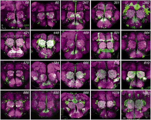
Table 1. Summary of genetic swap results.
Swapping GAL4 with LexA in olfactory neuron enhancer trap lines
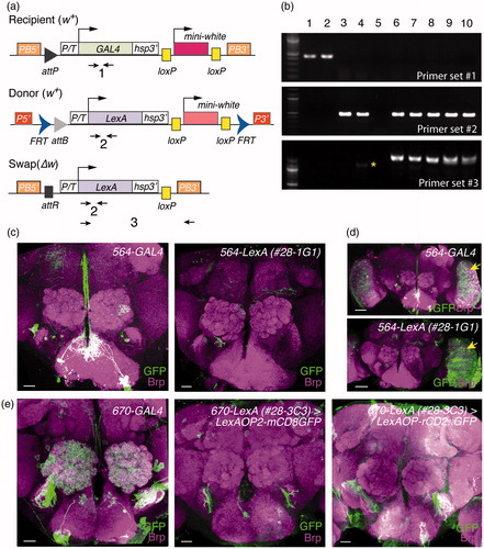
We conducted six genetic swap experiments to replace GAL4 with LexA in five GAL4 lines. From each of the five swap experiments, we successfully identified one to five true integrations. The frequencies of uncovering LexA-swap candidates and true integrations were 68.4 and 84.6%, respectively (, ). However, only 564-LexA partially recapitulated the expression patterns of 564-GAL4 in the optic lobe (arrows in ), but it did not recapitulate the GAL4 expression patterns in the central brain (). The other four examined LexA swap lines did not show expression patterns similar to the original GAL4 lines (, and data not shown). To address whether the LexA reporters would have any effect on the observed expression patterns, we used 670-lexA to drive two different reporters, LexAop2-mCD8GFP and LexAop-rCD2GFP, which contain LexA binding sites that were respectively derived from the binding motifs of colE1 and sulA (Lai & Lee, Citation2006; Pfeiffer et al., Citation2010) (). Neither of these reporters showed expression patterns that were similar to 670-GAL4. Therefore, the properties of reporters are unlikely to cause issues with expression reproducibility found in LexA swaps.
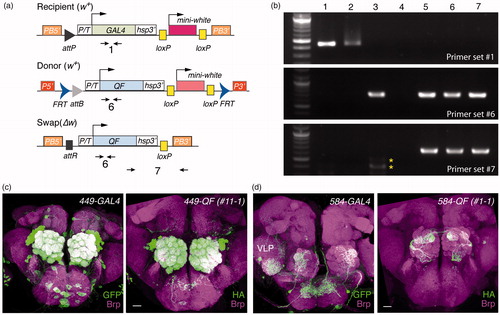
Figure 3. Genetic swap of GAL4 enhancer trap to LexA. (a) Primer sets used to verify the genetic conversion of GAL4 to LexA. (b) PCR analysis to confirm the swap results. Genomic DNA samples: lane 1, 584-GAL4; lane 2, 670-GAL4; lane 3, LexAL19.2L donor; lane 4, LexAL44.2 donor; lane 5, water; lane 6, 584-LexAL19.2L (2-4); lane 7, 584-LexAL44.2 (1-1); lane 8, 670-LexAL44.2 (28-3B8); lane 9, 670-LexAL44.2 (28-3C3); lane 10, 670-LexAL44.2 (28-3D3). The asterisk denotes non-specific PCR product. (c–e) Functional validation of swapped 564-LexA (c,d) and 670-LexA (e) lines. Adult brains were stained for mCD8 or GFP (green) and Bruchpilot (Brp, magenta). (d) Low magnification images of (c) are shown. Arrows indicate optic lobes. Image of 670-GAL4 is reproduced from . Scale bars, 20 μm (c, e) or 50 μm (d).
Swapping GAL4 with GAL80 in olfactory neuron enhancer trap lines
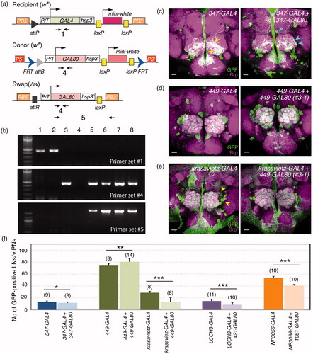
One efficient method to restrict GAL4 expression in a small subset of neurons is through intersectional interactions between GAL4 and GAL80 expressed in two partially overlapping neuronal populations. We therefore conducted 14 genetic swap experiments to replace GAL4 in the original enhancer trap lines with GAL80. The success rate of establishing swap candidate lines was 80.7% (). Among the candidate lines, we successfully identified one to 12 true integrations from 10 swap experiments. The frequency of true integrations was 73.2% (, ). When swapped 347-GAL80 was introduced to 347-GAL4 flies, the detectable innervation domain of 347-positive viPNs in the AL was mildly decreased (yellow arrowheads in , Supplementary Table 2), with some additional somas and innervations being detected (cyan arrowheads in ). 449-GAL80 likely did not suppress 449-GAL4 in the LNs around the AL (, Supplementary Table 2). 449-GAL4 is expressed in ∼60 lateral LNs (∼9 of them are GABA-negative) (Liou et al., Citation2018), and krasavietz-GAL4 is expressed in ∼16 lateral LNs (∼7 of them are GABA-negative) (Chou et al., Citation2010). Therefore, 449-GAL4-positive LNs may partially overlap with the krasavietz-GAL4 expressing LNs. If 449-GAL80 is expressed in the same population of LNs as 449-GAL4, we would expect to observe 449-GAL80 can suppress krasaviez-GAL4 in a subset of LNs. Indeed, the number of krasavietz-positive LNs was decreased when 449-GAL80 was introduced to the fly (, Supplementary Table 2). These results suggest genetically swapped GAL80 may have expression patterns close to or the same as those of the original GAL4 enhancer traps.
Figure 4. Genetic swap of GAL4 enhancer trap to GAL80. (a) Primer sets used to verify the genetic conversion of GAL4 to GAL80. (b) PCR analysis to confirm the swap results. Genomic DNA samples: lane 1, 347-GAL4; lane 2, 449-GAL4; lane 3, GAL80E22.1 donor; lane 4, water; lane 5, 347-GAL80 (1-3); lane 6, 347-GAL80 (4-4); lane 7, 449-GAL80 (2-1); lane 8, 449-GAL80 (3-1). (c–e) Functional validation of swapped GAL80 lines. Adult brains were stained for mCD8 (green) and Bruchpilot (Brp, magenta). (c) Compared to the processes of GFP-positive cells in the 347-GAL4 AL (yellow arrowheads, left panel), 347-GAL80 appears to have fewer GFP-positive processes marking 347-positive neurons in the AL, with additional somas and innervations observed (cyan arrowheads, right panel). Although 449-GAL80 did not obviously suppress 449-GAL4 expressions (d), it did suppress GAL4 activity in a subset of krasavietz-positive LNs (arrowheads) (e). Scale bars, 20 μm. (f) Quantification of GFP-positive LNs or viPNs in different GAL4-GAL80 combinations. Bar graphs show mean ± S.D. Numbers of analyzed ALs are indicated in the parenthesis. Student’s t-test was used to compare the groups. *p<.05, **p<.01, ***p<.001.
Swapping GAL4 with QF in olfactory neuron enhancer trap lines
In addition to GAL80 and LexA, a third binary system, QF-QUAS, has proven to be an efficient method for the intersectional restriction of GAL4 expression (Potter et al., Citation2010). We conducted 15 genetic swap experiments to replace the GAL4 with QF in 15 InSITE enhancer trap lines. We successfully uncovered one to nine true integrations from each of the eight swap experiments. The frequencies of finding QF-swap candidates and true integrations were 60.8 and 63.2%, respectively (, ). 449-QF exhibited very similar expression patterns compared to 449-GAL4 in the central brain, especially in LNs (). However, some other swapped QF lines showed very different expression patterns than the original GAL4 lines ( and data not shown). For instance, 584-GAL4 labels a single LN around the AL and a few LNs in the ventrolateral protocerebrum (VLP), but 584-QF is expressed in a subset of ORNs and not the AL LN nor the VLP LNs ().
Figure 5. Validation of GAL4 to QF enhancer trap swaps. (a) Primer sets used to verify the genetic conversion of GAL4 to QF. (b) PCR analysis to confirm the swap results. Genomic DNA samples: lane 1, 449-GAL4; lane 2, 584-GAL4; lane 3, QF.Q10B donor; lane 4, water; lane 5, 449-QF.Q10B (11-1); lane 6, 449-QF.Q10B (14-2); lane 7, 584-QF.Q10B (1-1). Asterisks denote non-specific PCR products. (c–d) Functional validation of swapped QF lines. Adult brains were stained for mCD8 (green, left panels) or mtdT-3xHA (green, right panels) and Bruchpilot (Brp, magenta). Images of 449-GAL4 and 584-GAL4 are reproduced from . Scale bars, 20 μm.
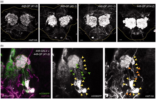
When we carefully compared the expression of 449-QF in the central brains of eight independent 449-QF swap lines, we noticed that the QF expression levels were quite variable in what was presumably same population of LNs (). We suspected some 449-GAL4-positive LNs may be excluded from the 449-QF-positive population during development when the 449-QF expression level is low in adult brains. We therefore examined the expression of 449-QF in 449-GAL4-positive larval LNs to ask whether weak 449-QF drivers may recapitulate 449-GAL4 expression patterns during development. We found that weak 449-QF indeed drives reporter expression in a partially overlapping subset of larval LNs labeled by 449-GAL4 (). These results suggest that generation and characterization of multiple independent swap lines may be required to identify a swap line that fully recapitulates the original GAL4 expression patterns.
Figure 6. Differential expression levels in independent QF swap lines. (a) The QF expression patterns of four representative 449-QF lines, showing weak LN-QF, medium LN-QF and strong LN-QF expression. All brains were imaged with the same confocal configurations for mtdT-3xHA. Yellow dashed lines show brain contours based on Brp staining (not shown). (b) 449-GAL4-driven mCD8GFP (green) and 449-QF-driven mtdT-3xHA (magenta) were co-stained in a late third instar larval brain. LNs labeled only by 449-GAL4, only by 449-QF and by both 449-GAL4 and 449-QF are indicated by green, orange and yellow arrowheads, respectively. Scale bars, 20 μm.
Discussion
Intersectional methods have made tracing and manipulating a single or small subset of defined neurons possible in Drosophila. A few large enhancer trap collections have been established to allow swapping or insertion of desired components to particular loci, however, the feasibility and potential limiting factors for successful swapping remain to be fully explored. In this work, we took advantage of the genetic swap feature in InSITE GAL4 enhancer trap lines and replaced the GAL4 component with LexA, GAL80 or QF. We found different swap efficiencies among LexA, GAL80 and QF. The limiting factor on experimental utility appeared to be the reproducibility of the expression patterns of swapped components. We hope that our efforts to validate some of these swapped lines will be useful to the community.
Different efficacy of swapped components
We conducted 35 genetic swap experiments to replace GAL4 with LexA, GAL80 or QF. In general, the frequencies of uncovering swap candidates and true integrations were 69 and 70%, respectively (). On average, we established fewer crosses for each swap experiment than the first InSITE report (Gohl et al., Citation2011), and we were able to uncover at least one swap candidate line (crosses with W+) in 25 out of 35 swap experiments. This success rate is partly due to the cross paradigm we designed, which excluded the donor chromosome before establishing swap candidate lines (, Supplementary Figure 2). Among those experiments with swap candidates, 22 out of 24 experiments yielded at least one true-integration line. Therefore, the performance of genetic swaps is feasible with a reasonably high success rate. However, only eight out of 17 verified cases reproduced major GAL4-expression patterns in the brain (). Thus, the feasibility of genetic swaps, substituting GAL4 with different elements, is limited by the recapitulation of GAL4 expression patterns in the original enhancer trap lines.
Possible factors that may influence the expression patterns of swapped lines
In the original InSITE work, Gohl et al. (Citation2011) used multiple primer sets to carefully verify true integrations. By this method, the authors observed the retention of the original donor transposon in which one FRT site was inactivated. With our crossing scheme, this situation could not occur because our method excludes the donor chromosome. The authors of the previous study also found a direct fusion between the recipient attP site and the donor attB site in a rare case. Our PCR verification strategy would not detect such an event, but we believe direct fusions of attP and attB sites are not likely to fully explain the differential expression patterns of swapped lines.
We attempted to ask whether the insertion site of the donor makes any contribution to the expression level of swapped LexA. In answer to this question, we did not observe any obvious differences in the swap efficiency between two LexA lines that received LexA from different donors (). We also ruled out that the failure to reproduce expression patterns may be at least partially due to the LexAop2 reporter, since the LexAop reporter also did not show expression patterns similar to that of the original GAL4 line (). Therefore, interactions between the donor flanking sequences and GAL4 insertions in addition to some feature(s) of the LexAop2 reporter cannot fully account for the differential expression patterns in swapped LexA lines. In agreement with our observations, LexA has been previously demonstrated to exhibit a more restricted expression pattern, or exhibit additional labeled cells, when driven by the same enhancer fragments as GAL4 counterparts (Dionne et al., Citation2018). However, the overall recapitulation of GAL4 expression patterns by such ‘molecular’ swapped LexA lines is better than the genetic swapped LexA lines in our hands (Supplementary Figure 3). Using alternative LexA variants (Pfeiffer et al., Citation2010; Yagi, Mayer, & Basler, Citation2010) as donors may be one potential solution for this issue.
Ideally, the expression patterns of swapped GAL80 lines could be examined through immunostaining with anti-GAL80 antibodies. However, our attempts to generate anti-GAL80 antibodies and our testing of commercially available anti-GAL80 antibodies have all failed to identify a reagent that is suitable for detection of GAL80 (data not shown). We therefore used indirect methods by introducing swapped GAL80 to the original GAL4 lines or to a second GAL4 line that shows partially overlapping expression patterns with the original GAL4 lines (). Although the GAL80 lines did not fully suppress GAL4 expression in the original lines, 449-GAL80 did effectively suppress krasavietz-GAL4 expression in a subset of GAL4-positive cells (). When introducing the swapped GAL80 to flies carrying the original GAL4, 347-GAL4/347-GAL80 fly brains showed additional labeled cells, as did 449-GAl4/449-GAL80 fly brains (). This lack of suppression and ectopic expression may be due to competition between GAL80 and the original GAL4 for the same set of transcription regulatory components and/or potential effects that originate from homozygous mutations at the inserted locus. In the future, a donor with multiple copies of GAL80 (e.g. tandem repeats of GAL80 or multiple GAL80 coding sequences separated by T2A or IRES) may be useful to create GAL80 lines with high enough expression levels to suppress GAL4.
We conducted 15 swap experiments to replace GAL4 with QF in 15 InSITE lines. Among eight swap events that we confirmed had at least one true integration, only three QF swaps demonstrated the same or similar expression patterns as those of the original GAL4 lines. Additionally, we often observed higher or lower expression in the tracheae of the swapped QF lines, a result that may be due to the QF itself (Potter et al., Citation2010). Several QF variants have recently been constructed and have been demonstrated to be less toxic than the original QF (Riabinina et al., Citation2015). These QF variants may be good candidates for developing new QF donors.
In addition to genetic swaps, InSITE GAL4 may be swapped by microinjecting donor plasmids (Gao et al., Citation2013). However, systematic analyses of microinjection-swapped InSITE lines with different donors are lacking. In the future, it will be beneficial to directly compare the two swap methods to test whether the differences in expression patterns may partly originate from the process of genetic swapping.
Variations of the expression patterns of successful swapped lines
The best genetic swap case we found was 449-QF (). Interestingly, we observed similar expression patterns but variable expression levels from independent 449-QF lines (). Such variable QF expression levels occurred as early as during larval development. Therefore, establishing multiple swap lines for a given swap experiment may be necessary and is certainly recommended.
InSITE is a genetic swappable enhancer trap collection that is complementary to the Janelia Farm GAL4 collection (http://flweb.janelia.org/cgi-bin/flew.cgi). The majority of InSITE lines and their insertion sites (Gohl et al., Citation2014) are available through the Bloomington Drosophila Stock Center (https://bdsc.indiana.edu/index.html) and FlyBase (http://flybase.org). Through modification of the donor cassettes to decrease the cryptic genetic interactions between flanking chromosome regions of the insertion site and the sequences of donor cassettes, the genetic swap of InSITE enhancer trap lines remains a feasible approach to expand the genetic toolbox of Drosophila.
Suplementary_information-rev1.pdf
Download PDF (3.3 MB)Acknowledgments
We thank Thomas Clandinin (Stanford U.) for providing InSITE enhancer trap lines and donor fly stock. We thank Marcus Calkins for invaluable comments.
Disclosure statement
No potential conflict of interest was reported by the authors.
Data availability
The authors declare that all data supporting the findings of this study are available within the article and its supplementary information files, or from the corresponding author upon reasonable request.
Additional information
Funding
References
- Bellen, H.J., Levis, R.W., Liao, G., He, Y., Carlson, J.W., Tsang, G., … Spradling, A.C. (2004). The BDGP gene disruption project: single transposon insertions associated with 40% of Drosophila genes. Genetics, 167, 761–781. doi:10.1534/genetics.104.026427
- Bohm, R.A., Welch, W.P., Goodnight, L.K., Cox, L.W., Henry, L.G., Gunter, T.C., … Zhang, B. (2010). A genetic mosaic approach for neural circuit mapping in Drosophila. Proceedings of the National Academy of Sciences of the United States of America, 107, 16378–16383. doi:10.1073/pnas.1004669107
- Brand, A.H., & Perrimon, N. (1993). Targeted gene expression as a means of altering cell fates and generating dominant phenotypes. Development, 118, 401–415. Retrieved from http://dev.biologists.org/content/118/2/401.long
- Chou, Y.H., Spletter, M.L., Yaksi, E., Leong, J.C., Wilson, R.I., & Luo, L. (2010). Diversity and wiring variability of olfactory local interneurons in the Drosophila antennal lobe. Nature Neuroscience, 13, 439–449. doi:10.1038/nn.2489
- Diao, F., Ironfield, H., Luan, H., Diao, F., Shropshire, W.C., Ewer, J., … White, B.H. (2015). Plug-and-play genetic access to drosophila cell types using exchangeable exon cassettes. Cell Repair, 10, 1410–1421. doi:10.1016/j.celrep.2015.01.059
- Dionne, H., Hibbard, K.L., Cavallaro, A., Kao, J.C., & Rubin, G.M. (2018). Genetic reagents for making split-GAL4 lines in Drosophila. Genetics, 209, 31–35. doi:10.1534/genetics.118.300682
- Dubnau, J., Chiang, A.S., Grady, L., Barditch, J., Gossweiler, S., McNeil, J., … Tully, T. (2003). The staufen/pumilio pathway is involved in Drosophila long-term memory. Current Biology, 13, 286–296. doi:10.1016/S0960-9822(03)00064-2
- Gao, X.J., Potter, C.J., Gohl, D.M., Silies, M., Katsov, A.Y., Clandinin, T.R., & Luo, L. (2013). Specific kinematics and motor-related neurons for aversive chemotaxis in Drosophila. Current Biology, 23, 1163–1172. doi:10.1016/j.cub.2013.05.008
- Gloor, G.B., Preston, C.R., Johnson-Schlitz, D.M., Nassif, N.A., Phillis, R.W., Benz, W.K., … Engels, W.R. (1993). Type I repressors of P element mobility. Genetics, 135, 81–95. Retrieved from http://www.genetics.org/content/135/1/81.long
- Gohl, D.M., Freifeld, L., Silies, M., Hwa, J.J., Horowitz, M., & Clandinin, T.R. (2014). Large-scale mapping of transposable element insertion sites using digital encoding of sample identity. Genetics, 196, 615–623. doi:10.1534/genetics.113.159483
- Gohl, D.M., Silies, M.A., Gao, X.J., Bhalerao, S., Luongo, F.J., Lin, C.C., … Clandinin, T.R. (2011). A versatile in vivo system for directed dissection of gene expression patterns. Nature Methods, 8, 231–237. doi:10.1038/nmeth.1561
- Gordon, M.D., & Scott, K. (2009). Motor control in a Drosophila taste circuit. Neuron, 61, 373–384. doi:10.1016/j.neuron.2008.12.033
- Hayashi, S., Ito, K., Sado, Y., Taniguchi, M., Akimoto, A., Takeuchi, H., … Goto, S. (2002). GETDB, a database compiling expression patterns and molecular locations of a collection of Gal4 enhancer traps. Genesis, 34, 58–61. doi:10.1002/gene.10137
- Jenett, A., Rubin, G.M., Ngo, T.T., Shepherd, D., Murphy, C., Dionne, H., … Zugates, C.T. (2012). A GAL4-driver line resource for Drosophila neurobiology. Cell Reports, 2, 991–1001. doi:10.1016/j.celrep.2012.09.011
- Kvon, E.Z., Kazmar, T., Stampfel, G., Yanez-Cuna, J.O., Pagani, M., Schernhuber, K., … Stark, A. (2014). Genome-scale functional characterization of Drosophila developmental enhancers in vivo. Nature, 512, 91–95. doi:10.1038/nature13395
- Lai, S.L., & Lee, T. (2006). Genetic mosaic with dual binary transcriptional systems in Drosophila. Nature Neuroscience, 9, 703–709. doi:10.1038/nn1681
- Lee, T., & Luo, L. (1999). Mosaic analysis with a repressible cell marker for studies of gene function in neuronal morphogenesis. Neuron, 22, 451–461. doi:10.1016/S0896-6273(00)80701-1
- Lin, C.C., & Potter, C.J. (2016). Editing transgenic DNA components by inducible gene replacement in Drosophila melanogaster. Genetics, 203, 1613–1628. doi:10.1534/genetics.116.191783
- Liou, N.F., Lin, S.H., Chen, Y.J., Tsai, K.T., Yang, C.J., Lin, T.Y., … Chou, Y.H. (2018). Diverse populations of local interneurons integrate into the Drosophila adult olfactory circuit. Nature Communications, 9, 2232. doi:10.1038/s41467-018-04675-x
- Luan, H., Peabody, N.C., Vinson, C.R., & White, B.H. (2006). Refined spatial manipulation of neuronal function by combinatorial restriction of transgene expression. Neuron, 52, 425–436. doi:10.1016/j.neuron.2006.08.028
- Lukacsovich, T., Asztalos, Z., Awano, W., Baba, K., Kondo, S., Niwa, S., & Yamamoto, D. (2001). Dual-tagging gene trap of novel genes in Drosophila melanogaster. Genetics, 157, 727–742. Retrieved from http://www.genetics.org/content/157/2/727.long
- McGuire, S.E., Le, P.T., & Davis, R.L. (2001). The role of Drosophila mushroom body signaling in olfactory memory. [see comments]. Science, 293, 1330–1333. doi:10.1126/science.1062622
- Pfeiffer, B.D., Ngo, T.T., Hibbard, K.L., Murphy, C., Jenett, A., Truman, J.W., & Rubin, G.M. (2010). Refinement of tools for targeted gene expression in Drosophila. Genetics, 186, 735–755. doi:10.1534/genetics.110.119917
- Potter, C.J., Tasic, B., Russler, E.V., Liang, L., & Luo, L. (2010). The Q system: a repressible binary system for transgene expression, lineage tracing, and mosaic analysis. Cell, 141, 536–548. doi:10.1016/j.cell.2010.02.025
- Riabinina, O., Luginbuhl, D., Marr, E., Liu, S., Wu, M.N., Luo, L., & Potter, C.J. (2015). Improved and expanded Q-system reagents for genetic manipulations. Nature Methods, 12, 219–222. doi:10.1038/nmeth.3250
- Simpson, J.H. (2016). Rationally subdividing the fly nervous system with versatile expression reagents. Journal of Neurogenetics, 30, 185–194. doi:10.1080/01677063.2016.1248761
- Stowers, R.S. (2011). An efficient method for recombineering GAL4 and QF drivers. Fly (Austin), 5, 371–378. doi:10.4161/fly.5.4.17560
- Venken, K.J., Schulze, K.L., Haelterman, N.A., Pan, H., He, Y., Evans-Holm, M., … Bellen, H.J. (2011). MiMIC: a highly versatile transposon insertion resource for engineering Drosophila melanogaster genes. Nature Methods, 8, 737–743. doi:10.1038/nmeth.1662
- Yagi, R., Mayer, F., & Basler, K. (2010). Refined LexA transactivators and their use in combination with the Drosophila Gal4 system. Proceedings of the National Academy of Sciences of the United States of America, 107, 16166–16171. doi:10.1073/pnas.1005957107