
Abstract
G protein-coupled receptors (GPCRs) represent a family of seven-pass transmembrane protein receptors whose ligands include neuropeptides and small-molecule neuromodulators such as dopamine and serotonin. These neurotransmitters act at long distances and are proposed to define the ground state of the nervous system. The Drosophila genome encodes approximately 50 neuropeptides and their functions in physiology and behavior are now under intensive studies. Key information currently lacking in the field is the spatiotemporal activation patterns of endogenous GPCRs. Here we report application of the Tango system, a reporter assay to detect GPCR activity, to endogenous GPCRs in the fly genome. We developed a method to integrate the sensor component of the Tango system to the C-terminus of endogenous genes by using genome editing techniques. We demonstrate that Tango sensors in the Sex-peptide receptor (SPR) locus allow sensitive detection of mating-dependent SPR activity in the female reproductive organ. The method is easily applicable to any GPCR and will provide a way to systematically characterize GPCRs in the fly brain.
Introduction
Exposure to a new environment as well as changes in the internal states of the body leaves traces in the nervous system of animals. Visualization of experience-dependent changes in the nervous system is thus a key step in identifying mechanisms underlying behavioral responses and adaptation. Various chemical and genetically encoded probes for neuronal activities have been developed to date (Deo & Lavis, Citation2018). These sensors offer high spatiotemporal resolutions, allowing the identification of single cells with response dynamics. To detect slower physiological changes ranging from minutes to hours, transcriptional assays including one based on the expression of immediate early genes have been developed in Drosophila (Fujita et al., Citation2013; Gao et al., Citation2015; Masuyama, Zhang, Rao, & Wang, Citation2012). Although these systems are useful for monitoring generic features of neuronal activity, such as intracellular Ca2+ and neurotransmission, they do not tell which neurotransmitter molecule actuated the responding cells.
The Tango system is a transcriptional reporter assay to detect activation of individual G protein-coupled receptors (GPCRs), which include receptors for most neuropeptides and some small molecule neurotransmitters. It was originally developed as an in vitro assay to monitor GPCR activation by converting ligand binding to the cleavage of a receptor-tethered transcription factor (Barnea et al., Citation2008) (), and later applied to the Drosophila brain to identify cell types undergoing activity-dependent changes through particular neuromodulator receptor signaling (Inagaki et al., Citation2012; Jagadish, Barnea, Clandinin, & Axel, Citation2014). The system comprises three components (): (1) a β-Arrestin-TEV protease fusion protein, (2) a transcription factor (TF), such as GAL4 or LexA, linked to a GPCR with a TEV protease cleavage sequence (TCS) and (3) a reporter transgene whose expression is under the control of the TF. Upon activation by ligand binding, the cytoplasmic tails of GPCRs are phosphorylated by G protein-coupled receptor kinase (GRK), to which β-Arrestin is recruited and block receptor activity as negative feedback regulation (Barnea et al., Citation2008; Luttrell & Lefkowitz, Citation2002). The Tango system takes advantage of this intermolecular association, whereby the recruited β-Arrestin-TEV cleaves and releases the TF from the GPCR (). The released TF then translocates into the nucleus and induces reporter gene expression (). In a previous implementation of the Tango system for Drosophila tissues, GPCR::TCS::LexA fusion protein was expressed broadly at high levels to visualize the spatiotemporal distribution of GPCR ligands by using reporter activity as a readout (Inagaki et al., Citation2012; Jagadish et al., Citation2014).
Figure 1. Schematic diagram of the Tango system. The system comprises three components: GPCR::TCS::LexA, β-Arrestin-TEV and lexAop-reporter. Activation of GPCR by ligand binding results in phosphorylation of its C-terminus by GRK. β-Arrestin-TEV is then recruited to the phosphorylated C-terminus of GPCR::TCS::LexA and cleaves the TCS to release LexA. Released LexA translocates into the nucleus and induces reporter expression from the lexAop-reporter transgene.
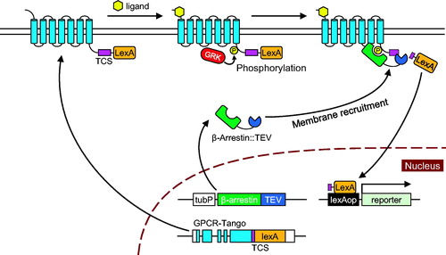
In the previous studies, the sensor component (GPCR::TCS::LexA in ) was overexpressed broadly in the brain including cells that do not express the target GPCR (Inagaki et al., Citation2012; Jagadish et al., Citation2014). Therefore, reporter expression could have been induced in those cells that normally do not respond to the particular GPCR ligand. Furthermore, neurotransmitters often have multiple cognate receptors and the previous Tango system for Drosophila do not tell which receptor types of receptors are activated in particular cells.
Here, we present a new method to apply the Tango system to detection of the activity of endogenous GPCRs. We use the CRISPR/Cas9 technique to integrate a Tango cassette (TCS::LexA) into the C-terminus of endogenous GPCR genes so that the resultant GPCR::TCS::LexA fusion protein is expressed in the same pattern as the endogenous GPCR gene. As a proof of principle, we demonstrate that the method can successfully visualize the activity of Sex-peptide receptor (SPR), a GPCR expressed in the brain and the female reproductive organ of Drosophila.
Results
Tango reporter system for endogenous GPCRs
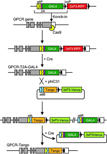
To apply the Tango system to monitoring endogenous GPCR activity in Drosophila, we developed a system that expresses GPCR::TCS::LexA at physiological levels from endogenous promoters by targeted integration of TCS::LexA cassettes (hereafter called Tango cassettes) into the C-terminus of endogenous GPCR genes. Integration of a Tango cassette was performed in two steps (). First, we inserted a transgene cassette that contains T2A-GAL4 (Diao & White, Citation2012) immediately in front of the stop codon of the GPCR gene by CRISPR/Cas9-mediated knock-in such that GAL4 is translated as an in-frame fusion with the endogenous GPCR protein (). The intervening T2A self-cleaving peptide induces ribosome skipping during translation (Donnelly et al., Citation2001), producing the GPCR and GAL4 proteins separately from the same transcript. Details of the T2A-GAL4 knock-in system will be described elsewhere (Kondo, unpublished). Then we took advantage of the attP and loxP recombinase sites to swap the T2A-GAL4 cassette for a Tango cassette by recombinase-mediated cassette exchange (RMCE).
Figure 2. Targeted integration of the Tango sensor. First, a transgene cassette containing T2A-GAL4 is inserted into the C-terminus of the target GPCR by CRISPR/Cas9-mediated knock-in. This results in in-frame fusion of the GPCR and T2A-GAL4. 3xP3-was used as a visible marker for selection of transformants. It was subsequently removed by transient expression of Cre recombinase. The resultant T2A-GAL4 cassette is flanked by a phiC31 attP site and a loxP site, which are used for exchange of T2A-GAL4 for a Tango sensor by RMCE. A plasmid vector that contains the Tango sensor flanked by attB and loxP is integrated into the attP site between the GPCR and the Tango sensor. RMCE is completed by removal of the 3xP3-venus selection marker and the vector backbone by treatment with Cre recombinase.
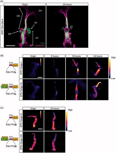
In previous studies, several variants of Tango sensors with differing sensitivities have been developed (Barnea et al., Citation2008; Hanson et al., Citation2009; Kroeze et al., Citation2015). One important variable is the cleavage efficiency of the TCS. Even without membrane recruitment after GPCR activation, β-Arrestin-TEV in the cytoplasm has the capacity to cleave the TCS albeit at low efficiency. This baseline activity could be problematic if β-Arrestin-TEV or GPCR-Tango is expressed at high levels. By changing the last glycine in the wild-type cleavage sequence (ENLYFQG) of TEV to leucine (ENLYFQL), the cleavage efficiency is greatly reduced, resulting in a reporter with minimal background (Hanson et al., Citation2009). Another variation is based on the activity-dependent phosphorylation of GPCR. Upon ligand binding, the cytoplasmic C-terminal tail of most GPCRs are phosphorylated by GRK, to which β-Arrestin is subsequently recruited. Some GPCRs are known to completely lack such phosphorylation sites and are not compatible with the Tango system (Barnea et al., Citation2008). Adding a seven-amino-acid peptide derived from the cytoplasmic tail of human arginine vasopressin receptor 2 (AVPR2) before the TCS significantly improves the sensitivity of Tango sensor for these GPCRs (Barnea et al., Citation2008). This peptide, called a V2 tail, contains multiple GRK phosphorylation sites and enhances the recruitment of β-Arrestin upon activation of the GPCR. To compare Tango variants with different sensitivities in vivo, we developed four Tango cassettes with different combinations of TCS sequences and a V2 tail or lack thereof: Tango-G and Tango-L have a wild-type and mutant TCS, respectively. In addition, Tango-VG and Tango-VL have a V2 tail ().
Figure 3. Spatiotemporal patterns of Tango activity in the female reproductive organ. (A) SPR-expressing cells in the female reproductive organ. SPR-expressing cells were visualized in flies that carry SPR-T2A-GAL4 and UAS-mCD8-GFP. Prominent expression was detected in the uterus (UT), the spermatheca (ST), part of the seminal receptacle (SR) and part of the oviduct (OD). No GFP expression was observed in the ovary (OV). Left: the reproductive organ of a virgin female. Right: the reproductive organ of a female fly 24 hours after mating. F-actin (magenta), mCD8-GFP (green). (B) Time-course analysis of reporter expression in SPR-Tango-L and -Tango-VL after mating. Left: Schematic designs. Each Tango sensor has a TEV cleavage site (TCS) and LexA. In addition, Tango-VL carries a V2 tail to promote β-Arrestin recruitment. Tango-L and Tango-VL carry a mutant TCS (amino-acid sequence: ENLYFQL) which is a less efficient substrate of TEV. Right: Strong mCD8-GFP was observed in the reproductive organ from 16 hours after mating in both Tango-L and Tango-VL. (C) Reporter expression in SPR-Tango-G and Tango-VG after mating. Left: Schematic designs. Tango-G and Tango-VG carry a wild-type TCS (ENLYFQG) that is cleaved by TEV by maximal efficiency. Right: Strong GFP expression was observed in the uterus and the oviduct regardless of mating status in both Tango-G and Tango-VG. The seminal receptacle and the spermatheca were removed from the reproductive organs before imaging. Scale bars in A, B, and C: 200 µm.
Detection of mating-dependent SPR activity using tango reporters
To validate the efficacy of the knock-in Tango system, we first chose to generate and evaluate Tango sensors for Sex peptide receptor (SPR). The spatial distribution and physiological function of SPR have been well documented (Tsuda, Peyre, Asano, & Aigaki, Citation2015; Yapici, Kim, Ribeiro, & Dickson, Citation2008). SPR is expressed in the oviduct and the spermathecae of the female reproductive organ, and is activated by its cognate ligand, Sex peptide (SP) in the seminal fluid upon mating. Several neurons in the ventral nerve cord that send processes to the oviduct also express SPR (Häsemeyer, Yapici, Heberlein, & Dickson, Citation2009). When female flies copulate, these neurons receive SP signals and relay the information to the central nervous system to induce post-mating behaviors such as courtship rejection and egg production.
We first generated a knock-in strain that carries T2A-GAL4 in the SPR locus by CRISPR/Cas9-mediated targeted integration. Subsequently, we replaced T2A-GA4 with either of the four Tango variants by RMCE to generate SPR-Tango strains. To test if the inserted transgene was expressed in the same pattern as the endogenous SPR gene, we first crossed SPR-T2A-GAL4 to UAS-CD8-GFP to visualize its expression in the female reproductive organ. Strong GFP fluorescence was observed in the uterus, the spermathecae and the seminal receptacle (). This confirms that transgenes inserted at the C-terminus of SPR faithfully recapitulate the expression pattern of SPR.
We then examined if the Tango sensors in the SPR locus could be activated by mating. We crossed each of the SPR-Tango knock-ins to a reporter strain that carries tubP-β-arrestin-TEV and 13xLexAop2-CD8::GFP. tubP-β-arrestin-TEV ubiquitously expresses β-Arrestin-TEV (Inagaki et al., Citation2012) while 13xLexAop2-CD8::GFP expresses membrane-targeted GFP in the presence of LexA. We collected virgin female flies that carry SPR-Tango, tubP-β-arrestin-TEV and 13xLexAop2-CD8::GFP for a few days. Half of them were mated to wild-type males while the other half were kept without males. We examined GFP fluorescence in the reproductive organs of virgin or mated females.
In SPR-Tango-VL and SPR-Tango-L, no GFP signals were observed in the reproductive organ of virgin females (). We detected strong mating-dependent GFP expression in all-female flies examined at 16 and 24 h after mating (). The results strongly suggest that these two Tango variants induced GFP expression in concordance with SPR activation. The spatial expression pattern of GFP in the female reproductive organ was essentially identical to that of endogenous SPR as visualized by SPR-T2A-GAL4: the oviduct, the spermathecae, and the seminal receptacle. This suggests that the SP in the seminal fluid had access to all of the cells that express SPR in the reproductive organ after it was transferred from the male. GFP expression in Tango-VL was generally stronger than in Tango-L, indicating that the phosphorylation sites from the V2 tail are also targeted by GRK in Drosophila and enhances β-Arrestin recruitment.
In contrast, we did not observe mating-dependent changes in GFP expression in Tango-G and Tango-VG (). Even without mating, female flies carrying these Tango variants exhibited strong GFP fluorescence in the uterus, the spermathecae, and the seminal receptacle, a pattern identical to that of SPR-T2A-GAL4 expression. Further, no significant change in signal intensity was observed upon mating. The results indicate that the cleavage efficiency of the wild-type TCS was so high in these cells that the basal activity of β-Arrestin-TEV was sufficient to cleave the TCS irrespective of receptor activation.
Evaluation of SPR-Tango sensors in the brain
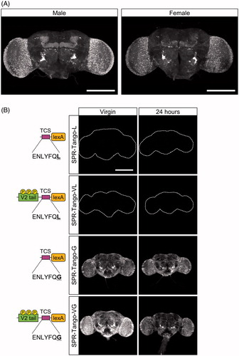
In addition to the female reproductive organ, SPR is expressed broadly in the brain (Yapici et al., Citation2008). Previous studies have shown that Myoinhibitory peptide (MIP), a neuropeptide involved in energy homeostasis in Drosophila (Jang, Chae, & Kim, Citation2017; Min S et al., Citation2016), activates SPR (Kim YJ et al., Citation2010), but it is not known whether SPR in the brain has any significant role in vivo. Using our SPR-Tango system, we sought to examine whether SPR has any activity in the brain.
First, we examined the expression pattern of endogenous SPR in the brain using the SPR-T2A-GAL4 line. When crossed to UAS-CD8-GFP, GFP fluorescence was widely observed in the brain, with elevated expression in the mushroom body and a subset of neurons beside the antennal lobe (). Expression patterns in male and female brains were very similar to each other. We then examined the reporter activity of the SPR-Tango variants by crossing them to tubP-β-arrestin-TEV, lexAop-CD8-GFP. We observed strong GFP expression in Tango-VG and Tango-G without mating (). Again, the distribution of the Tango signals with these variants was similar (). In contrast, GFP fluorescence was below the detection limit in Tango-L or Tango-VL (). No significant changes in GFP expression were observed in any of the Tango variants after mating (). Interestingly, while SPR-T2A-GAL4 is strongly expressed in Kenyon cells of the mushroom body, GFP expression induced by Tango-VG and -G was very low in these neurons (). Because tubP-β-arrestin-TEV is uniformly expressed in all neurons, we speculate that the observed reporter expression in Tango-G and Tango-VG was not simply caused by the basal activity of β-Arrestin-TEV, but it resulted from activity-dependent cleavage of the Tango sensors.
Figure 4. Spatiotemporal patterns of Tango activity in the brain. (A) SPR-expressing cells in male and female brains. SPR-expressing cells were visualized in flies that carry SPR-T2A-GAL4 and UAS-CD8-GFP. GFP was broadly expressed throughout the whole brains of both sexes. (B) Absence of mating-dependent changes in SPR-Tango activity. Left column: brains of virgin females. Right column: brains of females 24 hours after mating. The Tango variant used is indicated on the left. No GFP expression was observed in Tango-L or Tango-VL. Strong GFP signals were detected in Tango-G and Tango-VG in virgin females. No significant changes in the pattern or intensity were observed 24 hours after mating. Scale bars in A and B: 200 µm.
Discussion
Here we developed a method to detect the activity of endogenous GPCR proteins by inserting Tango sensors into the C-terminus of endogenous GPCR genes. Our Tango system comprises GPCR fused to the LexA transcription factor with a linker cleavable by TEV protease, a fusion protein of β-Arrestin and TEV protease and the lexAop-GFP reporter gene that is activated by LexA. To optimize the sensitivity of Tango sensors, we generated and compared four variants which differ in the cleavage efficiency and the presence of the V2 tail sequence that promotes recruitment of β-Arrestin upon GPCR activation. These two modifications have been previously used to adjust the sensitivity of Tango sensors in Drosophila and in cultured cells (Barnea et al., Citation2008; Hanson et al., Citation2009; Inagaki et al., Citation2012; Kroeze et al., Citation2015).
SPR is a GPCR that is expressed in the female reproductive organ and is activated by the seminal fluid transferred from the male fly at the time of mating. Mating-dependent activation of the reporter gene was observed for SPR-Tango-L and SPR-Tango-VL, variants with a linker sequence (ENLYFQL) that is not efficiently cleaved by TEV protease. SPR-Tango-VL has a V2-tail sequence and it indeed induced higher levels of reporter expression than SPR-Tango-L that lacks it. In contrast, SPR-Tango-G and SPR-Tango-VG, which have a linker sequence (ENLYFQG) that is efficiently cleaved by TEV protease, constitutively activated the lexAop-GFP reporter gene in the female reproductive organ even without mating. β-Arrestin-TEV is expressed at high levels from a constitutive α-tubulin promoter. We assume that this resulted in the random encounter of β-Arrestin-TEV with SPR-Tango and caused signal-independent cleavage of Tango-G and Tango-VG, while such transient interaction did not allow TEV to cleave the inefficient TCS in Tango-L and Tango-VL. Together, these results demonstrated that Tango-VL is the optimal configuration for detection of SPR activity in the female reproductive system.
We next evaluated the performance of the four SPR-Tango variants in the central nervous system. Although SPR in the brain was unlikely to be activated by SP after mating, we envisioned that MIP, another putative SPR ligand present in the brain (Kim et al., Citation2010), would activate SPR and induce reporter gene expression in certain cells in the brain. Unlike the mating-dependent signal, increase observed in the female reproductive organ, neither Tango-L nor Tango-VL induced reporter gene expression in any cells in the brain. On the contrary, Tango-VG and Tango-G induced reporter gene expression broadly in the brain with or without mating stimuli. If this were due to signal-independent cleavage by β-Arrestin-TEV, as observed in the female reproductive organ, the pattern of SPR-T2A-GAL4 expression and the pattern of the reporter expression induced by SPR-Tango would be identical. However, it was not the case. While SPR-T2A-GAL4 expression was highly enriched in the mushroom bodies, expression of the SPR-Tango reporter was conspicuously missing from them. Thus, we hypothesize that endogenous SPR is activated in many neurons of the brain but not in the mushroom body under normal conditions. Further studies are necessary to verify this hypothesis including identification of the ligand responsible for this activation.
The fly brain expresses more than 50 GPCRs that are receptors for neuropeptides or small-molecule neurotransmitters. Neuropeptides are implicated in various aspects of homeostasis and it is critical to understand under what conditions these peptides are released and which cells receive the signals. Tango is an ideal approach to address this problem. However, results from previous studies have indicated that the sensor needs to be optimized for each GPCR and target tissue, which is supported by our comparison of the four Tango variants with differing sensitivity. Our toolkit will thus provide a quick way to create and optimize Tango sensors for any GPCR in Drosophila.
Materials and methods
Fly stocks
The following transgenic strains were used in this study: tubP-β-Arrestin-TEV (Inagaki et al., Citation2012), 13xLexAop2-mCD8::GFP (Bloomington Stock Center #32203), 10xUAS-mCD8::GFP (Bloomington Stock Center #32184), nos-phiC31 (Bloomington Stock Center #34770), hs-Cre (Bloomington Stock Center #1092).
Generation of SPR-T2A-GAL4 and conversion into SPR-Tango by RMCE
Details of the system for generating T2A-GAL4 knock-ins and subsequent reporter gene replacement by RMCE will be described elsewhere. To generate SPR-T2A-GAL4, a donor vector with ∼800-bp homology arms and a gRNA expression vector were injected into the nos-Cas9 strain that maternally expresses Cas9 protein. The surviving flies were mated to y1 w1118 flies and their progeny were screened for transformants by RFP expression in the eye. Individual transformants were crossed to FM7 balancers to establish stocks and the 3xP3-RFP marker was removed by crossing them to hs-Cre.
The exchange vectors for replacing T2A-GAL4 knock-ins with Tango variants by RMCE were generated using basic molecular biology techniques. The exchange vectors carry each of the Tango variants, recombinase sites for RMCE and 3xP3-Venus as a selection marker. The maps and sequences of the vectors are provided in Supplementary Information. To generate transgenic Tango lines, each exchange vector was injected into fertilized eggs produced by y* w* nos-phiC31 females that had been mated to y1 w1118 SPR-T2A-GAL4 males. Note that both nos-phiC31 and SPR-T2A-GAL4 are transgenes on the X chromosome. Surviving female flies (genotype: y* w* nos-phiC31/y1 w1118 SPR-T2A-GAL4) were crossed to y1 w1118 males and their progeny were screened for transformants expressing Venus in the eye. Individual transformants were crossed to FM7 balancers to establish stocks. 3xP3-Venus and T2A-GAL4 were subsequently removed by hs-Cre to complete RMCE.
The sequences and maps of the vectors are provided in supplemental figure 1
Tissue imaging
Adult flies were anesthetized by CO2 and the brain and reproductive organs were dissected in ice-cold phosphate buffer saline (PBS, pH7.4). The harvested tissues were fixed in 4% paraformaldehyde in PBST (PBS + 0.3% Triton X-100) at 4˚C with gentle agitation. The fixed tissues were washed three times in PBST for 10 min each at 4˚C. They were mounted in glycerol by serial incubation in 50% and 86% glycerol in PBS, respectively, for one hour each. For images in , the fixed reproductive organs were permeablized for two hours in 2% TritonX-100 in PBS and washed in PBS for 10 min. They were stained with 100 nM Acti-Stain 555 Phalloidin (Cytoskelton, Inc) in PBS for two hours at room temperature with gentle agitation, followed by three washes in PBS for 10 min each at room temperature. They were mounted in glycerol as above.
The samples were imaged using LSM5 PASCAL (Carl Zeiss, Germany). The female reproductive organs were imaged using a 10x objective lens, the brains were imaged using a 20x objective lens. The imaging conditions of the reproductive organs were kept constant among different samples.
SuppInfo20190415.pdf
Download PDF (182.5 KB)Disclosure statement
No potential conflict of interest was reported by the authors.
References
- Barnea, G., Strapps, W., Herrada, G., Berman, Y., Ong, J., Kloss, B., … Lee, K.J. (2008). The genetic design of signaling cascades to record receptor activation. Proceedings of the National Academy of Sciences of the United States of America, 105, 64–69. doi:10.1073/pnas.0710487105
- Deo, C., & Lavis, L.D. (2018). Synthetic and genetically encoded fluorescent neural activity indicators. Current Opinion in Neurobiology, 50, 101–108. doi:10.1016/j.conb.2018.01.003
- Diao, F., & White, B.H. (2012). A novel approach for directing transgene expression in Drosophila: T2A-Gal4 in-frame fusion. Genetics, 190, 1139–1144. doi:10.1534/genetics.111.136291
- Donnelly, M.L., Luke, G., Mehrotra, A., Li, X., Hughes, L.E., Gani, D., & Ryan, M.D. (2001). Analysis of the aphthovirus 2A/2B polyprotein ‘cleavage' mechanism indicates not a proteolytic reaction, but a novel translational effect: a putative ribosomal 'skip'. Journal of General Virology, 82, 1013–1025. doi:10.1099/0022-1317-82-5-1013
- Fujita, N., Nagata, Y., Nishiuchi, T., Sato, M., Iwami, M., & Kiya, T. (2013). Visualization of neural activity in insect brains using a conserved immediate early gene, Hr38. Current Biology, 23, 2063–2070. doi:10.1016/j.cub.2013.08.051
- Gao, X.J., Riabinina, O., Li, J., Potter, C.J., Clandinin, T.R., & Luo, L. (2015). A transcriptional reporter of intracellular Ca(2+) in Drosophila. Nature Neuroscience, 18, 917–925. doi:10.1038/nn.4016
- Hanson, B.J., Wetter, J., Bercher, M.R., Kopp, L., Fuerstenau-Sharp, M., Vedvik, K.L., … Revankar, C. (2009). A homogeneous fluorescent live-cell assay for measuring 7-transmembrane receptor activity and agonist functional selectivity through beta-arrestin recruitment. Journal of Biomolecular Screening, 14, 798–810. doi:10.1177/1087057109335260
- Häsemeyer, M., Yapici, N., Heberlein, U., & Dickson, B.J. (2009). Sensory neurons in the Drosophila genital tract regulate female reproductive behavior. Neuron, 61, 511–518. doi:10.1016/j.neuron.2009.01.009
- Inagaki, H.K., Ben-Tabou de-Leon, S., Wong, A.M., Jagadish, S., Ishimoto, H., Barnea, G., … Anderson, D.J. (2012). Visualizing neuromodulation in vivo: TANGO-mapping of dopamine signaling reveals appetite control of sugar sensing. Cell, 148, 583–595. doi:10.1016/j.cell.2011.12.022
- Jagadish, S., Barnea, G., Clandinin, T.R., & Axel, R. (2014). Identifying functional connections of theinner photoreceptors in Drosophila using Tango-Trace. Neuron, 83, 630–644. doi:10.1016/j.neuron.2014.06.025
- Jang, Y.H., Chae, H.S., & Kim, Y.J. (2017). Female-specific myoinhibitory peptide neurons regulate mating receptivity in Drosophila melanogaster. Nature Communications, 8, 1630. doi:10.1038/s41467-017-01794-9
- Kim, Y.J., Bartalska, K., Audsley, N., Yamanaka, N., Yapici, N., Lee, J.Y., … Dickson, B.J. (2010). MIPs are ancestral ligands for the sex peptide receptor. Proceedings of the National Academy of Sciences of the United States of America, 107, 6520–6525. doi:10.1073/pnas.0914764107
- Kroeze, W.K., Sassano, M.F., Huang, X.P., Lansu, K., McCorvy, J.D., Giguère, P.M., … Roth, B.L. (2015). PRESTO-Tango as an open-source resource for interrogation of the druggable human GPCRome. Nature structural & molecular biology, 22, 362–369. doi:10.1038/nsmb.3014
- Luttrell, L.M., & Lefkowitz, R.J. (2002). The role of beta-arrestins in the termination and transduction of G-protein-coupled receptor signals. Journal of Cell Science, 115, 455–465.
- Masuyama, K., Zhang, Y., Rao, Y., & Wang, J.W. (2012). Mapping neural circuits with activity-dependent nuclear import of a transcription factor. Journal of Neurogenetics, 26, 89–102. doi:10.3109/01677063.2011.642910
- Min, S., Chae, H.S., Jang, Y.H., Choi, S., Lee, S., Jeong, Y.T., … Chung, J. (2016). Identification of a peptidergic pathway critical to satiety responses in Drosophila. Current Biology, 26, 814–820. doi:10.1016/j.cub.2016.01.029
- Tsuda, M., Peyre, J.B., Asano, T., & Aigaki, T. (2015). Visualizing molecular functions and cross-species activity of sex-peptide in drosophila. Genetics, 200, 1161–1169. doi:10.1534/genetics.115.177550
- Yapici, N., Kim, Y.J., Ribeiro, C., & Dickson, B.J. (2008). A receptor that mediates the post-mating switch in Drosophila reproductive behaviour. Nature, 451, 33–37. doi:10.1038/nature06483