
Abstract
Background: Lung ischemia-reperfusion injury (LIRI) remains the major cause of primary lung dysfunction after lung transplantation. Diabetes mellitus (DM) is an independent risk factor for morbidity and mortality following lung transplantation. Mitochondrial dysfunction is recognized as a key mediator in the pathogenesis of diabetic LIRI. Melatonin has been reported to be a safe and potent preserving mitochondrial function agent. This study aimed at investigating the potential therapeutic effect and mechanisms of melatonin on diabetic LIRI. Methods: High-fat-diet-fed streptozotocin-induced type 2 diabetic rats were exposed to melatonin, with or without administration of the SIRT3 short hairpin ribonucleic acid (shRNA) plasmid following a surgical model of ischemia-reperfusion injury of the lung. Lung function, inflammation, oxidative stress, cell apoptosis, and mitochondrial function were examined. Results: The SIRT3 signaling and mitophagy were suppressed following diabetic LIRI. Treatment with melatonin markedly induced mitophagy and restored SIRT3 expression. Melatonin treatment also attenuated subsequent diabetic LIRI by improving lung functional recovery, suppressing inflammation, decreasing oxidative damage, diminishing cell apoptosis, and preserving mitochondrial function. However, either administration of SIRT3 shRNA or an autophagy antagonist 3-methyladenine (3-MA) suppressing mitophagy, and compromised the protective action of melatonin. Conclusion: Data indicated that melatonin attenuates diabetic LIRI through activation of SIRT3 signaling-mediated mitophagy.
Introduction
Acute lung injury is commonly encountered in hospital and outpatient settings and remains a leading cause of patient morbidity and mortality. Many medical conditions, such as pulmonary embolism, acute respiratory distress syndrome (ARDS), cardiopulmonary bypass surgery, and lung transplantation can cause ischemia reperfusion–induced lung injuries.Citation1 The global prevalence of diabetes mellitus (DM) is significantly increasing across all age groups.Citation2 Emerging data suggest the lung is a target of diabetic injury, and DM is a significant risk factor for mortality at both 1 and 5 years after lung transplantation.Citation3 Previously, we reported that DM exacerbated lung ischemia-reperfusion injury (LIRI) in type 2 diabetic rats, and mitochondrial dysfunction play a central role in diabetic LIRI.Citation4,Citation5 To this end, preservation of mitochondrial function under type 2 DM represents a potential therapeutic target for treatment of LIRI.
Mitophagy, specifically responsible for the degradation of damaged mitochondria, is crucial for mitochondrial quality control.Citation6 Mitophagy is mediated by two pathways including Parkin-dependent pathway and mitophagy receptor-dependent pathway.Citation7 Prolonged hyperglycemia under the diabetic condition contribute to an over-generation of reactive oxygen species (ROS) by the mitochondria, which in turn may result in the development of diabetic complications.Citation8 Nonetheless, mitophagy signaling is impaired in diabetes in spite of extensive mitochondrial dysfunction, contributing to the accumulation of dysfunctional mitochondria and exacerbating the damage to tissue.Citation9 It is worthwhile noting that mitophagy plays a crucial role in the maintenance of mitochondrial function during diabetic ischemia reperfusion injury.Citation10 Our previous study showed that DM impaired mitophagy following lung transplantation.Citation5 However, the occurrence of mitophagy in warm diabetic LIRI is still not completely unknown.
Melatonin (N-acetyl-5-methoxytryptamine), is synthesized and secreted nocturnally by the pineal gland.Citation11 Convincing evidence demonstrated melatonin has protective effects against a variety of acute lung injuries especially in LIRI.Citation12 However, the effect and mechanism of melatonin treatment on LIRI under type 2 diabetic conditions are still unknown. SIRT3 is nicotinamide adenine dinucleotide (NAD+)-dependent enzymes, which is mainly located in the mitochondria.Citation13 In previous studies we found lung SIRT3 signaling was impaired in diabetic LIRI model, while normalizing SIRT3 signaling conferred pulmonary protective effect.Citation4 SIRT3 is documented to protect mitochondrial function through regulation of mitophagy and has been demonstrated to regulate a variety of cellular processes in both physiological conditions and pathological stress insults.Citation14 Moreover, melatonin is able to modulate SIRT3 to preserved mitochondrial function effectively.Citation15 However, it is currently unknown whether mitophagy could be regulated by melatonin in diabetic LIRI and its specific relationship with SIRT3 signaling in this circumstance.
We herein report that mitochondrial alterations in diabetic LIRI, mitochondrial abnormalities were associated with decreased mitophagy. Melatonin treatment improved mitochondrial function and less lung injury via the restoration mitophagy. Importantly, this mechanism appears a functional role for melatonin in preserving mitochondrial function by SIRT3 signaling-dependent mitophagy in diabetic LIRI experiments. These rescue mechanisms point the way toward a potential therapeutic target for the treatment of type 2 diabetic patients with acute lung injury induced by ischemia reperfusion injury.
Materials and methods
The study was conducted in accordance with the Basic & Clinical Pharmacology & Toxicology policy for experimental and clinical studies.Citation16
Animals
Pathogen-free male Sprague–Dawley weighing about 200 to 250 g were obtained from the animal care facility of Harbin Medical University. All animal experiments were performed under the Guidelines on National Institutes of Health Guidelines for the Care and Use of Laboratory Animals (NIH Publication No. 85–23, revised, 1996). The animal care and experimental protocols were approved by the Ethical Board of the Second Affiliated Hospital of Harbin Medical University (Harbin, China; No. SYDW2021-100).
Type 2 diabetic rat model
High-fat diet-fed streptozotocin-induced type 2 diabetic rat model was established as described previously.Citation4,Citation5,Citation17 Diabetes was defined as a fasting blood glucose level above 11.1 mmol/L 72 h after STZ injection. Rats fed the standard laboratory chow were used as the nondiabetic controls.
Rat LIRI model
Lung ischemia-reperfusion model was established as described previously.Citation4,Citation17 Briefly, the rats were anesthetized with sodium pentobarbital (30 mg/kg) administered intraperitoneally, intubated through a tracheostomy and ventilated with a tidal volume of 10 ml/kg, at a positive end-expiratory pressure of 2 cm H2O with 40% oxygen. The respiratory rate of 45–55 breaths/minute was adjusted to maintain arterial carbon dioxide tension (PaCO2) at 35 to 45 mmHg. Their right femoral vein and artery were cannulated with 24-gauge catheters for intravenous injection, blood gas analysis, and blood pressure monitoring. The rats underwent a left thoracotomy. The left hilum was injected with 50 IU heparin and clamped with a non-colliding micro clip to induce ischemia for 90 min with the tidal volume was adjusted to 6 ml/kg. The clip was removed for reperfusion and the ventilation was restored to the initial tidal volume with a positive end-expiratory pressure of 2 cm H2O, followed by suturing the thoracic cavity. During the observation period, animals were infused with pentobarbital sodium and rocuronium bromide to maintain stable anesthesia and muscle relaxation. After reperfusion for 4 h reperfusion, the animals were sacrificed. Rats in the sham groups underwent the same procedure except for the left lung hilum occlusion.
Experimental groups
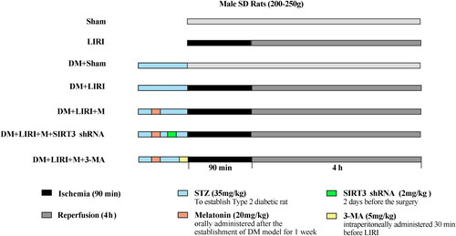
The rats were randomly assigned to the following groups (as depicted in ): sham group (Con + Sham), LIRI group (Con + IR), DM + sham group (DM + Sham), DM + LIRI group (DM + IR), DM + LIRI + melatonin-treated group (DM + IR + M), DM + LIRI + melatonin and SIRT3 shRNA treated group (DM + IR + M + S), and DM + LIRI + melatonin + 3-methyladenine (3-MA) treated group (DM + IR + M + 3MA). SIRT3 shRNA plasmid or scrambled shRNA plasmid was injected into the rat’s lung through 16-gauge oral catheter at a dose of 2 mg/kg 2 days before the surgery as our previously described.Citation4 Melatonin (20 mg/kg) was orally administered after the establishment of DM model for 1 week.Citation18 3-MA (15 mg/kg) was intraperitoneally administered 30 min before LIRI.Citation5
Figure 1. A schematic illustration of the experimental protocols. The left lung ischemia for 90 min and reperfusion for 4h. STZ, streptozotocin; 3-MA, 3-methyladenine.
The SIRT3 shRNA expression vector
The rat SIRT3 was treated as the target gene and shRNA oligonucleotides used pcDNATM6.2-GW/EmGFP-miR as expression vector were designed and synthesized by Gene Pharma (Shanghai, China) as our previously described.Citation4 The target-specific sequence was 5′-AATTCGGGTCCTTTGTATCAGCTACGGTTTTGGCCACTGACTGACCGTAGCTGATACAAAGGACCCA-3′ (sense) and
5′-GCCCAGGAAACATAGTCGATGCCAAAAC CGGTGACTGACTGGCATCGACTATGTTTCCTGGGTGGCC-3′ (antisense).
Real-time quantitative PCR
Real-time PCR was performed as described previously.Citation4,Citation17 Amplification was conducted using the following primers: 5′-GGAAAGTCGCACAGGAGATCATC-3′ (forward) and 5′-AGGTCCCTGGTCAGCCTTAACA-3′ for rat SIRT3. The following thermal cycling protocol was used 95 °C for 3 min, followed by 40 cycles of amplification at 95 °C for 30 s and 62 °C for 40 s.
Western blot analysis
Mitochondrial protein or tissue proteins were separated by 10% sodium dodecyl sulfate polyacrylamide gel electrophoresis and transferred onto polyvinyl difluoride membranes. After blocking, the membranes were incubated with primary antibodies against SIRT3 (2627, 1:1000, Cell Signaling Technology), Parkin (2132, 1:1000, Cell Signaling Technology), LC3-II (3868, 1:1000, Cell Signaling Technology), p62 (23214, 1:1000, Cell Signaling Technology), PGC-1α (MA5-32563, 1:1000, Thermo Fisher Scientific), GADPH (8884, 1:1000, Cell Signaling Technology), VDAC (4661, 1:1000, Cell Signaling Technology) at 4 °C overnight. Then, the membranes exposed to the corresponding horseradish peroxidase-conjugated secondary antibody (1:5000, Zhongshan Golden Bridge Biotechnology, Beijing, China) for 2 h. After being washed, the bound antibodies were detected with horseradish peroxidase (HRP)-conjugated secondary antibodies and visualized using an enhanced chemiluminescence reagents. The levels of target protein were quantified by densitometric analysis using Image J software.
Wet-to-dry weight (W/D) ratios and bronchoalveolar lavage fluid (BALF) protein concentrations
The upper section of lung tissues was dissected and weighed immediately. The lung tissues were desiccated at 80 °C for 72 h and weighed. The left lung was lavaged three times with 10 ml/kg of saline. The protein concentrations in BALF samples were measured using a BCA Protein Assay Kit (Beyotime, China).
Histological examination and scoring
The lung tissues were fixed in 4% paraformaldehyde and paraffin-embedded. The tissue sections (5 μm) were stained with hematoxylin and eosin (HE) and examined under a light microscope (BX41, Olympus, Tokyo, Japan), followed by photo imaged. The degrees of airway epithelial cell damage, interstitial edema, neutrophil infiltration, hemorrhage and hyaline membrane formation in individual sections were evaluated as the lung injury score (LIS) normal = 0; minimal change = 1; mild change = 2; moderate change = 3; and severe change = 4.Citation19 The lung injury was evaluated by 2 pathologists in a blinded manner.
Measurements of inflammatory and oxidative mediators
The levels of plasma tumor necrosis factor-α (TNF-α), interleukin-1β (IL-1β) and interleukin-10 (IL-10) were determined by Enzyme-linked immunosorbent assay (ELISA) using specific kits (R&D Systems, Minneapolis, MN, USA). In addition, the levels of myeloperoxidase (MPO) activity in lung tissue samples, malondialdehyde (MDA), superoxide dismutase (SOD) activity were measured using commercial kits (Nanjing Jiancheng, Nanjing, China) according to the manufacturers’ protocols.
Mitochondrial permeability transition pores (MPTPs) and mitochondrial membrane potential
MPTP opening was determined using a purified mitochondrial MPTP detection kit (GMS10095.3, Genmed Scientifics Inc, USA). Increased MPTP opening after more severe damage caused the mitochondria to swell, resulting in additional rapid decreased absorbance at 540 nm. The fluorescent signals were measured in a fluorescence microplate reader (Infinite M200 Pro, Tecan, Switzerland) at 540 nm. Mitochondrial membrane potential was assessed by a JC-1 staining using a kit from Sigma Aldrich (St. Louis, MO) as described previously.Citation4,Citation5
Terminal deoxynucleotidyl transferase dUTP nick end labeling (TUNEL) assay
Lung parenchymal cell apoptosis was detected by TUNEL using an In Situ Cell Death Detection kit (Roche Molecular Biochemicals, Mannheim, Germany) according to the manufacturer’s protocols. Cells with red nuclear staining were considered positive and all of the cells with DAPI (4′,6-diamidino-2-phenylindole) staining. The apoptosis index was expressed as the ratio of the number of apoptotic nuclei to the total number of nuclei counted.
mtDNA (mitochondrial DNA) quantification
The whole DNA was isolated from BALF and culture media by using the DNeasy Blood and Tissue Kit (Qiagen, Hilden, Germany) according to the manufacturer’s protocols. mtDNA levels were measured by SYBR-green dye-based RT-PCR assay. Rat NADH dehydrogenase 1 gene (mtDNA): forward 5′-CGCCTGACCAATAGCCATAA-3′ (forward), and 5′- ATTCGACGTTAAAGCCTGAGA-3′. The following thermal cycling protocol was used 95 °C for 3 min, followed by 40 cycles of amplification at 95 °C for 10 s and 55 °C for 30 s. Concentration of BALF mtDNA were converted to copy number via a DNA copy number calculator. mtDNA copy number was estimated according to the following formula: c = Q *VDNA/VPCR * 1/Vext. C means the concentration of mtDNA. VDNA means the total volume of DNA solution obtained from extraction, and 100 μl in this study; VPCR means the volume of DNA solution for RT-PCR, and 1 μl in this study; Vext means the volume of used for extraction, and 100 μl in this study.
Transmission electron microscopy (TEM)
One cubic millimeter fresh tissue was fixed in 2.5% glutaraldehyde at 4 °C overnight. The microstructural damages in the lungs of rats were evaluated by TEM. Mitochondria and osmiophilic multilamellar bodies in type II alveolar epithelial cells were imaged under an electron microscope. The degrees of mitochondrial injury were assessed with the Flameng score.Citation20 The scores corresponding to the criterion were listed as follows: 0, normal structures with intact particles; 1, normal structures with loss of particles; 2, swollen mitochondria but clear matrices; 3, broken cristae and concentrated matrices; 4, extensively destroyed cristae and ruptured membranes.
Statistical analysis
Data are expressed as median (interquartile range). Statistical comparisons of data among groups were compared by the non-parametric Kruskal-Wallis test. All statistical analyses were performed using Graph Pad Prism software version 5.0 (GraphPad Software, San Diego, CA, USA) and a P-value of <0.05 was considered statistically significant.
Results
Melatonin regulated mitophagy activity through SIRT3 signaling during diabetic lung ischemia reperfusion injury
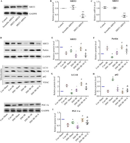
Initially, we detected SIRT3 expression by RT-PCR and western blot to evaluate whether SIRT3 could be silenced by the designed SIRT3 shRNA. As our previously report,Citation4 compared with control group, the expression level of SIRT3 protein was significantly decreased in SIRT3 shRNA group (p < 0.05, ). The mRNA expression of SIRT3 showed the same trend as the expression level of SIRT3 protein ().
Next, we measured the SIRT3 protein in both diabetic and nondiabetic rats by Immunoblotting analysis. As shown in , concomitant with our previous experiment of warm diabetic LIRI model, lung SIRT3 was significantly downregulated in type 2 diabetic rats, and it was further attenuated by LIRI. Melatonin treatment markedly restored the impaired lung SIRT3 expression caused by diabetic LIRI (p < 0.05, compared with the DM + IR group), while the lung SIRT3 expression was decreased in DM + IR + M + S group (p < 0.05, compared with the DM + IR + M group).
To investigate whether melatonin regulates diabetic lung ischemia-reperfusion induced mitophagy through SIRT3 signaling, the protein levels of mitochondrial LC3-II, Parkin, mitochondrial p62 were examined to assess mitophagy activation. As exhibited in , the Parkin protein levels were decreased in diabetic rats (p < 0.05, compared with the Con + Sham group), the expression level of Parkin attenuated in the DM + IR group (p < 0.05, compared with the Con + IR group). Melatonin treatment dramatically enhanced Parkin expression (p < 0.05, compared with the DM + IR group), while the effect of which was accentuated by SIRT3 knockdown. (p < 0.05, compared with the DM + IR + M group). The conversion of LC3-I to LC3-II is biochemical hallmarks of autophagy activation.Citation21 The p62 protein located on mitochondria were dependent on Parkin, and Parkin recruited p62 to mitochondria during mitophagy.Citation14,Citation22 Similar changes were observed in mitochondrial LC3-II, and mitochondrial p62 exhibited opposite changes compared with the previous levels. Thus, the elevated mitochondrial LC3-II and decreased p62 expression with administration of melatonin supports the hypothesis that melatonin enhances Parkin-mediated mitophagy through SIRT3 signaling during diabetic LIRI. Similar changes were observed in PGC-1α expression compared with the previous levels.
These results indicated Parkin-mediated mitophagy was decreased in response to DM during LIRI, melatonin inhibited diabetic LIRI-mediated mitophagy downregulation through SIRT3 signaling.
Melatonin improved pulmonary function through SIRT3-dependent mitophagy following diabetic LIRI
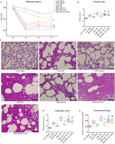
To determine the effect of melatonin on diabetic LIRI via regulation of mitophagy, autophagy inhibitor 3-MA was used. As presented in , there were no significant between-group differences with respect to PaO2/FiO2 at baseline. The PaO2/FiO2 in the Con + Sham group and the DM + Sham group showed no difference (p > 0.05). The PaO2/FiO2 in the DM + IR group was significantly lower than that in the Con + IR group during reperfusion at 240 min (p < 0.05). Melatonin treatment markedly increased PaO2/FiO2 (p < 0.05, compared with the DM + IR group). However, either SIRT3 knockdown or administration 3-MA abolished the effect of melatonin on the PaO2/FiO2 (p < 0.05). The wet weight-to-dry weight ratio showed opposite changes compared with the previous levels ().
Figure 2. Melatonin regulated mitophagy activity through SIRT3 signaling following diabetic lung ischemia reperfusion injury. (A) The downregulation of levels of SIRT3 were evaluated by western blots (n = 4 in each group). (B) SIRT3 expression. (C) mRNA expression of SIRT3 (n = 4 in each group). (D) Representative blots. (E) SIRT3 expression. (F) Parkin expression. (G) LC3-II expression. (H) p62 expression. LC3-II, microtubule-associated protein 1 light chain 3 beta. (I) PGC-1α expression. (**P < 0.05 versus Con group, *P < 0.05 versus Con + Sham group, ^P < 0.05 versus Con + IR group, #P < 0.05 versus DM + IR group, &P < 0.05 versus DM + IR + M group; n = 8 in each group).
As presented in , the grafts in the Con + IR group exhibited leukocyte infiltration, interstitial thickening, intra-alveolar hemorrhage and edema in the alveolar septa and spaces. The lung injury score was higher in the DM + IR group (p < 0.05, compared with the Con + IR group). Administration of melatonin reduced the lung injury scores (p < 0.05, compared with the DM + IR group), and this improvement was attenuated in the DM + IR + M + S group. The lung injury scores in the DM + IR + M + 3MA group were higher than that in the DM + IR + M group (p < 0.05). The concentrations of proteins in BALF exhibited the same trend as the lung injury score ().
These results indicated melatonin might protect the lung function by promoting mitophagy via SIRT3 during diabetic LIRI.
Melatonin alleviated inflammatory, oxidative stress through SIRT3-dependent mitophagy following diabetic LIRI
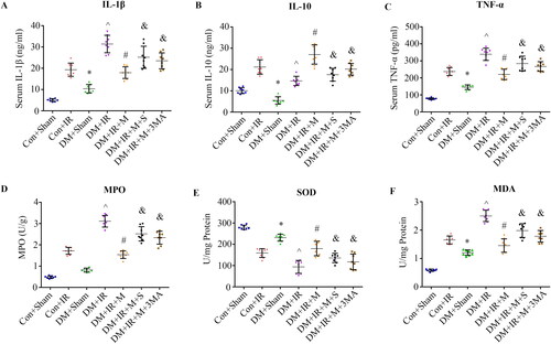
As shown in , the levels of serum IL-1β and TNF-α were significantly increased in the DM + IR group (p < 0.05, compared with the Con + IR group). Melatonin treatment markedly decreased the levels of serum IL-1β and TNF-α (p < 0.05, compared with the DM + IR group), whereas the effect of which was mitigated by SIRT3 knockdown (p < 0.05, compared with the DM + IR + M group). Administration of 3-MA also increased the levels of serum IL-1β and TNF-α (p < 0.05, compared with the DM + IR + M group). IL-10 showed the opposite changes compared with the previous levels (). The level of MPO, an indicator of neutrophil infiltration exhibited the same trend. Similar changes were observed in level of MDA, an indicator of the oxidative stress response (). Moreover, the antioxidative capacity (activities of SOD) showed the opposite changes compared with the previous levels ().
Figure 3. Melatonin improved pulmonary function through SIRT3-dependent mitophagy following diabetic LIRI. (A) Arterial blood gas analysis. T0–T4 represent the following time points: baseline, end of ischemia, 120 and 240 min after reperfusion. (B) Wet/dry weight ratio. (C) Histologic analysis of lung tissues (scale bars, 100 μm). (D) Lung injury score. (E) Protein concentrations in the BALF. BALF, bronchoalveolar lavage fluid; PaO2/FiO2, partial pressure of arterial oxygen (PaO2)/fraction of inspired oxygen (FiO2). (*P < 0.05 versus Con + Sham group, ^P < 0.05 versus Con + IR group, #P < 0.05 versus DM + IR group, &P < 0.05 versus DM + IR + M group; n = 8 in each group).
These results collectively indicated that the effects of melatonin on anti-inflammatory and anti-oxidative actions by promoting mitophagy might be partially mediated by SIRT3 signaling.
Melatonin attenuated cell apoptosis through SIRT3-dependent mitophagy following diabetic LIRI
The apoptosis in lung grafts of was evaluated by TUNEL assay. As shown in , the apoptotic index was significantly increased in the DM + IR group compared with apoptosis levels in the Con + IR group (p < 0.05). The percentages of apoptotic cells in DM + IR + M group were significantly lower than that in the DM + IR group (p < 0.05), but the antiapoptotic effect of melatonin was abolished either in the DM + IR + M + S group or the DM + IR + M + 3MA group.
Figure 4. Melatonin alleviated inflammatory, oxidative stress through SIRT3-dependent mitophagy following diabetic LIRI. (A) Serum concentrations of interleukin-1β (IL-1β). (B) Serum concentrations of interleukin-10 (IL-10). (C) Serum concentrations of TNF-α. (D) Lung concentrations of MPO. (E) Lung concentrations of SOD. (E) Lung concentrations of MDA. IL, interleukin; TNF-a, tumor necrosis factor-a; MPO, myeloperoxidase; MDA, malonaldehyde; SOD, superoxide dismutase. (*P < 0.05 versus Con + Sham group, ^P < 0.05 versus Con + IR group, #P < 0.05 versus DM + IR group, &P < 0.05 versus DM + IR + M group; n = 8 in each group).
These results indicated that the effects of melatonin on anti-apoptotic by promoting mitophagy might be partially mediated by SIRT3 signaling.
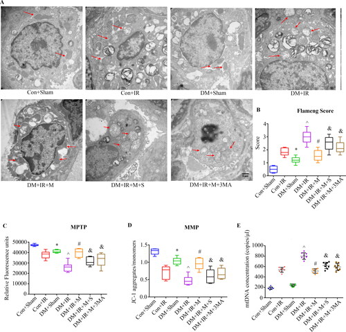
Melatonin limited mitochondrial damage and dysfunction through SIRT3-dependent mitophagy following diabetic LIRI
As shown in , the images of mitochondria morphology by electron microscopy showed mitochondrial structural integrity in the Con + IR group was damaged, while the mitochondrial Flameng scores of the DM + IR groups were greater than the scores of the Con + IR group (p < 0.05). Melatonin treatment significantly reduced the mitochondrial Flameng scores (p < 0.05, compared with the DM + IR group), whereas the effect of which was attenuated by SIRT3 knockdown (p < 0.05, compared with the DM + IR + M group). Similar, administration of 3-MA also partially abolished these effects (p < 0.05, compared with the DM + IR + M group). MPTP and JC-1 exhibited the same changes compared with the previous levels. mtDNA level was significantly increased in the DM + IR group compared with those in the Con + IR groups in BALF (p < 0.05). Melatonin treatment markedly reversed the elevated mtDNA level in BALF caused by diabetic LIRI (p < 0.05, compared with the DM + IR group). Of note, the benefits of melatonin on mtDNA level in BALF were also reversed by SIRT3 knockdown or 3-MA (p < 0.05, compared with the DM + IR + M group). These results suggested that the protective effects of melatonin against mitochondrial damage and dysfunction by promoting mitophagy might be partially mediated by SIRT3 signaling.
Figure 5. Melatonin attenuated cell apoptosis through SIRT3-dependent mitophagy following diabetic LIRI. (A) Representative in situ detection of lung parenchymal cell apoptosis by TUNEL staining (scale bars, 500 μm). (B) Percentage of TUNEL-positive nuclei. TUNEL, Terminal deoxynucleotidyl transferase dUTP nick end-labeling. (*P < 0.05 versus Con + Sham group, ^P < 0.05 versus Con + IR group, #P < 0.05 versus DM + IR group, &P < 0.05 versus DM + IR + M group; n = 8 in each group).
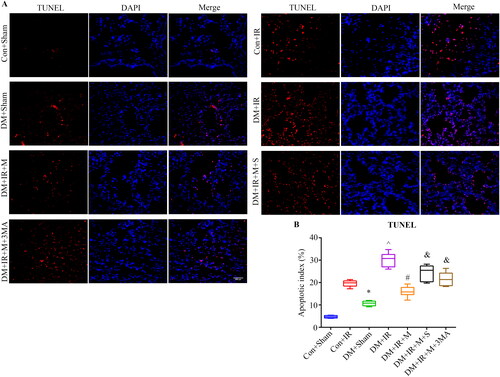
Figure 6. Melatonin limited mitochondrial damage and dysfunction through SIRT3-dependent mitophagy following diabetic LIRI. (A) Mitochondrial ultrastructure. Mitochondria (arrows) in type II alveolar epithelial cells were imaged with transmission electron micrographs (scale bars, 5 μm). (B) Flameng score. (C) Determination of MPTP opening. (D) Determination of mitochondrial membrane potential. (E) BALF mtDNA measurements. MPTP, mitochondrial permeability transition pores; MMP, mitochondrial membrane potential (*P < 0.05 versus Con + Sham group, ^P < 0.05 versus Con + IR group, #P < 0.05 versus DM + IR group, &P < 0.05 versus DM + IR + M group; n = 8 in each group).
Discussion
Here we found that in experimental diabetic LIRI model: (1) diabetic LIRI downregulated the SIRT3 signaling and suppressed mitophagy. (2) melatonin effectively preserved mitochondrial function via inducing the SIRT3-dependent mitophagy, thus ameliorating LIRI under type 2 diabetic conditions.
Melatonin is secreted by the pineal gland, with various therapeutic effects such as antioxidant, preserving mitochondrial function. Interestingly, lower melatonin secretion was independently associated with a higher risk of developing type 2 DM and its complications.Citation23 Therefore, melatonin has been encouraged to be administered in the treatment of ischemia reperfusion in diabetes.Citation24 We present data to show that melatonin protected against diabetic LIRI and that SIRT3 signaling-dependent mitophagy played a key role in this process. As melatonin membrane receptors (Mel1a receptor and Mel1b receptor) participate in different signaling pathways,Citation23 further study is required to evaluate the role and mechanism of melatonin membrane receptors in its protective action in diabetic LIRI.
Although the lung is one of the least studied organs in diabetes, numerous studies demonstrate that it is an inevitable target of diabetic complications.Citation8,Citation25 In diabetic patients, the risk of an exaggerated ischemia reperfusion injury is more serious, associated with worse outcomes than in the nondiabetic population.Citation26 LIRI remains a major factor for the early mortality of lung transplantations. Based on previous data of ours showing that DM aggravated LIRI and mitochondrial dysfunction played a key role in this process.Citation4,Citation5 Diabetic hyperglycemia result in over-generation of ROS that then may induce injury of mitochondrial DNA, respiratory complex proteins and membrane lipids. All these may contribute to a catastrophic feedforward cycle of oxidative stress and eventually cell death.Citation27 Thus, maintaining mitochondrial quality is critical for cell homeostasis in diabetic complication.Citation28 Clinical and experimental studies have shown that compared to individuals without diabetes, individuals with diabetes exhibit increased vulnerability to organ ischemia reperfusion.Citation29 Impaired lung mitochondrial function is the key elements of LIRI.Citation30 Hyperglycemia exacerbated mitochondrial dysfunction, which contributes substantially to development of LIRI.Citation4,Citation5 Our data identify the levels of MPTP and MMP, which were regarded as the markers of mitochondrial function. The level of MMP was decreased in diabetic state following LIRI surgery in comparison to non-diabetic state, and MPTP opening was also further increased in diabetic state following LIRI surgery. A plausible explanation could be that the DM induced mitochondrial dysfunction may make lung vulnerable to ischemia reperfusion injury. PGC-1α regulates mitochondrial biogenesis but also has effects on mitochondrial functions beyond biogenesis.Citation31,Citation32 Moreover, ample evidence has indicated PGC-1α also in the regulation of mitophagy.Citation33 More robust data concerning the PGC-1α functions as an upstream regulator of SIRT3,Citation34 while suppressing SIRT3 decreased PGC-1α expression and mitochondrial function.Citation35 In our current study, we found melatonin treatment markedly restored the impaired lung PGC-1α expression caused by diabetic LIRI through SIRT3 signaling, which indicate melatonin preserving mitochondrial function partially mediated by SIRT3 signaling.
Mitophagy is critical for mitochondrial quality control, which allows autophagosomes to selectively eliminate damaged mitochondria.Citation6 Abnormal mitochondria overwhelm the capacity of mitophagy at the stage of ischemia reperfusion injury, which may result in damaged mitochondria accumulate.Citation36–38 The accumulation of damaged mitochondria can lead to neighboring normal mitochondria injury by propagating injurious signals and inducing MPTP opening and mitochondrial dysfunction, which may contribute to excessive ROS production, and ultimately activation of cell apoptosis.Citation39 Various experimental models of ischemia reperfusion injury have shown beneficial effect of mitophagy on cell survival.Citation40–42 mtDNA are released by damaged mitochondria during LIRI contributing to primary graft dysfunction and lung injury.Citation43 Increased mtDNA is a consequence of impaired mitophagy.Citation44 The data presented in the current study showed that mtDNA level in BALF was significantly increased in diabetic state following LIRI surgery in comparison to non-diabetic state. Melatonin attenuated mtDNA level through SIRT3 signaling-dependent mitophagy following diabetic LIRI. Mitophagy can be regulated by several pathways, and Parkin-dependent pathway is one of the best characterized pathways mediating mitophagy.Citation45 Intriguingly, evidence is accumulating for the relationship between impaired mitophagy and mitochondrial dysfunction under diabetic condition.Citation46 In particular, recent studies have demonstrated mitophagy through Parkin degradation mechanisms is impaired under type 2 diabetic condition, which contribute to the accumulation of damaged mitochondria.Citation47 Concomitant with our previously diabetic lung transplantation model, we present data to show that Parkin-mediated mitophagy was suppressed associated with increased MPTP opening, decreased mitochondrial membrane potential, and mitochondrial damage cell death was increased in diabetic LIRI setting, which implied the relationship between the mechanisms underlying the diabetic state aggravating LIRI and impaired mitophagy, and the detailed mechanisms remains to be elucidated.
It has been well established that melatonin could preserve mitochondrial function in diabetic state in multiple organs.Citation48,Citation49 Melatonin presumably enters mitochondria through oligopeptide transporters.Citation50 Measurement of the subcellular distribution of melatonin showed that the concentration of melatonin in mitochondria significantly exceeds that in blood.Citation51 Moreover, melatonin also modulates Parkin-mediated mitophagy activity.Citation9 Herein, we observed melatonin treatment effectively rescued Parkin-mediated mitophagy and protected against diabetic LIRI. We further found that the pulmonary protective effect against diabetic LIRI of melatonin mediated mitophagy was compromised by 3-MA, a broad autophagy inhibitor. Taken together, these results provided evidence that melatonin exerts protective effects against diabetic LIRI by increasing mitophagy.
SIRT3, a nicotinamide adenine dinucleotide (NAD+)-dependent deacetylase, is mainly localized in mitochondria.Citation13 SIRT3 modulates global mitochondrial lysine acetylation, and preserving mitochondrial function.Citation52 We and others also found that SIRT3 signaling might represents a promising strategy to attenuated diabetic ischemia reperfusion insult.Citation4,Citation15 We found that lung SIRT3 signaling was dramatically decreased under type 2 diabetic condition, and it was further downregulated by the LIRI. Intriguingly, accumulating evidence suggests that melatonin can act as a SIRT3 inducer in multiple systems.Citation15,Citation53 Moreover, it was reported that a member of the Forkhead box, subgroup O (FoxO) transcription factors FoxO3, regulates PINK1 transcription.Citation54 SIRT3 is able to upregulate FoxO3a dependent gene expression via interacts with the daf-16 homolog Foxo3a in the mitochondria.Citation55 The network of SIRT3-FoxO3-PINK1-Parkin was demonstrated to control mitophagy.Citation56 Here, we found that melatonin treatment can rescue the impaired SIRT3 caused by diabetic LIRI. We also found SIRT3 inhibition impaired the Parkin-mediated mitophagy activity of melatonin and attenuated its protective action against diabetic LIRI. These data suggested that SIRT3-Parkin-mediated mitophagy reducing inflammation, oxidative stress, apoptosis and preserving mitochondrial function, which played a key role in the protective actions of melatonin in diabetic LIRI.
The present study has several limitations. First, we did not measure the levels of melatonin in plasma. Several studies found that circulating levels of melatonin are decreased in both patients with type 2 DM and diabetic animal models, which might contribute to diabetic complications.Citation57 Second, high fat-diet-fed streptozotocin-induced type 2 diabetic model was used to simulated pathophysiology condition of type 2 DM patients in present study, but whether these models are truly representative of diabetes requires further detailed elucidation. Third, although we demonstrate that melatonin enhanced LC3-II in the mitochondrial fraction, indicating that melatonin promotes mitophagy, we cannot formally exclude the possibility that increases in general autophagy may also contribute to the protective effect of melatonin during diabetic LIRI.
Our work supports the theory suggesting that melatonin treatment protected against diabetic LIRI by attenuating inflammation, oxidative stress, apoptosis and preserving mitochondrial function via SIRT3-mediated mitophagy. It also illustrates the potential for a personalized medicine approach where the presence of melatonin may identify as SIRT3 enhancement therapy for treating acute lung injury induced by ischemia reperfusion injury in patients with type 2 DM.
Disclosure statement
The authors declare that they have no conflict of interest.
Data availability statement
The datasets supporting the conclusions of this article are included within the article.
Additional information
Funding
References
- de Perrot M, Liu M, Waddell TK, Keshavjee S. Ischemia-reperfusion-induced lung injury. Am J Respir Crit Care Med. 2003;167(4):490–511. doi:10.1164/rccm.200207-670SO.
- Shaw JE, Sicree RA, Zimmet PZ. Global estimates of the prevalence of diabetes for 2010 and 2030. Diabetes Res Clin Pract. 2010;87(1):4–14. doi:10.1016/j.diabres.2009.10.007.
- Hackman KL, Bailey MJ, Snell GI, Bach LA. Diabetes is a major risk factor for mortality after lung transplantation. Am J Transplant. 2014;14(2):438–445. doi:10.1111/ajt.12561.
- Jiang T, Liu Y, Meng Q, et al. Hydrogen sulfide attenuates lung ischemia-reperfusion injury through SIRT3-dependent regulation of mitochondrial function in type 2 diabetic rats. Surgery. 2019;165(5):1014–1026. doi:10.1016/j.surg.2018.12.018.
- Jiang T, Liu T, Deng X, et al. Adiponectin ameliorates lung ischemia-reperfusion injury through SIRT1-PINK1 signaling-mediated mitophagy in type 2 diabetic rats. Respir Res. 2021;22(1):258. doi:10.1186/s12931-021-01855-0.
- Morales PE, Arias-Duran C, Avalos-Guajardo Y, et al. Emerging role of mitophagy in cardiovascular physiology and pathology. Mol Aspects Med. 2020;71:100822. doi:10.1016/j.mam.2019.09.006.
- Georgakopoulos ND, Wells G, Campanella M. The pharmacological regulation of cellular mitophagy. Nat Chem Biol. 2017;13(2):136–146. doi:10.1038/nchembio.2287.
- Wu J, Jin Z, Yan LJ. Redox imbalance and mitochondrial abnormalities in the diabetic lung. Redox Biol. 2017;11:51–59. doi:10.1016/j.redox.2016.11.003.
- Wang S, Zhao Z, Feng X, et al. Melatonin activates Parkin translocation and rescues the impaired mitophagy activity of diabetic cardiomyopathy through Mst1 inhibition. J Cell Mol Med. 2018;22(10):5132–5144. doi:10.1111/jcmm.13802.
- Yang YY, Gong DJ, Zhang JJ, Liu XH, Wang L. Diabetes aggravates renal ischemia-reperfusion injury by repressing mitochondrial function and PINK1/Parkin-mediated mitophagy. Am J Physiol Renal Physiol. 2019;317(4):F852–F864. doi:10.1152/ajprnal.00181.2019.
- Reiter RJ, Paredes SD, Manchester LC, Tan DX. Reducing oxidative/nitrosative stress: a newly-discovered genre for melatonin. Crit Rev Biochem Mol Biol. 2009;44(4):175–200. doi:10.1080/10409230903044914.
- Yip HK, Chang YC, Wallace CG, et al. Melatonin treatment improves adipose-derived mesenchymal stem cell therapy for acute lung ischemia-reperfusion injury. J Pineal Res. 2013;54(2):207–221. doi:10.1111/jpi.12020.
- Winnik S, Auwerx J, Sinclair DA, Matter CM. Protective effects of sirtuins in cardiovascular diseases: from bench to bedside. Eur Heart J. 2015;36(48):3404–3412. doi:10.1093/eurheartj/ehv290.
- Wang S, Zhao Z, Fan Y, et al. Mst1 inhibits Sirt3 expression and contributes to diabetic cardiomyopathy through inhibiting Parkin-dependent mitophagy. Biochim Biophys Acta Mol Basis Dis. 2019;1865(7):1905–1914. doi:10.1016/j.bbadis.2018.04.009.
- Yu L, Gong B, Duan W, et al. Melatonin ameliorates myocardial ischemia/reperfusion injury in type 1 diabetic rats by preserving mitochondrial function: role of AMPK-PGC-1alpha-SIRT3 signaling. Sci Rep. 2017;7:41337. doi:10.1038/srep41337.
- Tveden-Nyborg P, Bergmann TK, Jessen N, Simonsen U, Lykkesfeldt J. BCPT policy for experimental and clinical studies. Basic Clin Pharmacol Toxicol. 2021;128(1):4–8. doi:10.1111/bcpt.13492.
- Jiang T, Yang W, Zhang H, Song Z, Liu T, Lv X. Hydrogen sulfide ameliorates lung ischemia-reperfusion injury through SIRT1 signaling pathway in type 2 diabetic rats. Front Physiol. 2020;11:596. doi:10.3389/fphys.2020.00596.
- Yu L, Liang H, Dong X, et al. Reduced silent information regulator 1 signaling exacerbates myocardial ischemia-reperfusion injury in type 2 diabetic rats and the protective effect of melatonin. J Pineal Res. 2015;59(3):376–390. doi:10.1111/jpi.12269.
- Pirat A, Zeyneloglu P, Aldemir D, et al. Pretreatment with simvastatin reduces lung injury related to intestinal ischemia-reperfusion in rats. Anesth Analg. 2006;102(1):225–232. doi:10.1213/01.ane.0000189554.41095.98.
- Flameng W, Borgers M, Daenen W, Stalpaert G. Ultrastructural and cytochemical correlates of myocardial protection by cardiac hypothermia in man. J Thorac Cardiovasc Surg. 1980;79(3):413–424. doi:10.1016/S0022-5223(19)37950-4.
- Aggarwal S, Mannam P, Zhang J. Differential regulation of autophagy and mitophagy in pulmonary diseases. Am J Physiol Lung Cell Mol Physiol. 2016;311(2):L433–52. doi:10.1152/ajplung.00128.2016.
- Billia F, Hauck L, Konecny F, Rao V, Shen J, Mak TW. PTEN-inducible kinase 1 (PINK1)/Park6 is indispensable for normal heart function. Proc Natl Acad Sci USA. 2011;108(23):9572–9577. doi:10.1073/pnas.1106291108.
- Karamitri A, Jockers R. Melatonin in type 2 diabetes mellitus and obesity. Nat Rev Endocrinol. 2019;15(2):105–125. doi:10.1038/s41574-018-0130-1.
- Yu LM, Dong X, Xue XD, et al. Melatonin attenuates diabetic cardiomyopathy and reduces myocardial vulnerability to ischemia-reperfusion injury by improving mitochondrial quality control: role of SIRT6. J Pineal Res. 2021;70(1):e12698. doi:10.1111/jpi.12698.
- Zheng H, Wu J, Jin Z, Yan LJ. Potential biochemical mechanisms of lung injury in diabetes. Aging Dis. 2017;8(1):7–16. doi:10.14336/AD.2016.0627.
- Gregg EW, Sattar N, Ali MK. The changing face of diabetes complications. Lancet Diabetes Endocrinol. 2016;4(6):537–547. doi:10.1016/S2213-8587(16)30010-9.
- Koliaki C, Roden M. Alterations of mitochondrial function and insulin sensitivity in human obesity and diabetes mellitus. Annu Rev Nutr. 2016;36:337–367. doi:10.1146/annurev-nutr-071715-050656.
- Rovira-Llopis S, Bañuls C, Diaz-Morales N, Hernandez-Mijares A, Rocha M, Victor VM. Mitochondrial dynamics in type 2 diabetes: pathophysiological implications. Redox Biol. 2017;11:637–645. doi:10.1016/j.redox.2017.01.013.
- Qiu Z, Ming H, Lei S, et al. Roles of HDAC3-orchestrated circadian clock gene oscillations in diabetic rats following myocardial ischaemia/reperfusion injury. Cell Death Dis. 2021;12(1):43. doi:10.1038/s41419-020-03295-y.
- Cloer CM, Givens CS, Buie LK, et al. Mitochondrial transplant after ischemia reperfusion promotes cellular salvage and improves lung function during ex-vivo lung perfusion. J Heart Lung Transplant. 2023;42(5):575–584. doi:10.1016/j.healun.2023.01.002.
- Tan Y, Zhang Y, He J, et al. Dual specificity phosphatase 1 attenuates inflammation-induced cardiomyopathy by improving mitophagy and mitochondrial metabolism. Mol Metab. 2022;64:101567. doi:10.1016/j.molmet.2022.101567.
- Chang X, Liu R, Li R, Peng Y, Zhu P, Zhou H. Molecular mechanisms of mitochondrial quality control in ischemic cardiomyopathy. Int J Biol Sci. 2023;19(2):426–448. doi:10.7150/ijbs.76223.
- Liu L, Li Y, Wang J, et al. Mitophagy receptor FUNDC1 is regulated by PGC-1alpha/NRF1 to fine tune mitochondrial homeostasis. EMBO Rep. 2021;22(3):e50629. doi:10.15252/embr.202050629.
- Park SJ, Ahmad F, Philp A, et al. Resveratrol ameliorates aging-related metabolic phenotypes by inhibiting cAMP phosphodiesterases. Cell. 2012;148(3):421–433. doi:10.1016/j.cell.2012.01.017.
- Paku M, Haraguchi N, Takeda M, et al. SIRT3-mediated SOD2 and PGC-1alpha contribute to chemoresistance in colorectal cancer cells. Ann Surg Oncol. 2021;28(8):4720–4732. doi:10.1245/s10434-020-09373-x.
- Chang X, Toan S, Li R, Zhou H. Therapeutic strategies in ischemic cardiomyopathy: focus on mitochondrial quality surveillance. EBioMed. 2022;84:104260. doi:10.1016/j.ebiom.2022.104260.
- Jackson EK, Menshikova EV, Mi Z, et al. Renal 2’,3’-cyclic nucleotide 3’-phosphodiesterase is an important determinant of AKI severity after ischemia-reperfusion. J Am Soc Nephrol. 2016;27(7):2069–2081. doi:10.1681/ASN.2015040397.
- Zhou H, Ren J, Toan S, Mui D. Role of mitochondrial quality surveillance in myocardial infarction: from bench to bedside. Ageing Res Rev. 2021;66:101250. doi:10.1016/j.arr.2020.101250.
- Zhou H, Dai Z, Li J, et al. TMBIM6 prevents VDAC1 multimerization and improves mitochondrial quality control to reduce sepsis-related myocardial injury. Metabolism. 2023;140:155383. doi:10.1016/j.metabol.2022.155383.
- Zheng J, Chen L, Lu T, et al. MSCs ameliorate hepatocellular apoptosis mediated by PINK1-dependent mitophagy in liver ischemia/reperfusion injury through AMPKalpha activation. Cell Death Dis. 2020;11(4):256. doi:10.1038/s41419-020-2424-1.
- Yuan Y, Zheng Y, Zhang X, et al. BNIP3L/NIX-mediated mitophagy protects against ischemic brain injury independent of PARK2. Autophagy. 2017;13(10):1754–1766. doi:10.1080/15548627.2017.1357792.
- Tang C, Han H, Yan M, et al. PINK1-PRKN/PARK2 pathway of mitophagy is activated to protect against renal ischemia-reperfusion injury. Autophagy. 2018;14(5):880–897. doi:10.1080/15548627.2017.1405880.
- Mallavia B, Liu F, Lefrancais E, et al. Mitochondrial DNA stimulates TLR9-dependent neutrophil extracellular trap formation in primary graft dysfunction. Am J Respir Cell Mol Biol. 2020;62(3):364–372. doi:10.1165/rcmb.2019-0140OC.
- Sliter DA, Martinez J, Hao L, et al. Parkin and PINK1 mitigate STING-induced inflammation. Nature. 2018;561(7722):258–262. doi:10.1038/s41586-018-0448-9.
- Kane LA, Lazarou M, Fogel AI, et al. PINK1 phosphorylates ubiquitin to activate Parkin E3 ubiquitin ligase activity. J Cell Biol. 2014;205(2):143–153. doi:10.1083/jcb.201402104.
- Hoshino A, Ariyoshi M, Okawa Y, et al. Inhibition of p53 preserves Parkin-mediated mitophagy and pancreatic beta-cell function in diabetes. Proc Natl Acad Sci USA. 2014;111(8):3116–3121. doi:10.1073/pnas.1318951111.
- Tong M, Saito T, Zhai P, et al. Mitophagy is essential for maintaining cardiac function during high fat diet-induced diabetic cardiomyopathy. Circ Res. 2019;124(9):1360–1371. doi:10.1161/CIRCRESAHA.118.314607.
- Gao L, Zhao YC, Liang Y, et al. The impaired myocardial ischemic tolerance in adult offspring of diabetic pregnancy is restored by maternal melatonin treatment. J Pineal Res. 2016;61(3):340–352. doi:10.1111/jpi.12351.
- Kahya MC, Nazıroğlu M, Övey İS. Modulation of diabetes-induced oxidative stress, apoptosis, and Ca(2+) entry through TRPM2 and TRPV1 channels in dorsal root ganglion and hippocampus of diabetic rats by melatonin and selenium. Mol Neurobiol. 2017;54(3):2345–2360. doi:10.1007/s12035-016-9727-3.
- Huo X, Wang C, Yu Z, et al. Human transporters, PEPT1/2, facilitate melatonin transportation into mitochondria of cancer cells: an implication of the therapeutic potential. J Pineal Res. 2017;62(4):e12390. doi:10.1111/jpi.12390.
- Acuna-Castroviejo D, Escames G, Venegas C, et al. Extrapineal melatonin: sources, regulation, and potential functions. Cell Mol Life Sci. 2014;71(16):2997–3025. doi:10.1007/s00018-014-1579-2.
- Tseng AH, Shieh SS, Wang DL. SIRT3 deacetylates FOXO3 to protect mitochondria against oxidative damage. Free Radic Biol Med. 2013;63:222–234. doi:10.1016/j.freeradbiomed.2013.05.002.
- Zhai M, Li B, Duan W, et al. Melatonin ameliorates myocardial ischemia reperfusion injury through SIRT3-dependent regulation of oxidative stress and apoptosis. J Pineal Res. 2017;63(2):e12419. doi:10.1111/jpi.12419.
- Mei Y, Zhang Y, Yamamoto K, Xie W, Mak TW, You H. FOXO3a-dependent regulation of Pink1 (Park6) mediates survival signaling in response to cytokine deprivation. Proc Natl Acad Sci USA. 2009;106(13):5153–5158. doi:10.1073/pnas.0901104106.
- Jacobs KM, Pennington JD, Bisht KS, et al. SIRT3 interacts with the daf-16 homolog FOXO3a in the mitochondria, as well as increases FOXO3a dependent gene expression. Int J Biol Sci. 2008;4(5):291–299. doi:10.7150/ijbs.4.291.
- Das S, Mitrovsky G, Vasanthi HR, Das DK. Antiaging properties of a grape-derived antioxidant are regulated by mitochondrial balance of fusion and fission leading to mitophagy triggered by a signaling network of Sirt1-Sirt3-Foxo3-PINK1-PARKIN. Oxid Med Cell Longev. 2014;2014:345105. doi:10.1155/2014/345105.
- Abbott SM, Zee PC. Melatonin level and risk for type 2 diabetes. JAMA. 2013;310(5):536–537. doi:10.1001/jama.2013.7649.