Abstract
Background. Partial breast irradiation post-lumpectomy, with a balloon bearing a radioactive source in its center, is practiced as an alternative to whole breast irradiation in the treatment of breast cancer. The goal is to ablate residual malignant cells within 1 cm radius of the resected lumpectomy margin. We hypothesize that this goal may be achieved with a fluid-filled heated balloon.
Methods. Nubian-cross goats were treated under general anesthesia. The two mammary glands were sequentially bisected and a non-inflated balloon with a heating element was placed in the center of the gland which was re-sutured. Two series of experiments were conducted. In the first 22 goats (44 glands), the balloon was inflated with 5% dextrose to a pressure of 150 mmHg and heated at 87°C over selected time intervals of 1–24 minutes. In the second series (16 glands), the re-programmed device operated at 50–80 mmHg over selected time intervals of 5–20 minutes. The depth of necrosis was histologically determined after sacrificing the goats and excising the glands.
Results. In the first series, glandular necrosis was noted to extend to a depth of 3.2–9.6 mm for the above heating cycles. Corresponding figures for the second series ranged from 4.7–8.6 mm for treatment times of one minute ‘warm up’ to 20 minutes of heating at 90°C. The animals exhibited no systemic side effects post-treatment.
Conclusion. An experimental model describing a thermal technique causing necrosis of the goat mammary gland is described.
Introduction
With the advent of widespread screening mammography during the past two decades, breast cancer is increasingly detected at an earlier, often non-palpable stage resulting in 25–35% better survival Citation[1],Citation[2]. This development has led to a paradigm shift in treatment of this malignancy. Partial mastectomy and radiation therapy is replacing mastectomy with equally good 20-year disease free survival Citation[3],Citation[4]. This conservative change is also evident in radiation therapy. Partial breast irradiation (PBI) in lieu of whole breast irradiation (WBI) is gaining acceptance chiefly because 80–90% of the local recurrences appear within 1 cm of the lumpectomy margin post-irradiation even when the resection margins have been reported free of cancer Citation[5]. Radiation to the part of the breast where the cancer had been surgically removed can be given either intra-operatively Citation[6],Citation[7], or by a balloon bearing a radium seed inserted into the lumpectomy space Citation[8].
Hyperthermia as a means of cancer treatment has been traditionally used in conjunction with radiation. Heat (temperature range of 40–43°C) sensitizes the malignant cells and enhances the therapeutic effect of radiation Citation[9–13]. Thermal therapy as an independent method of ablating malignant tumors has been reported using laser, radiofrequency, microwave and focused ultrasound in organ systems such as liver, prostate and breast Citation[14–16].
Our approach to post-lumpectomy treatment of small cancers is a combination of hyperthermia and balloon brachytherapy. We tested a commercially available balloon/catheter device (Gynecare ThermaChoice, Ethicon), clinically used for treatment of abnormal uterine bleeding, to heat the goat mammary gland to cause necrosis and report the findings in this paper.
Methods
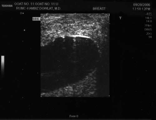
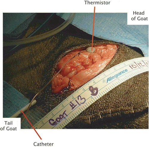
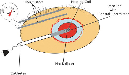
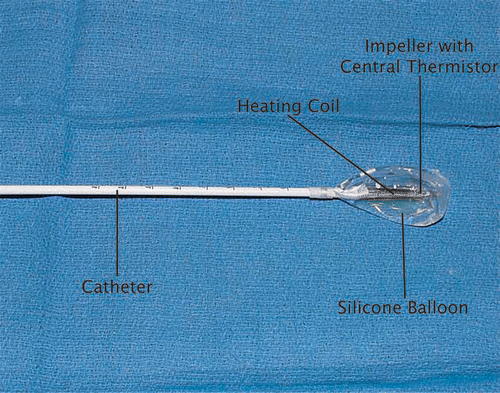
With approval of the Rush University Institutional Animal Care and Use Committee, thirty Nubian-cross goats (45–50 kg) in post-partum phase were selected. The selection was made after a literature search and consultation with the Institution's veterinary surgeons. The goat with two larger mammary glands was chosen in preference to the pig with multiple smaller glands. Under endo-tracheal anesthesia the two mammary glands were visualized by ultrasound (), mobilized through separate incisions and bisected longitudinally. Specific attention was paid to placement of the balloon in the proximal part of the gland where the lobules were plentiful and the lactiferous ducts were narrow. We deliberately excluded the terminal dilated ducts and sinuses by tying them off with sutures. At the earlier phase of this investigation, a small section (2–3 cm3) of the glandular tissue, equivalent to 20–30 cm3 in human breast, was excised to create a space for the balloon. At a later stage, the balloon was laid into the space and the incision was closed over it ensuring adequate tissue coverage (). The commercially available FDA approved device consists of a silicone balloon with a heating coil and an impeller. The latter ensures even distribution of heated 5% dextrose solution throughout the balloon ( and ). The device is programmed to work in 3 stages: ‘warm up’, ‘treatment’ and ‘cool-down’. The warm up stage starts when the balloon has been inserted into the tissue, inflated with 5% dextrose, and pressure is sustained above 150 mmHg. The heating coil warms the dextrose solution while the impeller circulates the fluid to maintain a uniform temperature of 87°C throughout the balloon. Once the balloon's central temperature reaches the preset temperature of 87°C, the ‘treatment’ phase begins. At this temperature, the treatment is given for 8 min. The heating element and impeller are automatically stopped and the cool down phase begins, lasting 20–45 seconds Citation[17].
Series I
The treatment cycle, as described above, was conducted once (8 minutes) in the first group of four animals, twice (16 minutes) in the second, and three times (24 minutes) in the third group; thus the collected data represent information from eight glands per group for increasing time intervals. In a fourth group of animals the heating device was manually halted at Principal Investigator (PI) determined time intervals so as to overcome the time restrictions imposed by the device's software. In addition, three animals were used as controls whereby all the surgical and manipulative steps were taken but thermal therapy was not given.
The external temperature of the mammary gland was continuously monitored and recorded with two thermistors (Omega Inc., CT) inserted into the subcutaneous tissue; one on each side of the balloon approximately at its midpoint (). The distance from the skin was 2–3 mm and to the balloon 10–15 mm depending on the thickness of the gland of that particular animal as determined by ultrasound. The primary reason for placement of the subcutaneous thermistors was to monitor the skin temperature and prevent scalding/burning of the skin. The incision was temporarily closed with staples, in order to prevent heat loss during treatment. The peak subcutaneous temperatures recorded ranged from 45–55°C with no clinically detected skin burn. Upon completion of the experiment, the balloon was deflated and the volume of the fluid was measured and recorded.
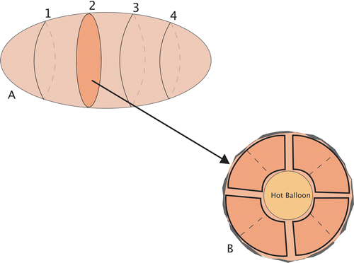
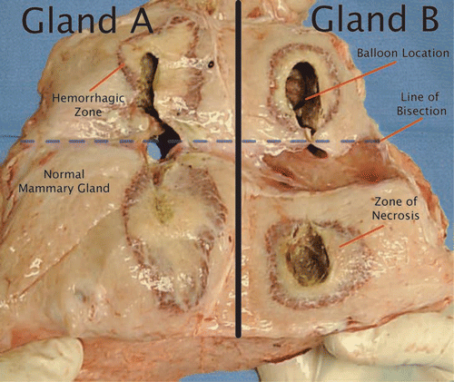
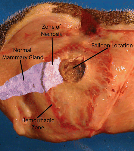
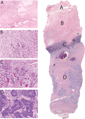
One week post-therapy, the animals were euthanized and the glands were excised for histological examination Citation[18]. Each gland was sectioned at 3–4 mm intervals perpendicular to the longitudinal axis of the balloon yielding 4–5 blocks of tissue (). The blocks were preserved in 10% formalin before representative sections were taken radially at 3, 6, 9 and 12 o’clock (). The sections were stained with Hematoxylin and Eosin. The zone of necrosis was determined microscopically (). Previous studies have shown consistent histological changes caused by heat Citation[18],Citation[19]. Thermally treated tissue shows both a central coagulum and a concentric peripheral red zone of damage Citation[18],Citation[16]. We observed a central rim of fibrosis and immediately adjacent necrotic glandular tissue with an intact light microscopic appearance (). Peripheral to this zone is an area of granulation tissue and necrosis (). The extent of the lesion was determined by a microscopic measurement from the inner zone to the nearest viable glandular tissue. Adipose or other non-glandular tissue was excluded from any measurements.
Series II
Having demonstrated the proof of the concept, the firmware of the device was modified to simulate its application for treatment of patients with breast cancer after lumpectomy. The manufacturer was requested to reduce the operating pressure of the balloon to 50 mmHg, as breast tissue, unlike the muscular uterine wall, is primarily fatty and does not require high pressure for its conformity with the balloon surface. Additionally, during the first series of the experiments it was noted that the high balloon pressure resulted in herniation of the balloon and pressure drop in several occasions, necessitating further inflation of the balloon. The time restriction of the device was also removed to allow the investigator the freedom of choosing the duration of thermal therapy. The uniformity of the fluid temperature in the balloon was maintained by an impeller located next to the heating coil Citation[17].
Figure 6. Histologic section of treated mammary gland. (A) Completely adjacent glandular structure immediately adjacent to the hot balloon. Underlying necrotic glands with normal appearing nuclei. (B) Glandular tissue with prominent new vessel formation, polymorphonuclear cells, and karyorrhectic debris. (C) Prominent new vessel formation in a background of myxoid stroma with mostly mononuclear cell infiltrate. No residual ductal glands are seen. (D) Morphologically normal mammary gland.
It should be pointed out that with regard to extra-capsular air/fluid entrapment interfering with heat propagation, we did not have imaging means, apart from ultrasound, to assess the non-conformity of the balloon with respect to the encapsulating glandular tissue.
A second series of experiments were conducted at balloon pressures of 50–80 mmHg and heating temperature of 90°C at increasing time intervals in successive animals. The first three experiments were performed each lasting 30, 60, and 90 seconds respectively, to evaluate the impact of heat during the warm up period before the balloon temperature reached 90°C. Next, thermal therapy experiments were conducted on pairs of mammary glands at intervals of 1, 2, 3, 5, 10, 15 and 20 minutes. Two glands in two animals received no thermotherapy and served as controls. Sham operations with placement of balloons and thermistors were performed without heating. At the completion of treatment at 90°C, a 4-minute cool down period was allowed for the central temperature to reach approximately 60°C before deflating the balloon.
The timing is illustrated for five glands heated for 15 minutes (). Two temperature sensors/thermistors were inserted into the subcutaneous tissue, one on each side of the balloon. Temperatures in tissue near the cavity were measured from warm up through cool down. The sensors provided six measures per minute. The recorded temperatures from the five glands were averaged for each 10-second interval. A time of 0 is the start of treatment, so negative times indicate the warm up period.
Figure 7. Average thermistor (sensor) readings (°C) for five glands treated for 15 minutes at 90°C balloon/central temperature.
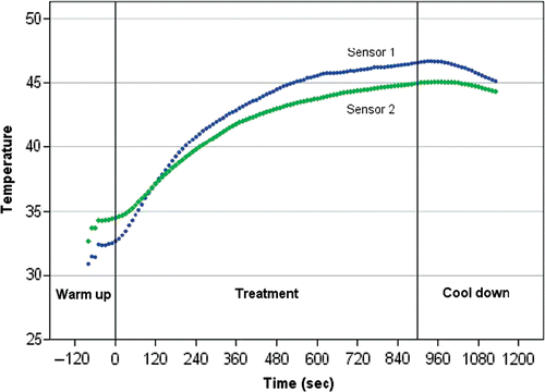
Sensor temperatures for other groups of treatment times show the same pattern. It was common for temperatures to reach a plateau after eight minutes of treatment (480 seconds).
The goats were euthanized 7 days post-treatment. Similar to series I, the glands were excised, serially sectioned and examined (, and ).
Results
During the series I experiments, circumferential necrosis was noted to increase in depth with increasing treatment time (). Microscopically, the thermally induced necrosis consisted of a zone of coagulum in contact with the balloon, followed by a segment of recognizable ducto-glandular elements and deeper still, a zone of necrosis where the normal glandular topography had disintegrated. Finally, a zone of hemorrhagic ring separates the heat ablated inner zones from the normal unaffected glandular tissue. Principal Investigator had previously encountered this concentric zones of necrosis in interstitially laser treated breast cancers Citation[20]. Others have described it with application of radiofrequency as the ablative thermal modality Citation[21–23].
Similarly, during the second series, tissue necrosis around the balloon was noted ranging from 4.7 to 8.6 mm ().
Table I. Data of treated goat mammary glands with treatment time and resulting mean necrosis during series I (FDA-approved ThermaChoice device).
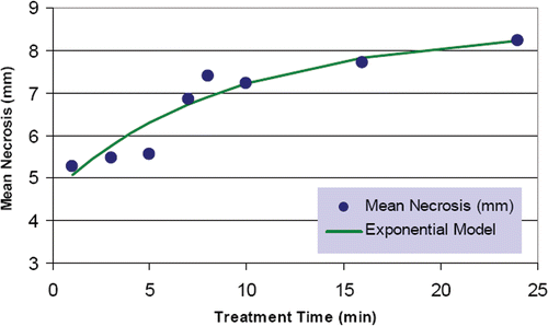
Significant variability in the depth of glandular necrosis was observed and was attributed to varying tissue thermal properties, presence of blood vessels and dilated milk ducts acting as a heat sink, and possible non-conformity of contact between the balloon and the mammary gland; a limitation of this model. However, the overall trend was that of increased depth of necrosis with time up to approximately 16 minutes, beyond which minimal increase was noted.
Table II. Data of treated goat mammary glands with treatment time and resulting mean necrosis during series II (modified ThermaChoice device).
The animals exhibited no systemic side effects post-treatment. Due to technical complications such as balloon rupture/leak or herniation of the balloon into a dilated duct, 7 out of the 60 experiments were excluded from the data set. The sections from five control experiments revealed normal mammary glandular and ductal anatomy with minimal necrosis attributed to trauma of handling ().
Modeling necrosis depth as a function of treatment time
It was expected that there would be a direct relationship between heating time and depth of necrosis. If necrosis is proportional to temperature of affected tissues, then necrosis depths should follow the patterns in . The data points can be modeled by Newton's law of cooling/heating, which states that the rate of temperature change in an object is proportional to the difference between the temperature of the object and the temperature of the surrounding medium. In this study, there is a reversal: the object is tissue which surrounds the probe, a medium with higher temperature than the tissue. The transfer of heat through tissue might not be as smooth as an exponential model would imply. As the tissue heats up, it coagulates as it loses water due to heating and may not be as effective in heat transfer. Further, if the tissue carbonizes, it acts like a ceramic material in conducting heat Citation[19]. Since the heat reaching the probes did follow a pattern implied by Newton's law of heating, it appears that the tissue changes in these experiments were not sufficient to interrupt patterns of heat flow. Under the assumption of necrosis depth being proportional to temperature, it was reasonable to build an exponential model to relate depth to time of treatment.
Data from series I was aggregated by treatment time. In cases where there was only one gland subjected to a particular treatment time, that gland was binned with an adjacent singleton (). An exponential model is fitted to the necrosis data under the assumption of a horizontal asymptote at 8.5 mm. The model is d = 3.8130 * 0.8969t + 8.5 where d is the depth of necrosis and t is minutes of treatment time. The model is a better fit to data than an ordinary linear regression, with 94% of necrosis variation accounted for by the model, compared to 88% from linear regression.
There were too few time intervals to build a similar exponential model for data in series II.
Discussion
Minimally invasive techniques are emerging for diagnosis and treatment of breast cancer. The principal reason for this evolution is early detection of the cancer through annual screening examination. Mammography has played a major role in detecting breast tumors, which are often non-palpable, thus creating a cascade of changes during the past two decades. For example, needle instead of knife, and breast conserving therapy (BCT) instead of mastectomy. This trend has extended to the adjuvant treatments as well. Partial breast irradiation with insertion of a balloon bearing a radioactive source into the lumpectomy cavity to deliver a one week treatment (brachytherapy), instead of whole breast irradiation in six weeks has gained acceptance by patients and physicians Citation[24]. The early results of brachytherapy in terms of morbidity and local recurrence are comparable to those achieved by conventional WBI Citation[24–27]. Introduction of intra-operative radiation therapy (IORT) during the past decade is yet another way to shorten and to streamline breast cancer treatment. Currently IORT is given as a boost to the tumor bed, but several trials are evaluating the treatment as a one-time radiation to the breast after lumpectomy Citation[6],Citation[7]. These reports indicate good to excellent cosmetic results, patient satisfaction, minimal morbidity and local recurrence rate equivalent to WBI.
The concept of thermal therapy instead of radiation for ablation of cancer, either as a primary means of ablating the malignant tumor or as an adjunct to surgery, has been developed and tested in the recent years. Treatment of the lumpectomy cavity with a hot balloon simplifies the process further. In radiation therapy, reliance is placed on ionizing radiation to devitalize cancer cells. Radiation damages the genes of the malignant cells, making it impossible for them to grow and divide. Radiation also damages the normal cells, most of which may recover and function normally Citation[28]. Thermal ablation, on the other hand, disrupts the entire structure of the cell irreversibly. The necrotic tissue is gradually removed by phagocytosis and replaced with fibrosis Citation[29],Citation[30]. The concept and process are relatively simple and appeal to women who are fearful of the long term side effects of radiation.
The in situ ablation techniques include radiofrequency ablation (RFA), interstitial laser therapy (ILT), microwave thermotherapy, and focused ultrasound (FUS) ablation. These treatment modalities utilize thermal energy/heat to destroy breast cancer Citation[14],Citation[15],Citation[31],Citation[32]. They require imaging guidance to direct energy to the target.
Direct heating of the breast tissue after surgical resection of the tumor consists of placing an electrically heated device in the lumpectomy cavity and delivering heat to its wall. Shafirstein et al. developed and tested a device with a metal sphere at its tip for this purpose. The conductive interstitial thermal therapy technique caused 10–15 mm of tissue necrosis when the device was heated to 70–150°C for 20 minutes Citation[19],Citation[33]. Klimberg et al. reported on post-lumpectomy treatment of breast cancer patients with radiofrequency and noted 10 mm (range 2–18 mm) zone of necrosis. The extended margins have the potential to reduce the number of returns to the operating room for re-excision of residual cancer Citation[34].
In the experimental model described in this paper, we have demonstrated that the glandular tissue in contact with the hot balloon exhibits acute necrosis to a distance of 3–20 mm (and , and , and Tables and ) depending on the length of treatment. In our search for an experimental model to resemble the human breast, goat mammary gland in the post-partum phase came closest. The porcine model was also considered but rejected because of excessive adipose tissue and scant lactiferous ducts and glands. In practice however, in the goat model, either the ducts were too dilated with milk or the glands were not large enough to accommodate the inflated balloon in a symmetrical fashion Citation[18]. The presence of milk in the dilated glands would serve as a heat sink and interfere with optimal heat distribution to the glandular tissue. We did not have access to CT imaging to check for conformity of the balloon embedded in the gland. Ultrasound was employed to visualize the location of the balloon in the gland. In the first series, the commercially available balloon was programmed to function at high pressure suitable for uterine application and restricted heating time (8 minutes per cycle). These restrictions were removed for the second series. Consequently, the experiments ran at a lower pressure (50 versus 150 mmHg), less fluid volume (15–20 mL versus 60–80 mL) and shorter pre-heating period (1 versus 4–5 minutes).
Our data () demonstrates a direct relationship between the length of heating and the depth of necrosis; up to a maximum point, after which the depth of necrosis did not increase significantly; indicating equilibrium between heat gain and heat loss.
Based upon the data derived from the two series of experiments, the provisional criteria for inclusion of patients in a pilot clinical study are as follows: (1) patients with histologically proven T1 invasive ducal carcinoma without evidence of multicentricity, (2) microscopically free margin of resection. The treatment time of 16 minutes at 87°C will be considered. The breast under investigation will be examined by MRI before and one week after thermal therapy. All patients will receive their standard chemo/radiation therapy per protocol. Thus their treatment will not be compromised by this investigation.
Summary
An experimental model deploying a thermal technique for ablation of the goat mammary gland is described. It demonstrates the possibility of ablating occult cancer cells in the immediate zone of surgical resection which otherwise may cause future local recurrence. Its simple design, which can be given at the time of lumpectomy after removal of breast cancer, makes it an attractive option for patients who reject adjuvant radiation therapy because of the concern about its long term side effects and logistical issues.
Acknowledgement
This project was partially supported by a grant from Chicago Bears and McCormick Breast and Ovarian Cancer Research Foundation.
Declaration of interest: The authors report no conflicts of interest. The authors alone are responsible for the content and writing of the paper.
References
- Ferrini R, Mannino E, Ramsdell E, Hill L. Screening mammography for breast cancer: American College of Preventive Medicine practice policy statement. Am J Prev Med 1996; 12(5)340–341
- Elmore JG, Armstrong K, Lehman CD, Fletcher SW. Screening for breast cancer. JAMA 2005; 293(10)1245–1256
- Veronesi U, Cascinelli N, Mariani L, Greco M, Saccozzi R, Luini A, Aguilar M, Marubini E. Twenty-year follow-up of a randomized study comparing breast-conserving surgery with radical mastectomy for early breast cancer. N Engl J Med 2002; 347(16)1227–1232
- Fisher B, Anderson S, Bryant J, Margolese RG, Deutsch M, Fisher ER, Jeong JH, Wolmark N. Twenty-year follow-up of a randomized trial comparing total mastectomy, lumpectomy, and lumpectomy plus irradiation for the treatment of invasive breast cancer. N Engl J Med 2002; 347(16)1233–1241
- Gage I, Recht A, Gelman R, Nixon AJ, Silver B, Bornstein BA, Harris JR. Long-term outcome following breast-conserving surgery and radiation therapy. Int J Radiat Oncol Biol Phys 1995; 33(2)245–251
- Veronesi U, Gatti G, Luini A, Intra M, Ciocca M, Sanchez D, et al. Full-dose intraoperative radiotherapy with electrons during breast-conserving surgery. Arch Surg 2003; 138(11)1253–1256
- Holmes DR, Baum M, Joseph D. The TARGIT trial: Targeted intraoperative radiation therapy versus conventional postoperative whole-breast radiotherapy after breast-conserving surgery for the management of early-stage invasive breast cancer (a trial update). Am J Surg 2007; 194(4)507–510
- Polgar C, Sulyok Z, Fodor J, Orosz Z, Major T, Takacsi-Nagy Z, Mangel LC, Somogyi A, Kásler M, Nemeth G. Sole brachytherapy of the tumor bed after conservative surgery for T1 breast cancer: Five-year results of a phase I-II study and initial findings of a randomized phase III trial. J Surg Oncol 2002; 80(3)121–128
- Li G, Mitsumori M, Ogural M, Horii N, Kawamural S, Masunaga S, Nagata Y, Hiraoka M. Local hyperthermia combined with external irradiation for regional recurrent breast carcinoma. Int J Clin Oncol 2004; 9(3)179–183
- McCormick B. Counterpoint: Hyperthermia with radiation therapy for chest wall recurrences [Comment]. J Nat Comp Cancer Network 2007; 5(3)345–348
- Wiedemann G, Mella O, Coltart RS, Schem BC, Dahl O. Hyperthermia improves local tumour control in locally advanced breast cancer. Klinische Wochenschrift 1988; 66(20)1034–1038
- Ben-Yosef R, Vigler N, Inbar M, Vexler A. Hyperthermia combined with radiation therapy in the treatment of local recurrent breast cancer. Israel Med Assoc J 2004; 6(7)392–395
- Feyerabend T, Wiedemann GJ, Jager B, Vesely H, Mahlmann B, Richter E. Local hyperthermia, radiation, and chemotherapy in recurrent breast cancer is feasible and effective except for inflammatory disease. Int J Radiat Oncol Biol Phys 2001; 49(5)1317–1325
- Huston TL, Simmons RM. Ablative therapies for the treatment of malignant diseases of the breast. Am J Surg 2004; 189: 694–701
- van Esser S, van den Bosch MAAJ, van Diest PJ, Mali WTM, Borel R, Inne HM, van Hillegersberg R. Minimally invasive ablative therapies for invasive breast carcinomas: an overview of current literature. World J Surg 2007; 31: 2284–2292
- Gianfelice D, Khiat A, Amara M, Belblidia A, Boulanger Y. MR imaging-guided focused US ablation of breast cancer: Histopathologic assessment of effectiveness-initial experience. Radiology 2003; 227(3)849–855
- http://www.endheavyperiods.com/downloads/thermachoice_508.pdf, Ethicon Inc. Gynecare Thermachoice* III: Uterine balloon therapy with fluid circulation. 2003
- Thomsen S, Ryan T, Freeman L, DiFrancesco M. Influence of different anatomies on healing of interstitial thermal lesions in goat and pig breasts. Proc SPIE 2001; 4247: 69–76
- Shafirstein G, Novak P, Moros EG, Siegel E, Hennings L, Kaufmann Y, Ferguson S, Myhill J, Swaney M, Spring P. Conductive interstitial thermal therapy device for surgical margin ablation: In vivo verification of a theoretical model. Int J Hyperthermia 2007; 23(6)477–492
- Bloom KJ, Dowlat K, Assad L. Pathologic changes after interstitial laser therapy of infiltrating breast carcinoma. Am J Surg 2001; 182(4)384–388
- Bland KL, Gass J, Klimberg VS. Radiofrequency, cryoablation, and other modalities for breast cancer ablation. Surg Clin North Am 2007; 87(2)539–550
- Singletary SE. Radiofrequency ablation of breast cancer. Am Surg 2003; 69(1)37–40
- Izzo F, Thomas R, Delrio P, Rinaldo M, Vallone P, DeChiara A, Gerardo B, D'Aiuto G, Cortino P, Curley Steven. Radiofrequency ablation in patients with primary breast carcinoma: A pilot study in 26 patients. Cancer 2001; 92(8)2036–2044
- Benitez PR, Keisch ME, Vicini F, Stolier A, Scroggins T, Walker A, White J, Hedberg P, Hebert M, Arthur D, Zannis V, Quiet C, Streeter O, Silverstein M. Five-year results: The initial clinical trial of MammoSite balloon brachytherapy for partial breast irradiation in early-stage breast cancer. Am J Surg 2007; 194(4)456–462
- Polgar C, Major T, Fodor J, Nemeth G, Orosz Z, Sulyok Z, Udvarhelyi N, Somogyi A, Takacsi-Nagy Z, Lövey K, Agoston P, Kasler M. High-dose-rate brachytherapy alone versus whole breast radiotherapy with or without tumor bed boost after breast-conserving surgery: Seven-year results of a comparative study. Int J Radiat Oncol Biol Phys 2004; 60(4)1173–1181
- Kuske RR, Winter K, Arthur DW, Bolton J, Rabinovitch R, White J, Hanson W, Wilenzick RM. Phase II trial of brachytherapy alone after lumpectomy for select breast cancer: Toxicity analysis of RTOG 95-17. Int J Radiat Oncol Biol Phys 2006; 65(1)45–51
- Chen PY, Vicini FA, Benitez P, Kestin LL, Wallace M, Mitchell C, Pettinga J, Martinez AA. Long-term cosmetic results and toxicity after accelerated partial-breast irradiation: A method of radiation delivery by interstitial brachytherapy for the treatment of early-stage breast carcinoma. Cancer 2006; 106(5)991–999
- Burnet NG, Wurm R, Nyman J, Peacock JH. Normal tissue radiosensitivity – How important is it?. Clin Oncol (R Coll Radiol) 1996; 8(1)25–34
- Dick EA, Joarder R, De Jode MG, Wragg P, Vale JA, Gedroyc WM. Magnetic resonance imaging-guided laser thermal ablation of renal tumours. BJU Int 2002 Dec; 90(9)814–822
- Rossi S, Di Stasi M, Buscarini E, Quaretti P, Garbagnati F, Squassante L, Paties CT, Silverman DE, Buscarini L. Percutaneous RF interstitial thermal ablation in the treatment of hepatic cancer. Am J Roentgenol 1996; 167(3)759–768
- Dowlatshahi K, Dieschbourg JJ, Bloom KJ. Laser therapy of breast cancer with 3-year follow-up. Breast J 2004; 10(3)240–243
- Dowlatshahi K, Francescatti DS, Bloom KJ. Laser therapy for small breast cancers. Am J Surg 2002; 184(4)359–363
- Shafirstein G, Hennings L, Kaufmann Y, Novak P, Moros EG, Ferguson S, Siegel E, Klimberg SV, Waner M, Spring P. Conductive interstitial thermal therapy (CITT) device evaluation in VX2 rabbit model. Tech Cancer Res Treat 2007; 6(3)235–245
- Klimberg VS, Kepple J, Shafirstein G, Adkins L, Henry-Tillman R, Youssef E, Brito J, Talley L, Korourian BS, Korourian S. eRFA: Excision followed by RFA – A new technique to improve local control in breast cancer. Ann Surg Oncol 2006; 13(11)1422–1433

