Abstract
Purpose: To demonstrate the efficacy and safety of percutaneous microwave coagulation treatment (PMCT) by a new microwave delivery system (Forsea Microwave™) in large hepatocellular carcinomas (HCC) (≥5 cm).
Materials and methods: Four patients with 4 HCC lesions measuring ≥6 cm in the greatest dimension underwent PMCT by means of the Forsea Microwave™ microwave delivery system. Final therapeutic efficacy was evaluated with dynamic computer tomography (CT) scans performed within one month after PMCT. During and after PMCT, patients’ complaints and any abnormal physical signs were recorded for safety assay. CT or ultrasound scan (US) performed immediately after the treatment was used to detect acute complications related to the treatment. Repeated dynamic CT scans were performed every three to four months thereafter to detect local disease recurrence and/or other recurrences.
Results: Three of these patients achieved a complete ablation of the cancer nodules (two patients with two treatment sessions and one patient with three treatment sessions). One of these patients obtained a complete ablation of the cancer nodule with two treatment sessions except the lesion of portal vein tumour thrombus (PVTT). No obvious symptomatic complication was observed except abdominal pain during and after the treatment in two of these patients. All the patients remained asymptomatic and no recurrent tumour was observed during their follow-up (1-19 months).
Conclusions: PMCT by the Forsea Microwave™ microwave delivery system could offer a satisfactory therapeutic effect and is applicable to the treatment of large HCC.
Introduction
Hepatocellular carcinomas (HCC) are the fifth most common malignancy worldwide, accounting for 5.4% of new cancer cases annually Citation[1],Citation[2]. Surgical resection has been referred to as the most effective method for the treatment of HCC. However, the majority of newly diagnosed HCC patients in China are not suitable for surgical resection because the tumour lesions are usually confirmed to exceed 5 cm in the greatest dimension and/or are accompanied by tumour vascular invasion or insufficient hepatic function Citation[3–5]. Therefore, most patients are not eligible for surgery. The patients with suitability for curative resection only represent a proportion of 30% or less in the whole HCC population. Hence non-operative therapies are playing increasingly important roles in the clinical treatment of HCC Citation[6–8].
Percutaneous microwave coagulation treatment (PMCT) is widely used as a less invasive modality for the local treatment of small HCC cases in China Citation[9],Citation[10]. PMCT is similar to surgical resection and can offer favourable outcomes to patients with HCC lesions measuring ≤3 cm in the greatest dimension Citation[11]. As for the patients with HCC lesions measuring ≥5 cm in the greatest dimension, transcatheter chemoembolisation (TACE) is regularly recommended Citation[12]. However, the survival benefits conferred by TACE treatment in these patients still remain controversial Citation[13]. Substantial improvements have been achieved in the attempt to expand the necrosis volume of coagulation therapy in the last decade due to the rapid development of microwave antenna technology. PMCT can become a more effective treatment and can potentially be applied to the treatment of HCC lesions measuring ≥5 cm in the greatest dimension Citation[14],Citation[15]. Here we report the efficacy and safety studies of PMCT with a new microwave delivery system in four patients with large HCC (larger than 5 cm in the greatest dimension).
Materials and methods
Patients
Four patients, including three men and one woman, with four HCC lesions measuring ≥6 cm in the greatest dimension were referred to our hospital between 12 January 2007 and 24 June 2008.
All patients tested positive for the hepatitis B surface antigen. The liver function reserves of these patients were evaluated to be Child-Pugh classification A. This was the initial treatment for these patients. In all patients, the histological diagnosis was confirmed by ultrasound scan (US) guided, fine-needle biopsy. Exclusion criteria were as follows: (1) serious blood coagulation dysfunction (prothrombin time: PT > 23 s, platelet count <40,000 ml−1); (2) serious cirrhosis (Child-Pugh classification C). This study was approved by the Medical Ethical Committee of the Xijing Hospital. Written informed consent was provided by all patients or their families.
PMCT therapy
The microwave delivery system (Forsea Microwave™; Forsea Microwave & Electronic Research Institute, Nanjing, China) used in this study consisted of a microwave generator which emits a 2450-MHz microwave signal, a soft coaxial cable and a 14-gauge microwave antenna 15 cm in length () Citation[15].
The microwave coagulator was approved according to FDA regulations. The microwave antenna was connected to the microwave generator with a coaxial cable. This device can produce a coagulated lesion with a diameter of 5 cm in liver at a power output of 60 W for 5 min. Bucinnazine (50 mg) and diazepam (5 mg) were intramuscularly injected 10 min before PMCT as pre-medications for analgesia and sedation. After the skin surface was disinfected and local anaesthesia was induced with 1% lidocaine, the microwave antenna was introduced with real-time US guidance into the tumour nodules. The antenna was percutaneously introduced at 5 mm beyond the deep margin of the nodule. To achieve a coagulation area as large as possible, the antenna was successively withdrawn every 10 mm to repeat the treatment until the antenna was 5 mm beyond the superficial margin of the nodule. The orientation of the needle track was then changed to repeat the treatment.
When the antenna was removed, the needle track was coagulated with microwaves for 10 s to prevent liver haemorrhaging and tumour seeding. Patients who experienced severe pain during or immediately after the treatment received 10 mg of morphine hydrochloride intramuscularly.
Imaging equipment
An ultrasound (US) scanner (Acuson Sequoia 512; Siemens, Berlin, Germany) with a 2–5-MHz vector transducer was used to estimate the maximum extent of the tumours and monitor the microwave ablation procedures and follow-up examinations. Tumour dimensions were estimated by a designated author (J. Shao) with US. A spiral computed tomography (CT) scanner (Xpress/SX; Toshiba, Tokyo, Japan) was used to measure the size of the coagulated lesion. 1.5 mL/kg of iopromide (Ultravist 300; Schering, Berling, Germany) power was injected at 3 mL/s, and the following parameters were used to acquire contrast material-enhanced images: 10-mm thick sections, 10-mm collimation, and 1-s scan acquisition, pitch of 1 : 1, 120 kV, and 250 mA.
Assessment of therapeutic efficacy
To examine the final effect, abdominal CT scans were obtained within one month of treatment. The CT studies were interpreted independently by two experienced radiologists. Complete ablation of the tumour was defined such that no contrast-enhanced portion was discovered within the coagulated lesion on both the hepatic arterial and portal venous phase images. Repeated dynamic CT scans were performed every three to four months thereafter to detect local disease recurrences and/or other recurrences.
During and after treatment, patients’ complaints and any abnormal physical signs were recorded for safety analysis. After the procedure, all patients were hospitalised for at least 72 hours. CT and ultrasound scans performed immediately after the procedure were used to detect acute complications related to the treatment. If no complications occurred, the patients were then discharged.
Results
Case presentation
Case 1
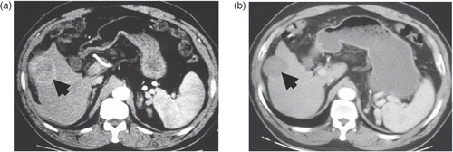
A 57- year-old man with a history of chronic hepatitis B-related liver cirrhosis had symptoms of abdominal pain and abdominal distention for two weeks before he was admitted to our hospital. Transverse contrast-enhanced CT scans showed a 6.1 × 5.2 cm cancer nodule in the right lobe of the liver (). This patient was assigned to receive one session of PMCT therapy with one microwave antenna. Microwave energy was applied for a total of 23 min by two antenna insertions (13 min for the first insertion and 10 min for the second) with a power output of 60 W. He experienced severe abdominal pain during and after the treatment, which was alleviated with analgesic medication. No any other symptomatic complication was observed. The final effect was evaluated as a complete ablation by the abdominal CT scan two weeks after the treatment, which was illustrated with a 4.9 × 4.3 cm coagulation zone without an uncoagulated portion as shown in . Abdominal CT scans showed that no recurrent tumour was observed after a follow-up of 19 months.
Figure 2. Transverse contrast-enhanced CT scans in patient 1. (a) Pre-ablation scan showed a 6.1 × 5.2 cm cancer nodule (indicated by the wide arrow) in the right lobe. (b) Scan obtained two weeks after one session of ablation showed a complete ablation with a 4.9 × 4.3-cm coagulation zone (indicated by the wide arrow).

Case 2
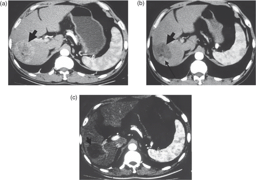
A 59- year-old man had a symptom of right upper abdominal distention for one year and was referred to our unit. Transverse contrast-enhanced CT scans showed a 6.4 × 6.1 cm cancer nodule in the right lobe of the liver (). He was recommended to receive two sessions of the PMCT therapy. One microwave antenna was used in the first session, and microwave energy was applied for a total of 8 min by two antenna insertions (5 min for the first insertion and 3 min for the second) with a power output of 60 W. An abdominal CT scan showed there was an uncoagulated portion around the coagulation zone (indicated by the narrow arrow in ), which implied that the cancer lesion was incompletely ablated and that a second session was needed for this patient. One microwave antenna was used in the second session, and microwave energy was applied for a total of 13 min by three antenna insertions (6 min for the first insertion, 5 min for the second insertion and 2 min for the third) with a power output of 60 W. This patient experienced severe abdominal pain during and after the treatment and received analgesic medication. No other symptomatic complication was observed. The abdominal CT scan two weeks after the second treatment showed that the uncoagulated portion around the coagulation zone had disappeared, and the final effect was therefore evaluated as a complete ablation, as shown in . This patient remains asymptomatic, and no recurrent tumour was observed after a follow-up of 17 months.
Figure 3. Transverse contrast-enhanced CT scans in patient 2. (a) Pre-ablation scan showed a 6.4 × 6.1 cm cancer nodule (arrows) in the right lobe. (b) Scan obtained one month after the first session of ablation showed an incomplete ablation (residual area indicated by the narrow arrow) with a 6.4 × 5.3-cm coagulation zone (indicated by the wide arrow). (c) Scan obtained two weeks after the second session of ablation showed a complete ablation with a 5.8 × 5.2-cm coagulation zone (indicated by the wide arrow).

Case 3
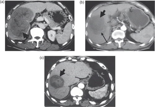
A 59-year-old man was symptomatic of right upper abdominal distention and anorexia for one month before he was admitted to our hospital. Transverse contrast-enhanced CT scans showed a 13.8 × 9.2 cm cancer nodule in the right lobe of the liver with concomitance of portal vein tumour thrombus (PVTT) (). This patient underwent two sessions of the PMCT therapy. One microwave antenna was used in the first session, and microwave energy was applied for a total of 32 min by six antenna insertions (6 min for the first insertion, 5 min for the second insertion, 8 min for the third insertion, 4 min for the fourth insertion, 4 min for the fifth insertion, and 5 min for the last) with a power output of 60 W. Abdominal CT scans showed that the lesion was incompletely ablated due to the existence of an uncoagulated portion around the coagulation zone (indicated by the narrow arrow in ) and a second session was needed for this patient. One microwave antenna was used in the second session, and microwave energy was applied for a total of 10 min by two antenna insertions (4 min for the first insertion and 6 min for the second) with a power output of 60 W. No symptomatic complication was observed during and after the treatments. The final effect was evaluated as a complete ablation of the tumour by the abdominal CT scan two weeks after the second treatment, as demonstrated by the disappearance of the uncoagulated portion (). This patient remains asymptomatic and no recurrent tumour was observed after a follow-up of 14 months.
Figure 4. Transverse contrast-enhanced CT scans in patient 3. (a) Pre-ablation scan showed a 13.8 × 9.2-cm cancer nodule (indicated by the wide arrow). (b) Scan obtained two weeks after the first session of ablation showed an incomplete ablation (residual area indicated by the narrow arrow) with a 10 × 6.5-cm coagulation zone (indicated by the wide arrow). (c) Scan obtained two weeks after the second session of ablation showed a complete ablation (except the PVTT lesion) with a 7.3 × 6.1 cm coagulation zone (indicated by the wide arrow).

Case 4
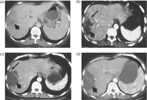
A 44-year-old woman was symptomatic of right upper abdominal distention and pain for two weeks and was referred to our unit. Transverse contrast-enhanced CT scans showed a 9.8 × 5.2 cm cancer nodule in the right lobe of the liver (). This patient was given three sessions of PMCT therapy, and her symptoms disappeared after the third treatment. One microwave antenna was used in the first session, and microwave energy was applied for a total of 23 min by three antenna insertions (6 min for the first insertion, 5 min for the second insertion, and 12 min for the last) with a power output of 60 W. Abdominal CT scans showed that there was an uncoagulated portion around the coagulation zone (indicated by the narrow arrow in ), which implied that the lesion was incompletely ablated and that a second session was needed for this patient. One microwave antenna was used in the second session, and microwave energy was applied for a total of 20 min by four antenna insertions (6 min for the first insertion, 5 min for the second, 6 min for the third, and 3 min for the last) with a power output of 60 W. One more abdominal CT scan showed that the uncoagulated portion still remained around the coagulation zone (indicated by the narrow arrow in ), which indicated that the lesion was still incompletely ablated, and a third session was given to this patient. One microwave antenna was used in the third session, and microwave energy was applied for a total of 13 min by three antenna insertions (6 min for the first insertion, 4 min for the second, and 3 min for the last) with a power output of 60 W. No symptomatic complication was observed during and after the treatments. The final effect was evaluated as a complete ablation of the tumour by an abdominal CT scan two weeks after the third treatment (). Her first follow-up was performed 1 month ago.
Figure 5. Transverse contrast-enhanced CT scans in patient 4. (a) Pre-ablation scan showed a 9.8 × 5.2-cm cancer nodule (indicated by the wide arrow). (b) Scan obtained one week after the first session of ablation showed an incomplete ablation (residual area indicated by the narrow arrow) with a 8.9 × 4.8-cm coagulation zone (indicated by the wide arrow). (c) Scan obtained two weeks after the second session of ablation showed an incomplete ablation (residual area indicated by the narrow arrow) with a 8.3 × 5.2-cm coagulation zone (indicated by the wide arrow). (d) Scan obtained two weeks after the third session of ablation showed a complete ablation with a 7.4 × 4.8-cm coagulation zone (indicated by the wide arrow).

Discussion
There are three widely used image-guided ablation methods, including PMCT, radiofrequency (RF) ablation, and cryotherapy, for the treatment of small liver cancers Citation[16]. Tumour thermoablative techniques such as RF ablation and microwave ablation have been widely applied as minimally invasive approaches to the management of small liver tumours, especially in patients with significant co-morbidities or those who refuse surgical resection Citation[17],Citation[18]. However, a key limitation of these methods is the unsatisfactorily small size of the coagulation area in one treatment session. Although the local effectiveness of RF ablation has been greatly improved by using the cooled needle during the treatment, the ablative area was not yet large enough to meet the requirements of many clinical settings Citation[19]. It was evidenced that the tissue temperature in the heat field of microwave was notably higher than that of RF in terms of the same energy output, and that PMCT was more efficacious than RF ablation in treatments of some highly vascularised tumours Citation[20],Citation[21]. However, severe skin burns could arise as a common complication due to the unexpected temperature increase of the antenna shaft. As a result, the application of microwave energy is usually limited to 60 W for 5 min, which could only generate in vivo coagulation necrosis with a short axis less than 3 cm. Two or more treatment sessions are needed for complete ablation of one small tumour. Kuang et al. adopted a new microwave delivery system (FORSEA; Qinghai Microwave Electronic Institute, Nanjing, China), which employed a cooled-shaft antenna that overcame the obstacles of conventional microwave ablation, to acquire a microwave energy output of 80 W for more than 20 min and obtain a complete ablation in some HCC with diameters up to 5 cm in one treatment session Citation[15]. In this study, we applied this microwave ablation system to the treatment of HCC patients with cancer nodules exceeding 5 cm. The treatment outcomes of these four patients achieved in this study revealed that PMCT is an ideal option for large HCC, which demonstrated that tumour sizes ≥5 cm are not contra-indicative to PMCT.
According to the National Comprehensive Cancer Network (NCCN) Clinical Practice Guidelines in Oncology, TACE was regularly recommended to the patients in Cases 1, 2, and 4. However, whether they could achieve some clinical benefit from the TACE treatment still remains undefined. In this study, these patients were alternatively assigned to receive the PMCT with the Forsea Microwave™ microwave delivery system. A complete ablation was usually achieved in one or two treatment sessions. Abdominal pain was observed as the main complication post-treatment because the tumour location was adjacent to the liver capsule. To date, no recurrent cancer has been detected in the follow-up examinations. As for the patient of Case 3, this patient was at the advanced stage of the disease because of the concomitance of the PVTT lesion. Few methods are available in this case. He received two sessions of PMCT and obtained an amazing result, a complete ablation (except the PVTT lesion) of the cancer nodules, as shown in . From the aforementioned cases, we obtained the following experience in the treatment of large HCC by PMCT (1) Tumour size is not a negative determinant to the effectiveness of PMCT; (2) Even if the cancer lesion could not be completely ablated in one session of PMCT, the capillary bed within the cancer nodule could be diminished and blood flow of the feeding vessel could be slowed down, which facilitated the effectiveness of the following PMCT procedure; (3) The destruction or embolism of the feeding artery induced by PMCT also contributed to the effectiveness of PMCT in some large HCC cases.
Conclusion
PMCT by the Forsea Microwave™ microwave delivery system was applicable to the treatment of large HCC and achieved a satisfactory therapeutic effect. Further clinical trials are needed to verify the effectiveness and safety of PMCT by Forsea Microwave™ microwave delivery system, especially compared with the TACE treatment in large HCC.
Declaration of interest: The authors report no conflicts of interest. The authors alone are responsible for the content and writing of the paper.
References
- Blum HE, Spangenberg HC. Hepatocellular carcinoma: An update. Arch Iran Med 2007; 10: 361–371
- Lau WY, Lai EC. Hepatocellular carcinoma: Current management and recent advances. Hepatobiliary Pancreat Dis Int 2008; 7: 237–257
- Cormier JN, Thomas KT, Chari RS, Pinson CW. Management of hepatocellular carcinoma. J Gastrointest Surg 2006; 10: 761–780
- Hasegawa K, Kokudo N, Makuuchi M. Surgical management of hepatocellular carcinoma. Liver resection and liver transplantation. Saudi Med J 2007; 28: 1171–1179
- Rougier P, Mitry E, Barbare JC, Taieb J. Hepatocellular carcinoma (HCC): An update. Semin Oncol 2007; 34: S12–20
- Crocetti L, Lencioni R. Thermal ablation of hepatocellular carcinoma. Cancer Imag 2008; 27: 19–26
- Ohmoto K, Yoshioka N, Tomiyama Y, Shibata N, Kawase T, Yoshida K, Kuboki M, Yamamoto S. Thermal ablation therapy for hepatocellular carcinoma: Comparison between radiofrequency ablation and percutaneous microwave coagulation therapy. Hepatogastroenterol 2006; 53: 651–654
- Llovet JM, Bruix J. Novel advancements in the management of hepatocellular carcinoma in 2008. J Hepatol 2008; 48: S20–37
- Zhou P, Liu X, Li R, Nie W. Percutaneous coagulation therapy of hepatocellular carcinoma by combining microwave coagulation therapy and ethanol injection. Eur J Radiol 2008, [E-pub ahead of print]
- Dong BW, Zhang J, Liang P, Yu XL, Su L, Yu DJ, Ji XL, Yu G. Sequential pathological and immunologic analysis of percutaneous microwave coagulation therapy of hepatocellular carcinoma. Int J Hyperthermia 2003; 19: 119–133
- Horigome H, Nomura T, Nakao H, Saso K, Takahashi Y, Akita S, Sobue S, Mizuno Y, Nojiri S, Hirose A, et al. Treatment of solitary small hepatocellular carcinoma: Consideration of hepatic functional reserve and mode of recurrence. Hepatogastroenterol 2000; 47: 507–511
- Georgiades CS, Hong K, Geschwind JF. Radiofrequency ablation and chemoembolization for hepatocellular carcinoma. Cancer J 2008; 14: 117–122
- Marelli L, Stigliano R, Triantos C, Senzolo M, Cholongitas E, Davies N, Tibballs J, Meyer T, Patch DW, Burroughs AK. Transarterial therapy for hepatocellular carcinoma: Which technique is more effective? A systematic review of cohort and randomized studies. Cardiovasc Intervent Radiol 2007; 30: 6–25
- Yu NC, Lu DS, Raman SS, Dupuy DE, Simon CJ, Lassman C, Aswad BI, Ianniti D, Busuttil RW. Hepatocellular carcinoma: Microwave ablation with multiple straight and loop antenna clusters–Pilot comparison with pathologic findings. Radiology 2006; 239: 269–275
- Kuang M, Lu MD, Xie XY, Xu HX, Mo LQ, Liu GJ, Xu ZF, Zheng YL, Liang JY. Liver cancer: Increased microwave delivery to ablation zone with cooled-shaft antenna–Experimental and clinical studies. Radiol 2007; 242: 914–924
- Orlacchio A, Bazzocchi G, Pastorelli D, Bolacchi F, Angelico M, Almerighi C, Masala S, Simonetti G. Percutaneous cryoablation of small hepatocellular carcinoma with US guidance and CT monitoring: Initial experience. Cardiovasc Intervent Radiol 2008; 31: 587–594
- Xu HX, Lu MD, Xie XY, Yin XY, Kuang M, Chen JW, Xu ZF, Liu GJ. Prognostic factors for long-term outcome after percutaneous thermal ablation for hepatocellular carcinoma: A survival analysis of 137 consecutive patients. Clin Radiol 2005; 60: 1018–1025
- Lu MD, Xu HX, Xie XY, Yin XY, Chen JW, Kuang M, Xu ZF, Liu GJ, Zheng YL. Percutaneous microwave and radiofrequency ablation for hepatocellular carcinoma: A retrospective comparative study. J Gastroenterol 2005; 40: 1054–1060
- Shibata T, Shibata T, Maetani Y, Isoda H, Hiraoka M. Radiofrequency ablation for small hepatocellular carcinoma: Prospective comparison of internally cooled electrode and expandable electrode. Radiol 2006; 238: 346–353
- Liang P, Wang Y. Microwave ablation of hepatocellular carcinoma. Oncol 2007; 72: S124–131
- Shock SA, Meredith K, Warner TF, Sampson LA, Wright AS, Winter TC, III, Mahvi DM, Fine JP, Lee FT Jr. Microwave ablation with loop antenna: In vivo porcine liver model. Radiol 2004; 231: 143–149

