
Abstract
Purpose: To evaluate the clinical efficacy of ultrasound-guided percutaneous microwave ablation (PMWA) therapy for symptomatic uterine fibroids in a multicentre study.
Materials and methods: Patients with symptomatic uterine fibroids who underwent PMWA at multiple treatment centres in China between January 2013 and August 2015 were prospectively studied to compare the reduction rate of uterine fibroids, haemoglobin level and uterine fibroid symptom and health-related quality of life questionnaire (UFS-QOL) scores before and at 3, 6 and 12 months after ablation.
Results: A total of 311 patients (405 leiomyomas) from eight treatment centres underwent the treatment (age, 29–55 years; mean ± SD, 41 ± 5.11 years). The mean diameter of the myomas ranged from 2.03 to 12.50 cm (mean, 5.10 ± 1.28 cm) and the volume ranged from 4.40 to 1022.14 cm3 (mean, 95.01 ± 70.29 cm3). Forty-eight myomas were identified as FIGO type 1/2 fibroids, 256 as type 3/4 fibroids and 101 as type 5/6 fibroids. The mean ablation rate was 86.6% (54.0–100%). The mean reduction rate was 63.5%, 78.5% and 86.7% at 3, 6 and 12 months posttreatment, respectively. The haemoglobin level increased significantly from 88.84 ± 9.31 g/L before treatment to 107.14 ± 13.32, 116.05 ± 7.66 and 117.79 ± 6.51 g/L at 3, 6 and 12 months posttreatment, respectively (p = .000). The symptom severity score (SSS) and health-related quality of life (HRQL) scores were also significantly improved posttreatment compared with before treatment (p = .000).
Conclusion: PMWA is an effective, minimally invasive treatment for symptomatic leiomyomas that can significantly improve the quality of life of patients.
Introduction
Uterine leiomyoma is the most common gynaecologic benign tumour. Uterine fibroids are the most common myomas in women of childbearing age, with a reported incidence of 20–77% [Citation1,Citation2]. Most myomas are asymptomatic, but they can cause certain significant symptoms such as menorrhagia, secondary anaemia and pelvic pressure. At present, the primary treatment methods are administration of gonadotropin-releasing hormone analogues, myomectomy, hysterectomy and uterine artery embolisation. In addition, as an ever-expanding alternative, thermal ablation therapy includes microwave ablation, high-intensity focussed ultrasonography and radiofrequency ablation [Citation1,Citation3]. Patients who wish to preserve the uterus opt for minimally invasive therapies such as uterine artery embolisation and in situ ultrasound (US)-guided thermal ablation.
US-guided percutaneous microwave ablation (PMWA) therapy utilises electromagnetic microwave heat to induce cellular death via coagulation necrosis. Single-centre research studies have confirmed the safety and efficacy of this treatment; it has been shown that this treatment is effective, minimally invasive and uterus-conserving with effective relief of clinical symptoms after therapy [Citation4–11]. However, no multicentre study with a large sample has been conducted to confirm the conclusions of these single-centre investigations. To fill this gap, we have conducted a prospective study across multiple treatment centres in China to evaluate the clinical efficacy of PMWA for the treatment of uterine fibroids.
Materials and methods
This study has been approved by the Ethics Committee of the Chinese PLA General Hospital (ratification No. 2012036, registration No. ChiCTR-ONC-12002968).
Enrolment criteria for the treatment centres
One of the criteria was that the staff at the hospital should be trained in standard operating procedures and data collection at the Interventional Ultrasound Department of the Chinese PLA General Hospital (the standard procedures are set by multiple centres together). The second criterion was that the annual number of uterine fibroid cases treated at each centre should be at least 20.
Patient enrolment criteria
Patients diagnosed with uterine fibroids through MRI and US at each treatment centre were recruited between January 2013 and August 2015. The inclusion criteria were (1) leiomyoma-related symptoms such as menorrhagia, secondary anaemia and urinary frequency; (2) a strong desire to preserve the uterus; (3) no response to medication or other conservative treatment; (4) no use of hormonal drugs within the first 3 months of ablation; (5) absence of peri-menopausal signs; and (6) availability of a safe abdominal puncture path. The exclusion criteria were (1) menstruation, pregnancy or lactation; (2) plans for future pregnancy; (3) fibroids that grew rapidly in the short term; (4) cervical intraepithelial neoplasia (CIN) III; (5) malignant neoplasm of any organ; (6) uncontrolled acute pelvic inflammation; (7) dysfunction of vital organs (e.g. heart, brain, liver or kidney); and (8) severe coagulation disorder.
Instruments
The instruments used were a KV2000 MW tumour coagulator (Nanjing Kangyou Medical Instruments Co., Nanjing, China) and an ECO-100C Microwave System (Nanjing ECO Microwave System Co. Ltd, Nanjing, China).
Sonography system
The Acuson Sequoia 512 (Siemens AG, Munich, Germany), Acuson S2000 (Siemens AG, Munich, Germany), LOGIQ E9 (General Electric Company, Fairfield, CT), MyLab_Twice (Esaote S.p.A., Genoa, Italy) and HI_VISION_Preirus (Hitachi, Ltd, Tokyo, Japan) were used.
Contrast agent
SonoVue (Bracco, Milan, Italy) was used.
Preparation for the procedure
(1) Before ablation, all the patients were required to understand the principles, plan, expected effects and complications of the treatment and to provide their consent by signing an informed consent form. (2) Patients underwent routine blood, urine and stool tests; a test to measure bleeding and clotting time; electrocardiography; chest radiography; contrast-enhanced MRI; and the thin-cytologic test. (3) Conventional ultrasonography and contrast-enhanced ultrasonography (CEUS) were performed to assess the position, size and blood supply of the myomas.
Methods used to induce anaesthesia
Anaesthesia was induced via intravenous conscious sedation (induction: midazolam 1.0 mg, sulfentanyl 10 μg, propofol 1.0 mg/kg; maintenance: propofol 4–10 mg/kg·h) and local infiltration (2% lidocaine 3.0–5.0 mL).
Treatment procedures
Patients adopted a supine position for the treatment. Under US guidance, one or two microwave antennas were inserted into the fibroid, depending on its size. The output energy of the microwave was set to induce ablation. The entire ablation process was monitored via real-time ultrasonography. The ablation procedure was discontinued when the hyper-echo (caused by microbubbles during microwave emission and roughly representing the ablation field [Citation12]) covered the entire nodule. CEUS was performed immediately to preliminarily evaluate the ablation effect. If the contrast enhancement was found to be within the lesion, supplemental ablation was immediately performed.
Effectiveness assessment
Assessment of the ablation effect: the myoma length, width and height were measured via CEUS before ablation and immediately after ablation; then, the volume of the fibroid and the ablation zone were calculated. The formulae were as follows:
Assessment of the clinical effect:
Assessment times: 3, 6 and 12 months after ablation.
Assessment indicators:
Fibroid volume: calculated via conventional ultrasonography according to the above formulae for Mean diameter and Volume.
Haemoglobin levels before and after treatment.
The uterine fibroid symptom and health-related quality of life questionnaire (UFS-QOL) as reported by Spies et al. in 2002 [Citation13]. This instrument includes a symptom severity score (SSS) and a health-related quality of life (HRQL) score. The patients completed the questionnaire by themselves before and after treatment.
Statistical analysis
Each treatment centre uploaded monthly data of the eligible via email. All statistical analyses were performed using SPSS version 17.0 (SPSS Inc., Chicago, IL). The quantitative data were described as means ± SD and were tested using the t-test if the data followed a normal distribution; if not, the Wilcoxon signed rank sum test was used. A value of p < .05 was considered to indicate statistical significance.
Results
General characteristics
Eight Chinese treatment centres treating a total of 344 patients were included. Six patients were excluded due to the lack of a safe window. Twenty-three of the enrolled patients were lost to follow-up. Therefore, 311 patients were ultimately included in the statistical analysis. The age of the patients ranged from 29 to 55 years (mean, 41 ± 5.11 years). Two hundred and thirty-two patients had one fibroid, 48 patients had two fibroids and 31 patients had three or more fibroids. A total of 405 leiomyomas were treated. Thirty-seven leiomyomas were not treated because their mean diameter was less than 2 cm. The mean diameter ranged from 2.03 to 12.50 cm (mean ± SD, 5.10 ± 1.28 cm) and the volume ranged from 4.40 to 1022.14 cm3 (mean ± SD, 95.01 ± 70.29 cm3). Among all leiomyomas, 48 were identified as FIGO type [Citation14] 1/2 fibroids, 256 as type 3/4 fibroids and 101 as type 5/6 fibroids. Two patients with large fibroids (mean diameters of 12.5 and 10.8 cm) and 2 patients with more than 10 fibroids were treated twice to ensure that the ablation was successful. The other patients received only one round of treatment, during which the lesion was completely ablated. The CEUS examination conducted after ablation showed no enhancement in the ablation area, but a circular enhancement was observed at the periphery of the fibroid (). The time to discharge ranged from 4 to 7 days (mean ± SD, 5.43 ± 0.89 days). For 46 patients, discharge of necrotic masses from the vagina occurred 7–270 days after ablation. Among these patients, 21 had type 1/2 fibroids and 25 had type 3/4 fibroids. The mean volume of the mass was 6.91 ± 8.73 cm3 and the volume of the largest mass was 32.97 cm3. The masses were discharged entirely in 10 patients with type 1/2 fibroids and 4 patients with type 3/4 fibroids, while masses were discharged partially in the other patients.
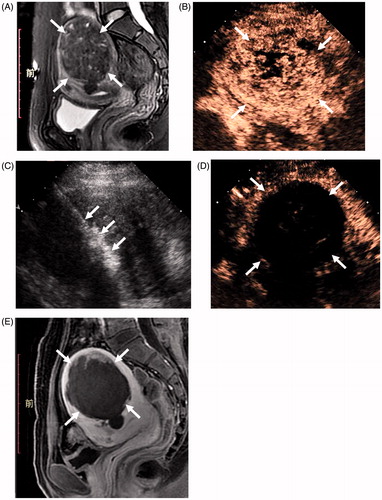
Figure 1. Examinations of a patient with fibroids before and after ablation. (A) MRI (T2WI) before ablation showed the fibroid. (B) Before ablation, the contrast-enhanced ultrasonography (CEUS) image showed non-homogeneous enhancement within the fibroid. (C) During ablation, tissue effects of ablation. (D) After ablation, no contrast enhancement was observed in the ablation area. (E) Contrast-enhanced MRI 3 days after ablation showed the ablation zone.
Effectiveness of the ablation
The mean ablation rate of the myomas was 86.6% (54.0–100%).
Clinical efficacy
The fibroid volumes, haemoglobin levels and UFS-QOL scores are shown in . The differences in these parameters between baseline and posttreatment were statistically significant (p < .05). The mean myoma reduction rate was 63.5%, 78.5% and 86.7% at 3, 6 and 12 months after treatment, respectively.
Table 1. Fibroid volumes, haemoglobin levels and uterine fibroid symptom and health-related quality of life questionnaire (UFS-QOL) scores before and after ablation.
Complications and side effects
No major complications occurred during ablation and follow-up. Twenty-seven patients (8.68%) experienced grade 1 (Common Terminology Criteria for Adverse Events (CTCAE) v4.0 of the National Cancer Institute) lower abdominal pain, which can be tolerated and which disappeared within 12 h. Nineteen patients (6.11%) had a small amount of vaginal secretion. These secretions resolved within 20 days after ablation.
Discussion
PMWA is a minimally invasive treatment that involves in situ inactivation of tumours. In response to the applied electrical field of microwaves, the rotation of dipole molecules and ionic polarisation produce friction and heat, thus inducing cellular death via coagulation necrosis [Citation15]. This technology has been widely used to treat solid tumours of the liver, spleen, thyroid and other organs [Citation16–24]. In 2011, Zhang et al. [Citation5] studied PMWA therapy for uterine fibroids and confirmed its safety and efficacy.
To avoid the subjective bias of a single-centre study, this study summarises data from multiple treatment centres and objectively evaluates the clinical efficacy of PMWA. The effectiveness of ablation was evaluated via CEUS. At present, contrast-enhanced MRI is considered the gold standard for evaluating the effect of thermal ablation. A pathological study showed that the coagulated necrotic area corresponded to the zone of non-contrast enhancement [Citation15,Citation25]. CEUS can objectively reflect perfusion of the microcirculation in tissue. Studies have shown high consistency between CEUS and contrast-enhanced MRI with regard to evaluation of the ablation effect, and CEUS could therefore be used as an alternative to contrast-enhanced MRI for evaluation of the ablation zone [Citation26,Citation27]. Moreover, CEUS is a convenient, real-time, dynamic procedure, so residual lesions can be found immediately after the treatment, after which further ablation treatment can be performed.
Uterine leiomyoma is a benign lesion, so the main purpose of treatment is to relieve clinical symptoms and improve patient quality of life. In this study, the mean ablation rate was 86.6%. In some fibroids, the ablation rate was less than 60%. This is because only partial ablation was performed in some cases depending on the location and size of the fibroid; in particular, complete ablation was not performed in the case of fibroids that were in a relatively unsafe position such as close to the bowel or bladder. Partial ablation was primarily undertaken to ease clinical symptoms and avoid serious complications.
The volume of the myomas decreased significantly after treatment. The reduction rate at 6 months after ablation was 78.5%, which is consistent with that of a previous single-centre study [Citation5]. The reduction rate at 12 months was 86.7%, which is slightly lower than the rate of 93.1% reported previously but higher than that of 74.5% reported over the same period for intramural leiomyomas [Citation11]. This is because the percentage of patients with complete sarcoid discharges in the present study was 4.50%, which is lower than the percentage of 10.0% reported in a previous study [Citation5]. Type 1/2 myomas have a higher reduction rate because necrotic tissue can be discharged through the natural passage of the uterine cavity (the vagina), which is not possible for type 5/6 and most type 3/4 myomas, as there is no route of passage outside the body.
Improvement in clinical symptoms and quality of life is an important indication of treatment efficacy. Abnormal uterine bleeding is reported to be primarily relevant to the position of the myomas, especially type 1/2 myomas that are in or partially intruding into the endometrial cavity. Pelvic pressure increases with an increase in the size of a myoma, and decreases in urinary frequency and urgency are observed following a decrease in myoma volume after therapy [Citation1,Citation2,Citation28–30]. After PMWA treatment, the fibroid volume decreased significantly and the area of the endometrium of the uterus also decreased, resulting in the relief of clinical symptoms such as menorrhagia. Thus, the haemoglobin level was significantly increased, pelvic pressure was reduced and the SSS scores were significantly decreased. Therefore, patient quality of life was improved and the HRQL scores were significantly increased. At 12 months after treatment, the SSS and HRQL scores of the patients were similar to those of healthy women (22.5 ± 21.1 and 86.4 ± 17.7, respectively) [Citation13]. This finding confirms the findings of single-centre studies, as the clinical efficacy of PMWA has been shown to be reliable.
During ablation and 12 months of follow-up, no major complications occurred. The discharge of necrotic tissue was considered to be a cure for the disease [Citation7]. Vaginal secretion could be caused by liquefaction of the necrotic tissue and by irritation and inflammation of the endometrium.
In summary, PMWA is a convenient, effective and minimally invasive therapy for FIGO type 1–6 uterine fibroids. PMWA has the added advantage of rapid recovery, and it significantly improves patient quality of life. These characteristics make PMWA therapy worthy of popularisation and application as a minimally invasive treatment method for uterine fibroids.
Disclosure statement
The authors alone are responsible for the content and writing of the paper.
References
- Khan AT, Shehmar M, Gupta JK. (2014). Uterine fibroids: current perspectives. Int J Womens Health 6:95–114.
- Stewart EA. (2001). Uterine fibroids. Lancet 357:293–8.
- Quinn SD, Gedroyc WM. (2015). Thermal ablative treatment of uterine fibroids. Int J Hyperthermia 31:272–9.
- Zhang J, Feng L, Zhang BS, et al. (2011). The study of follow up of percutaneous microwave ablation for uterine fibroids treatment. Zhonghua Yi Xue Za Zhi 91:48–50.
- Zhang J, Feng L, Zhang B, et al. (2011). Ultrasound-guided percutaneous microwave ablation for symptomatic uterine fibroid treatment – a clinical study. Int J Hyperthermia 27:510–16.
- Wang F, Zhang J, Han ZY, et al. (2012). Imaging manifestation of conventional and contrast-enhanced ultrasonography in percutaneous microwave ablation for the treatment of uterine fibroids. Eur J Radiol 81:2947–52.
- Yang Y, Zhang J, Han ZY, et al. (2014). Ultrasound-guided percutaneous microwave ablation for submucosal uterine fibroids. J Minim Invasive Gynecol 21:436–41.
- Zhao WP, Han ZY, Zhang J, Liang P. (2015). A retrospective comparison of microwave ablation and high intensity focussed ultrasound for treating symptomatic uterine fibroids. Eur J Radiol 84:413–17.
- Xia M, Jing Z, Han ZY, et al. (2014). Research of dose–effect relationship parameters of percutaneous microwave ablation for uterine leiomyomas – a quantitative study. Sci Rep 4:6469.
- Xia M, Jing Z, Zhi-Yu H, et al. (2014). Feasibility study on energy prediction of microwave ablation upon uterine adenomyosis and leiomyomas by MRI. Br J Radiol 87:20130770.
- Hao Y, Zhang J, Han Z, et al. (2014). Follow-ups of mid-term and long-term outcomes for uterine intramural myomas after percutaneous microwave ablation therapy. Zhonghua Yi Xue Za Zhi 94:664–6.
- Dong B, Liang P, Yu X, et al. (2003). Percutaneous sonographically guided microwave coagulation therapy for hepatocellular carcinoma: results in 234 patients. AJR Am J Roentgenol 180:1547–55.
- Spies JB, Coyne K, Guaou Guaou N, et al. (2002). The UFS-QOL, a new disease-specific symptom and health-related quality of life questionnaire for leiomyomata. Obstet Gynecol 99:290–300.
- Munro MG, Critchley HO, Fraser IS. (2011). The FIGO classification of causes of abnormal uterine bleeding in the reproductive years. Fertil Steril 95:2204–8.
- Liang P, Yu J, Lu MD, et al. (2013). Practice guidelines for ultrasound-guided percutaneous microwave ablation for hepatic malignancy. World J Gastroenterol 19:5430–8.
- Ren C, Liang P, Yu XL, et al. (2016). Percutaneous microwave ablation of adrenal tumours under ultrasound guidance in 33 patients with 35 tumours: a single-centre experience. Int J Hyperthermia 32:517–23.
- Yue W, Chen L, Wang S, et al. (2015). Locoregional control of recurrent papillary thyroid carcinoma by ultrasound-guided percutaneous microwave ablation: a prospective study. Int J Hyperthermia 31:403–8.
- Li M, Yu X, Liang P, et al. (2015). Ultrasound-guided percutaneous microwave ablation for hepatic malignancy adjacent to the gallbladder. Int J Hyperthermia 31:579–87.
- Liang P, Dong B, Yu X, et al. (2003). Prognostic factors for percutaneous microwave coagulation therapy of hepatic metastases. AJR Am J Roentgenol 181:1319–25.
- Duan YQ, Gao YY, Ni XX, et al. (2007). Changes in peripheral lymphocyte subsets in patients after partial microwave ablation of the spleen for secondary splenomegaly and hypersplenism: a preliminary study. Int J Hyperthermia 23:467–72.
- Wang Y, Liang P, Yu X, et al. (2009). Ultrasound-guided percutaneous microwave ablation of adrenal metastasis: preliminary results. Int J Hyperthermia 25:455–61.
- Liang P, Gao Y, Zhang H, et al. (2011). Microwave ablation in the spleen for treatment of secondary hypersplenism: a preliminary study. AJR Am J Roentgenol 196:692–6.
- Qi C, Yu XL, Liang P, et al. (2012). Ultrasound-guided microwave ablation for abdominal wall metastatic tumors: a preliminary study. World J Gastroenterol 18:3008–14.
- Feng B, Liang P, Cheng Z, et al. (2012). Ultrasound-guided percutaneous microwave ablation of benign thyroid nodules: experimental and clinical studies. Eur J Endocrinol 166:1031–7.
- Onishi H, Matsushita M, Murakami T, et al. (2004). MR appearances of radiofrequency thermal ablation region: histopathologic correlation with dog liver models and an autopsy case. Acad Radiol 11:1180–9.
- Lei F, Jing Z, Bo W, et al. (2014). Uterine myomas treated with microwave ablation: the agreement between ablation volumes obtained from contrast-enhanced sonography and enhanced MRI. Int J Hyperthermia 30:11–18.
- Zhou XD, Ren XL, Zhang J, et al. (2007). Therapeutic response assessment of high intensity focused ultrasound therapy for uterine fibroid: utility of contrast-enhanced ultrasonography. Eur J Radiol 62:289–94.
- Parker WH. (2007). Aetiology, symptomatology, and diagnosis of uterine myomas. Fertil Steril 87:725–36.
- Pron G, Bennett J, Common A, et al. (2003). The Ontario Uterine Fibroid Embolization Trial. Part 2. Uterine fibroid reduction and symptom relief after uterine artery embolization for fibroids. Fertil Steril 79:120–7.
- Yang JH, Chen MJ, Chen CD, et al. (2011). Impact of submucous myoma on the severity of anemia. Fertil Steril 95:1769–72.