
Abstract
Purpose: The purpose of this study is to retrospectively evaluate the outcomes of radiofrequency ablation (RFA) of low-risk small papillary thyroid carcinomas (PTCs) in patients who were ineligible for surgery.
Materials and methods: Between 2005 and 2009, six PTCs (mean diameter, 0.92 cm; range, 0.6–1.3 cm) in six patients were treated with RFA by three radiologists in two hospitals. The inclusion criteria for this study were (1) pathologically confirmed PTC without cytological aggressiveness, (2) single PTC without extrathyroidal extension, (3) no metastatic tumours and (4) ineligibility for surgery. RFA was performed using a radiofrequency generator and an 18-gauge internally cooled electrode. The medical records were reviewed and analysed, focussing on the procedural profiles of RFA, symptoms and complications during and after RFA, and changes in tumours on follow-up ultrasonography.
Results: Before and after RFA, the results of thyroid function tests were normal in all patients. During 48.5 ± 12.3 months (range, 36–65 months) of follow-up, along with a significant reduction in the mean volume (98.5 ± 3.3%), four ablation zones (4/6, 66.7%) completely disappeared. Two ablation zones exhibited only small calcified residues with nearly complete disappearance of the corresponding non-calcified solid portions, and in one of them, malignant cells were absent as assessed by fine-needle aspiration and core-needle biopsy. Transient hypertension with mild headache (n = 1) and mild neck pain (n = 1) developed during the procedure and subsided without any treatment.
Conclusion: Besides surgery and active surveillance, which are conflicting currently used management plans, RFA might represent an effective and a safe alternative for managing low-risk small PTCs, especially in patients ineligible for surgery.
Introduction
The standard treatment of differentiated thyroid cancer is surgery [Citation1]. However, given the increasing incidence of differentiated thyroid cancer and a common belief that most small papillary thyroid carcinomas (PTCs) are indolent [Citation2–5], the suggested approach to treating low-risk papillary thyroid microcarcinoma (PTMC) has become more conservative, amounting to thyroid lobectomy rather than total thyroidectomy [Citation1,Citation6,Citation7].
In addition, active surveillance of this type of cancer has been in the spotlight recently, according to the results of a Japanese observational study [Citation1,Citation5,Citation8]. Yet up to date, “low-risk” has been described as the absence of clinically evident metastases, local invasion and aggressive cytological evidence [Citation1].
However, a detailed procedure of active surveillance remains poorly characterised, with regard to risk factors for significant progression, frequency of ultrasonography (US) evaluation during follow-up, the role of serum thyroid stimulating hormone and thyroglobulin and socio-psychological factors for refusal of active surveillance [Citation1,Citation8]. Regarding the size criteria, although PTMCs are mainly considered for active surveillance, there has been no discussion on whether we should exclude low-risk small PTCs that were smaller than 1.5 cm but larger than 1 cm from active surveillance. Meanwhile, along with the recent medical progress and increased life expectancy, the incidence of PTCs has increased in patients who are ineligible for surgery owing to their age or medical condition, as well as in patients who refuse surgery even if surgery is possible [Citation9]. For these patients, management protocols have not been standardised, surgery seems to be somewhat excessive even if it is possible, and active surveillance may not be easily accepted by all.
In this regard, minimally invasive therapy can be a potentially useful alternative for treating these patients. A variety of interventional modalities have been successfully applied for treating patients with malignant tumours such as the lung, kidney and liver cancer [Citation10–12]. As for thyroid diseases including benign nodules, recurrent and primary cancers, successful application of interventional modalities has been consistently reported [Citation13–24]. However, with regard to RFA, reported as a more effective modality among the various interventional modalities in the treatment of benign thyroid nodules [Citation25,Citation26], there were only a few case reports of RFA for primary thyroid cancer [Citation27–29].
Therefore, in this retrospective study, we evaluated RFA outcomes of low-risk small PTCs in patients who were ineligible for surgery.
Materials and methods
The institutional review boards of two hospitals approved this retrospective study and waived informed consent. Written informed consent to treatment had been obtained from all patients before each procedure. The clinical characteristics and outcomes of RFA in the six patients with small PTCs are summarised in .
Table 1. The clinical characteristics and outcomes of RFA in the 6 patients with low risk small papillary thyroid carcinoma.
Patients
Between 2005 and 2009, when thyroid lobectomy was routinely recommended for treating low-risk small PTCs and the concept of active surveillance in low-risk PTMCs had not been widely introduced, six thyroid low-risk small PTCs (maximal dimensions of 6, 6, 9, 10, 11 and 13 mm; a mean dimension of 9.2 mm) in six consecutive patients (four women, two men; mean age, 72 years; range, 64–79 years) were RFA treated by three radiologists (J. H. K., D. G. N. and J. H. B., radiologists with 1, 3 and 4 years of experience, respectively, in the field of thyroid intervention) at two hospitals. The criteria for RFA in primary thyroid cancer were as follows: (1) PTC confirmed with US-guided fine-needle aspiration (FNA) or core-needle biopsy (CNB), (2) one cancer without extrathyroidal extension on US, (3) no metastatic tumours in the neck on US and CT examinations and beyond the neck on chest radiography and physical examination. (4) Ineligibility for surgery owing to a high risk associated with general anaesthesia and/or refusal of surgery as follows: poor pulmonary function (n = 1) and refusal to undergo surgery (n = 5) on the background of diverse diseases, including osteoporosis (n = 4), bronchial asthma (n = 3), cervical spinal stenosis (n = 2), hypertension (n = 1), bronchiectasis (n = 1), fatty liver with hyperlipidaemia (n = 1), medical history of radical hysterectomy and bilateral salpingo-oophorectomy owing to endometrial cancer (n = 1) and brain surgery owing to cerebral haemorrhage (n = 1).
RFA procedure
Since the patients were requested for RFA after the PTCs were diagnosed by US-guided FNA or CNB [Citation30,Citation31], pre-ablation and post-ablation assessment were performed, in addition to RFA, by the same radiologist among the three radiologists (J. H. K., D. G. N. and J. H. B.), respectively. A 10–14 MHz linear probe and a real-time US system (IU22 US scanner, Philips Healthcare, Andover, MA; Aplio SSA-770A, Toshiba Medical Systems, Tokyo, Japan) were used in all procedures. Before and after RFA, routine laboratory tests including thyroid function test, complete blood cell count and blood coagulation test were performed. All patients were treated on an outpatient basis. Conscious sedation was not performed because verbal communication with the patients was necessary for monitoring possible injury of important structures such as recurrent laryngeal nerve and trachea during RFA [Citation32]. For those patients who could not tolerate ablation-related pain, the power was reduced or turned off for several seconds. RFA treatments were delivered using a radiofrequency generator (Cool-Tip, Radionics, Burlington, MA) and a thyroid-dedicated straight-type internally cooled electrode (Well-Point RF Electrode, Taewoong Medical, Korea); the latter was developed and modified (a 7 cm-long 18-gauge electrode with a 0.5 cm-long active tip) for use in the thyroid gland with an internal cooling system. The patients were treated with 2% lidocaine for local anaesthesia at the puncture site in the supine position with the neck extended.
We used a trans-isthmic method and a moving-shot technique similar to those used for treating benign thyroid lesions or recurrent thyroid cancers [Citation33,Citation34]. Because the most PTCs in our study were close to the thyroid capsule and were not round in shape, prolonged fixation of the electrode for complete ablation might be dangerous to surrounding critical structures. An electrode was inserted into a lesion under US guidance, with the electrode tip initially positioned in the deepest and most remote region of the lesion. Ablation started by supplying a power of 10–15 W to a 0.5 cm-long active tip and ended when all parts of the tumour had changed to transient hyperechoic zones. If a transient hyperechoic zone did not form at the electrode tip within 5–10 s, radiofrequency power was increased in 5–10 W steps, up to 20 W. All ablation procedures were monitored with continuous real-time US for reporting symptoms and possible complications. After ablation, the patients were observed for 30–60 min in the hospital while we assessed any discomfort or complications associated with RFA. The follow-up US sessions were performed at 1, 6 and 12 months after treatment and every 1-year thereafter.
Image analysis
All medical records including US images were reviewed by one radiologist (J. Y. S., radiologist with 2 years of experience in the field of thyroid intervention) who did not perform any US or RFA on the enrolled patients. Before RFA, the locations of the PTCs were categorised as followed; left and right location, cephalocaudal location (upper, middle and lower), anteroposterior location (anterior, central and posterior) and shortest distance between PTC and thyroid capsule. After RFA, any US-detectable changes of PTCs in addition to clinical characteristics during the follow-up period of 48.5 ± 12.3 months (range, 36–65 months) were analysed. One patient (patient 3) is still under observation but the remainings are lost to follow up. For each thyroid cancer case, the pre- and post-treatment volumes were calculated as V = πabc/6 (where V is the volume, a is the largest diameter and b and c are the two other perpendicular diameters). The volume reduction was calculated from the following equation: volume reduction (%) = [(initial volume − final volume) × 100]/initial volume.
Statistical analysis
Data were analysed using statistical software (SPSS for Microsoft Windows version 21.0; SPSS Inc., Chicago, IL). A paired t-test was used for comparing the largest diameter and volume of PTCs before RFA and at the last follow-up visit. The level of significance was defined as p < 0.05.
Results
The locations of PTCs were as followed: one left and five right, cephalocaudal location (one upper, four middle and one lower), anteroposterior location (two anterior and four central). The distance between PTC and thyroid capsule was 1 ± 0.9 mm (range, 0–2 mm). Before and after RFA, the results of routine laboratory tests including thyroid function tests were normal in all patients. The energy delivered per cubic millimetre of pre-treatment thyroid cancer ranged from 13.9 to 172.0 J (mean, 57.1 J).
Five of the six PTCs were ablated in 1 session. In the remaining patient (patient 5), although most of the tumour was ablated by 1st session, 2nd session of RFA was performed without FNA or CNB to ensure complete ablation of the tumour because the tumour had an ill-defined and infiltrative margin and there was a concern of incomplete ablation of the tumour margin after 1st session of ablation. The mean diameter of the PTCs was 9.2 ± 2.8 mm (range, 6–13 mm), decreasing significantly to 1.3 ± 2.2 mm (range, 0–5 mm) of the ablation zones (p = .0006) by the end of the follow-up period. The mean volume of the PTCs was 0.260 ± 0.177 mL (range, 0.047 ± 0.381 mL), also decreasing significantly to 0.006 ± 0.013 mL (range, 0.000–0.031 mL) of the ablation zones (p = .0154) by the end of the follow-up period, with the mean volume reduction rate of 98.5 ± 3.3% (range, 91.8–100%).
After the RFA, on US, the volume of ablation zones compared with that of PTCs increased by the time of 1 month follow-up and gradually decreased by the end of the follow-up period, as follows: 215.5 ± 178.8% (range, 75–550%) at 1 month, 52.6 ± 26.5% (range, 27.2–97.2%) at 6 months, 15.3 ± 9.4% (range, 0–23.4%) at 1 year, 9.2 ± 8.7% (range, 0–17.8%) at 2 years and 4.2 ± 3.9% at 3 years (). Of the six ablation zones, 4 (4/6, 66.7%; patients 1, 2, 3 and 5) ablation zones disappeared on US at 1 year (n = 1), 2 years (n = 1) and 4 years (n = 2) after the RFA (). One ablation zone (patient 3) exhibited a residue (a volume reduction of 93.4%) but the residue demonstrated no malignant cells on FNA and CNB at 2 years follow-up. The residue (patient 3) disappeared on the 4 years follow-up (). The two ablation zones (patients 4 and 6) that showed calcified residues on the last follow-up visits also demonstrated volume reductions (99.4% and 91.8%) on US at the time of the corresponding last follow-up visits (48 and 36 months). One of these zones (patient 4) underwent FNA and CNB at the residue (83.3% volume reduction) and demonstrated no malignant cells at the 3 years follow-up after RFA (). During the follow-up periods, neither lymph node nor distant metastasis were observed. Transient hypertension with headache (n = 1) and mild neck pain (n = 1) developed and subsided without requiring any medication during procedure. All the patients were tolerable during the procedure. No patients required painkiller during or after RFA.
Figure 1. Volume changes of ablation zones after radiofrequency ablation of papillary thyroid carcinomas.
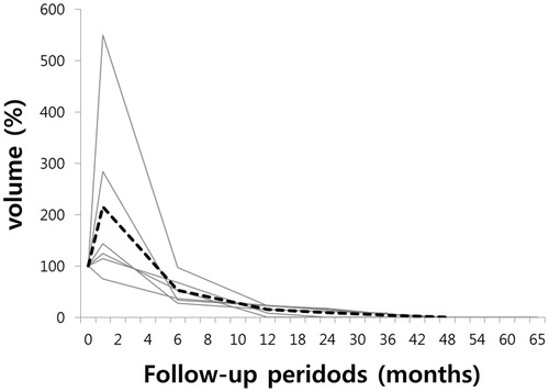
Figure 2. (A, B and C) Complete disappearance of papillary thyroid carcinoma after radiofrequency ablation. (A) Ultrasonography of a 64-year-old woman revealed a 10 mm mass proven as a papillary thyroid carcinoma on both fine-needle aspiration and core-needle biopsy in the right thyroid gland. (B) At 1-month follow-up after radiofrequency ablation, ultrasonography revealed a 12 mm ablation zone. (C) At 2-year follow-up, the ablation zone completely disappeared on ultrasonography.
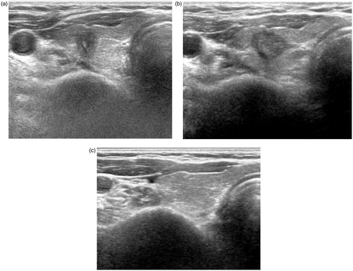
Figure 3. (A–E) Gradual reduction and complete disappearance of papillary thyroid carcinoma after radiofrequency ablation. (A) Ultrasonography of a 76-year-old woman revealed a 6-mm mass in the left thyroid gland. (B) FNA cytology of the mass revealed papillary thyroid carcinoma cells with nuclear atypia, including nuclear grooves, high N/C ratio and nuclear pseudoinclusion (Papanicolaou stain, original magnification ×400). (C) At 2-year follow-up after radiofrequency ablation, US revealed a smaller but persistent, 4-mm ill-defined ablation zone in the left thyroid gland. (D) FNA cytology of the ablation zone demonstrated benign follicular epithelial cells with hemosiderin pigment-laden macrophages (× 100). (E) At 4-year follow-up, US revealed complete disappearance of the ablation zone.
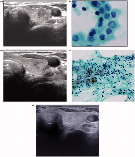
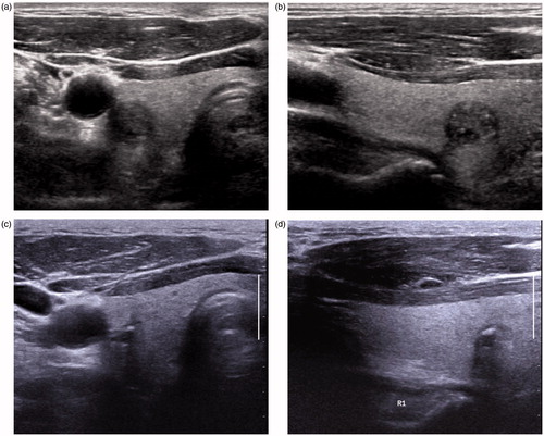
Figure 4. (A–D) Gradual reduction and resultant calcified residues of papillary thyroid carcinoma after radiofrequency ablation. (A and B) Transverse and longitudinal ultrasonography of a 79-year-old man revealed an 11-mm mass proven as a papillary thyroid carcinoma on-fine needle aspiration in the right thyroid gland. (C and D) At 4-year follow-up after radiofrequency ablation, transverse and longitudinal ultrasonography revealed a 3-mm calcified residue in the ablation zone. At 3 year, fine-needle aspiration and core-needle biopsy of the ablation zone revealed no tumour cells (results not shown).
Discussion
This study was the first case series to describe PTC management by RFA and presented the longest follow-up periods among the various interventional modalities for PTC. In this study, RFA was demonstrated to be a potentially effective and safe method for controlling low-risk small PTCs that are not size-limited to PTMC, for patients who are not eligible for surgery. All the patients in this study were tolerable to RFA during the procedure. There were neither major complications including hypothyroidism nor recurrence during follow-up.
The increased volume of ablation zone at 1 month after RFA was concordant with the results of microwave ablation study for primary PTMC, reported by Yue et al., which must be also due to the ablation of sufficient amount of normal tissue surrounding the thyroid cancer tissue for obtaining the margins free of cancer cells [Citation17]. Complete ablation including the safety margin could result in complete pathologic eradication as shown at the laser ablation study by Valcavi et al. [Citation16] and might hinder marginal recurrence after RFA as this study presented. However, incomplete ablation might cause residues of viable tumours, as shown by Kim et al. [Citation27]. In this regard, patient 5’s PTC abutting on the thyroid capsule with an ill-defined and infiltrative margin might not be a good indication for RFA. However, for this patient who was risky for surgery because of poor pulmonary function test, performing RFA was inevitable, fortunately, the ablation zone disappeared at 1 year after RFA and remained to be stable during 3-year follow up. In addition, the resultant calcific residue after RFA should be observed carefully; however, additional diagnostic procedures or repeated RFA could be waived with care, because residues did not present any malignant cells on FNA or CNB and remained stable in the present study and in other previous studies on recurrent thyroid cancers [Citation14,Citation35].
Incidental PTC is a more common problem in elderly people compared with young people and is usually associated with very indolent course, as revealed by a variety of autopsy studies [Citation36]. Many aged people may live and die from unrelated causes in the setting of de novo PTC [Citation37]. In addition, elderly people are usually associated with other comorbidities, as this study presented. For these reasons, some elderly people tended to refuse surgery even before active surveillance has become widely adopted and even if general anaesthesia was possible, as argued in the present study.
Active surveillance can be an important management alternative for these patients. However, considering that over 10% of patients stopped active surveillance and underwent surgery without progression of disease in observational trials for PTMC, active surveillance does not seem to be a perfect strategy for all patients [Citation5]. In addition, the patients enrolled for active surveillance should be followed on a regular basis for early detection of rarely occurring disease progression [Citation9]. In this regard, our study suggested that RFA could be safely and effectively applied to low-risk small PTCs, and was not confined only to PTMCs, for patients ineligible for surgery. However, considering the high frequency of the incidental thyroid cancers, cautious consideration about the necessity of the interventional management should precede RFA treatment itself for avoiding excessive overuse of this relatively easily accessible procedure. In addition, before the decision to perform RFA, careful evaluation of concomitant thyroid cancers and presence of lymph node metastases using imaging modalities such as US or CT is very important because of the tendency toward the multiplicity of thyroid cancers and frequent association with lymph node metastases [Citation38].
Apart from the intrinsic limitations of any retrospective study, several other limitations of our study and future issues should be mentioned. Because the patients underwent only chest radiography and physical examination as a routine protocol for the work-up of metastasis beyond the neck, hidden metastasis could not be excluded but the possibility was very low in the patients with low-risk small PTCs. Even when ablation zones completely disappear on US, it is difficult to determine that cancer cells are fully eliminated. Owing to the indolent nature of differentiated thyroid cancer, hasty inference should not be made concerning the effect of RFA on long-term outcomes or efficacy of treating patients with PTCs. As a detailed protocol of active surveillance remains poorly defined, the indication of RFA for primary PTCs should be studied with regard to the size and imaging criteria of PTCs, as well as the patient’s age. In the future, a study for determining such indication between active surveillance and interventional management such as RFA may be necessary. To generalise the outcomes of RFA and to evoke an optimal inclusion for RFA, a well-designed prospective study with larger number of patients and longer duration of follow-up should be warranted.
In conclusion, besides surgery and active surveillance, which are conflicting current management plans, RFA might represent an effective and safe alternative for managing low-risk small PTCs, especially in patients ineligible for surgery.
Disclosure statement
Jung Hwan Baek's financial activities related to the present article: none to disclose. Jung Hwan Baek's financial activities not related to the present article: patent holder of unidirectional ablation electrode. Other relationships of Jung Hwan Baek: none to disclose. All the other authors have nothing to disclose.
References
- Haugen BR, Alexander EK, Bible KC, et al. (2016). 2015 American Thyroid Association management guidelines for adult patients with thyroid nodules and differentiated thyroid cancer: the American Thyroid Association guidelines task force on thyroid nodules and differentiated thyroid cancer. Thyroid 26:1–133.
- Shin JH, Baek JH, Chung J, et al. (2016). Ultrasonography diagnosis and imaging-based management of thyroid nodules: revised Korean Society of thyroid radiology consensus statement and recommendations. Korean J Radiol 17:370–95.
- Russ G. (2016). Risk stratification of thyroid nodules on ultrasonography with the French TI-RADS: description and reflections. Ultrasonography (Seoul, Korea) 35:25–38.
- Chen AY, Jemal A, Ward EM. (2009). Increasing incidence of differentiated thyroid cancer in the United States, 1988–2005. Cancer 115:3801–7.
- Ito Y, Miyauchi A, Inoue H, et al. (2010). An observational trial for papillary thyroid microcarcinoma in Japanese patients. World J Surg 34:28–35.
- Cooper DS, Doherty GM, Haugen BR, et al. (2006). Management guidelines for patients with thyroid nodules and differentiated thyroid cancer: the American Thyroid Association guidelines taskforce. Thyroid 16:109–42.
- Cooper DS, Doherty GM, Haugen BR, et al. (2009). Revised American Thyroid Association management guidelines for patients with thyroid nodules and differentiated thyroid cancer. Thyroid 19:1167–214.
- Brito JP, Ito Y, Miyauchi A, Tuttle RM. (2016). A clinical framework to facilitate risk stratification when considering an active surveillance alternative to immediate biopsy and surgery in papillary microcarcinoma. Thyroid 26:144–9.
- Papini E, Guglielmi R, Gharib H, et al. (2011). Ultrasound-guided laser ablation of incidental papillary thyroid microcarcinoma: a potential therapeutic approach in patients at surgical risk. Thyroid 21:917–20.
- Dodd GD III, Soulen MC, Kane RA, et al. (2000). Minimally invasive treatment of malignant hepatic tumours: at the threshold of a major breakthrough. Radiographics: Rev Publ Radiol Soc N Am 20:9–27.
- Pandharipande PV, Gervais DA, Mueller PR, et al. (2008). Radiofrequency ablation versus nephron-sparing surgery for small unilateral renal cell carcinoma: cost-effectiveness analysis. Radiology 248:169–78.
- Dupuy DE. (2011). Image-guided thermal ablation of lung malignancies. Radiology 260:633–55.
- Gharib H, Hegedus L, Pacella CM, et al. (2013). Clinical review: nonsurgical, image-guided, minimally invasive therapy for thyroid nodules. J Clin Endocrinol Metab 98:3949–57.
- Kim JH, Yoo WS, Park YJ, et al. (2015). Efficacy and safety of radiofrequency ablation for treatment of locally recurrent thyroid cancers smaller than 2 cm. Radiology 276:909–18.
- Cakir B, Topaloglu O, Gul K, et al. (2007). Ultrasound-guided percutaneous laser ablation treatment in inoperable aggressive course anaplastic thyroid carcinoma: the introduction of a novel alternative palliative therapy – second experience in the literature. J Endocrinol Investig 7:624–5.
- Valcavi R, Piana S, Bortolan GS, et al. (2013). Ultrasound-guided percutaneous laser ablation of papillary thyroid microcarcinoma: a feasibility study on three cases with pathological and immunohistochemical evaluation. Thyroid 23:1578–82.
- Yue W, Wang S, Yu S, Wang B. (2014). Ultrasound-guided percutaneous microwave ablation of solitary T1N0M0 papillary thyroid microcarcinoma: initial experience. Int J Hyperthermia: Off J Eur Soc Hyperthermic Oncol N Am Hyperthermia Group 30:150–7.
- Yue W, Chen L, Wang S, Yu S. (2015). Locoregional control of recurrent papillary thyroid carcinoma by ultrasound-guided percutaneous microwave ablation: a prospective study. Int J Hyperthermia: Off J Eur Soc Hyperthermic Oncol, N Am Hyperthermia Group 31:403–8.
- Lee SJ, Jung SL, Kim BS, et al. (2014). Radiofrequency ablation to treat loco-regional recurrence of well-differentiated thyroid carcinoma. Korean J Radiol 15:817–26.
- Negro R, Salem TM, Greco G. (2016). Laser ablation is more effective for spongiform than solid thyroid nodules. A four-years retrospective follow up study. Int J Hyperthermia 32:822–8.
- Kohlhase KD, Korkusuz Y, Gröner D, et al. (2016). Bipolar radiofrequency ablation of benign thyroid nodules using a multiple overlapping shot technique in a 3-month follow-up. Int J Hyperthermia 32:511–6.
- Heck K, Happel C, Grünwald F, Korkusuz H. (2015). Percutaneous microwave ablation of thyroid nodules: effects on thyroid function and antibodies. Int J Hyperthermia 31:560–7.
- Pacella CM, Mauri G, Achille G, et al. (2015). Outcomes and risk factors for complications of laser ablation for thyroid nodules: a multicenter study on 1531 patients. J Clin Endocrinol Metab 100:3903–10.
- Mauri G, Cova L, Ierace T, et al. (2016). Treatment of metastatic lymph nodes in the neck from papillary thyroid carcinoma with percutaneous laser ablation. Cardiovasc Intervent Radiol 39:1023–30.
- Ha EJ, Baek JH, Kim KW, et al. (2015). Comparative efficacy of radiofrequency and laser ablation for the treatment of benign thyroid nodules: systematic review including traditional pooling and bayesian network meta-analysis. J Clin Endocrinol Metab 100:1903–11.
- Ahn HS, Kim SJ, Park SH, Seo M. (2016). Radiofrequency ablation of benign thyroid nodules: evaluation of the treatment efficacy using ultrasonography. Ultrasonography (Seoul, Korea) 35:244–52.
- Kim HY, Ryu WS, Woo SU, et al. (2010). Primary papillary thyroid carcinoma previously treated incompletely with radiofrequency ablation. J Cancer Res Ther 6:310–12.
- Jeon EJ, Shon HS, Dal Jung E. (2015). Radiofrequency ablation for the papillary thyroid micro-carcinoma in the high risk surgical patient. J Thyroid Disorders Therapy 4:1–3.
- Sun J, Liu X, Zhang Q, et al. (2016). Papillary thyroid carcinoma treated with radiofrequency ablation in a patient with hypertrophic cardiomyopathy: a case report. Korean J Radiol 17:558–61.
- Lee YH, Baek JH, Jung SL, et al. (2015). Ultrasound-guided fine needle aspiration of thyroid nodules: a consensus statement by the Korean Society of thyroid radiology. Korean J Radiol 16:391–401.
- Na DG, Kim JH, Sung JY, et al. (2012). Core-needle biopsy is more useful than repeat fine-needle aspiration in thyroid nodules read as nondiagnostic or atypia of undetermined significance by the Bethesda system for reporting thyroid cytopathology. Thyroid 22:468–75.
- Ha EJ, Baek JH, Lee JH. (2015). Ultrasonography-based thyroidal and perithyroidal anatomy and its clinical significance. Korean J Radiol 16:749–66.
- Ha EJ, Baek JH, Lee JH. (2014). Moving-shot versus fixed electrode techniques for radiofrequency ablation: comparison in an ex-vivo bovine liver tissue model. Korean J Radiol 15:836–43.
- Na DG, Lee JH, Jung SL, et al. (2012). Radiofrequency ablation of benign thyroid nodules and recurrent thyroid cancers: consensus statement and recommendations. Korean J Radiol 13:117–25.
- Lim HK, Baek JH, Lee JH, et al. (2015). Efficacy and safety of radiofrequency ablation for treating locoregional recurrence from papillary thyroid cancer. Eur Radiol 25:163–70.
- Ito Y, Uruno T, Nakano K, et al. (2003). An observation trial without surgical treatment in patients with papillary microcarcinoma of the thyroid. Thyroid 13:381–7.
- Udelsman R. (2010). Treatment of persistent or recurrent papillary carcinoma of the thyroid-the good, the bad, and the unknown. J Clin Endocrinol Metab 95:2061–3.
- Yeh MW, Bauer AJ, Bernet VA, et al. (2015). American thyroid association statement on preoperative imaging for thyroid cancer surgery. Thyroid 25:3–14.