
Abstract
Purpose: To determine the factors affecting the recurrence of the solitary benign thyroid nodules (BTN) after microwave ablation (MWA).
Materials and methods: Between January 2013 and January 2015, a total of 110 patients with at least one solid thyroid nodule (solid component ≥ 80%) were enrolled. MWA was performed under continuous ultrasound (US) guidance. Before and during the follow-up, the thyroid nodule volume, thyroid function and cosmetic complaints were evaluated. Recurrence is defined by the new blood flow in the total ablation area or/and > 50% increase in nodule volume.
Results: Almost all thyroid nodules were significantly decreased in size after MWA. After 12 months, the average volume of thyroid nodules was decreased from 12.6 ± 15.1 to 3.2 ± 5.7 ml. Of the total 110 patients, 16 cases had recurrence 12 months after MWA, and these patients had a larger initial volume than that of the non-recurrence patients (11.6 ± 14.9 vs. 23.9 ± 12.5, p < 0.01). The recurrence group also demonstrated more irregular blood vessels (1.8 8 ± 1.1 vs. 2.8 5 ± 1.3, p < 0.05), and a lower energy (1575.5 ± 674.3 J/ml vs. 1172.3 ± 454.2 J/ml, p < 0.01). In addition, 81.2% (13/16) of the patients in the recurrence group were adjacent to the vital structures, which is significantly higher than that of the non-recurrence group 28.7% (27/94) (p < 0.01).
Conclusions: The US-guided MWA results in a satisfactory long-term outcome of the patients with a benign solitary thyroid nodule. We identified three risk factors for recurrence: initial volume, vascularity and the energy per 1 ml reduction in nodular volume.
Introduction
Thyroid nodules were detected by palpation in 4–8% of the adults and by ultrasound (US) in 10–70% of the adults [Citation1–3]. Most of the thyroid nodules are benign, and the incidence of thyroid cancer is reported to be 2.2–12.4/100 000 [Citation4]. Benign thyroid nodules (BTN) may cause nodule-related cosmic problems and symptoms. Previously, subtotal thyroidectomy was the primary treatment for large goitre, and most patients who underwent the surgery had a permanent scar in the neck. In addition, these patients need to take levothyroxine after surgery to prevent hypothyroidism, and might develop adverse effects such as atrial fibrillation and reduction of bone density [Citation5]. Recently, thermal ablation provides an option to avoid hypothyroidism in patients reluctant to surgery. These treatments, including laser ablation (LA), radiofrequency ablation (RFA) and high intensity focussed ultrasound (HIFU), have been proved clinically effective [Citation6–9].
Microwave ablation (MWA) is another minimally invasive technique used for thermal tissue ablation. Although it was used to treat both malignant and benign tumour nodules [Citation10–15], this technique has been proved to be effective and safe for reducing the volume in BTN with local pressure symptoms [Citation16–20]. Thus, it may represent a promising new approach for the management of BTN [Citation21–23]. However, a few studies reported that the volume of some nodules did not change significantly after MWA [Citation24,Citation25]. In this study, we investigated the effectiveness of the treatment of the microwave on the BTN. In addition, we analysed the causes of post-MWA recurrence in patients with nodular goitre ().
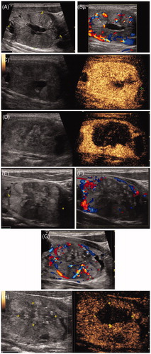
Figure 1. MWA treatment of a 35-year old man with a left thyroid solid nodule. (A) Two-dimensional ultrasonic image of the thyroid nodule. (B) The colour Doppler image of the thyroid nodule. (C) The contrast enhanced ultrasound image of the thyroid nodule before treatment. (D) The contrast enhanced ultrasound image of the nodule on day 3 after the ablation. (E) Two-dimensional ultrasonic image of the nodule three month after the ablation. (F) The colour Doppler image of the nodule three month after the ablation. Vascularity was shown around the nodule. (G) The colour Doppler image of the nodule six month after the ablation. Vascularity was shown inside the nodule in ablation area. (H) The contrast enhanced ultrasound image of the nodule six month after the ablation. Reperfusion was shown in the ablation area of the nodule.
Materials and methods
Patients
This observational study was retrospective and was approved by the Ethics Committee of the PLA General Hospital. A written informed consent was obtained from each patient before the procedure. From January 2013 to January 2015, a total of 110 consecutive patients with BTNs were treated by MWA at PLA General Hospital in China.
The inclusion criteria were: 1) solid nodule or mixes nodules with a solid component >80% of total volume; 2) reported symptomatic and/or cosmetic problems; 3) refusal of or ineligible for surgery; 4) normal serum thyroid hormones, thyrotropin and calcitonin levels; 5) benign pathological results from two separate US-guided fine-needle aspiration (FNA); 6) no suspicious malignant feature observed from US imaging; 7) anxiety about a malignant transformation. The exclusion criteria were: 1) malignancy nodules on US imaging; 2) cytological evidence for malignancy [Citation24,Citation26].
Pre-ablative assessment
MWA equipment: The microwave instrument (KU-2000 Kangyou Medical, Nanjing, China) including a microwave generator, a flexible low-loss coaxial cable and a cooled shaft antenna were used to deliver microwave energy. The generator is capable of producing 1 to 100 W of power at 2450 MHz. The internally cooled needle microwave antenna is 16-gauge, and coated with polytetrafluoroethylene to prevent adhesion. It is 1.6 mm in diameter and 3 mm in length. In order to prevent overheating of the shaft and to avoid skin injury, chilled distilled water was circled by a peristaltic pump through dual channels inside the shaft.
Before ablation, all patients were evaluated by 2D-US examination, colour Doppler US, US contrast, laboratory tests, FNA cytology and clinical examination. Three orthogonal diameters of the nodules and the proportion of solid component were measured by US before MWA. Nodule internal vascularity was classified using a 1–4 point scale [Citation8,Citation27]. Each nodule’s volume was calculated as V=πabc/6. All the patients were received a laryngoscopy for assessing vocal cord function before ablation. The laboratory tests included the measurements of TSH, FT3, FT4, antithyroperoxidase antibodies (TPOAb), calcitonin concentration, a blood routine examination, the activated partial thromboplastin time and the prothrombin time.
Before treatment, the physicians recorded the cosmetic score: 1) no palpable mass; 2) no cosmetic problem but a palpable mass; 3) cosmetic problem only on swallowing and 4) readily detected cosmetic problem [Citation27–30]. The patients with anticoagulant treatment stopped taking the treatment for 7–10 days.
Procedure
Each patient was placed in the supine position with the neck mild extension. A venous catheter was inserted into a forearm vein before ablation. Parameters such as PO2, continuous electrocardiograms, breath rate and blood pressure were monitored by a multi-parametric monitor connected to the patient.
When the best puncture site was determined by the US, local anaesthesia with 2% lidocaine was performed subcutaneously at the location. Then the internal cooled microwave antenna (16-gauge) was inserted from the isthmus and placed in the targeted thyroid nodule along its longest axis with the guidance of US.
For the microwave procedure, a power output of 20–30 W was used during the MWA. Under US guidance, we performed MWA and the moving shot technique which has been proved effective in ablation of thyroid nodules [Citation8]. We divided the thyroid nodules into small ablation elements in imagination, and performed MWA one-by-one by moving the electrode following the reported procedure [8].
By US monitoring, the tip of antenna was always depicted as a hyperechoic spot, and the echo variations were monitored within the thyroid nodule using the real-time US. The MWA was terminated when the targeted nodule was covered by hyperechoic zones. For patients with bilateral BTN, MWA was first performed for the larger nodule and then for the contralateral one on a later day.
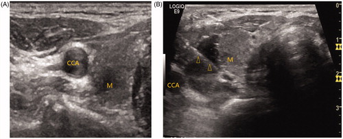
Because many important nerves and blood vessels are located in the neck, the injuries of neurovascular structures may lead to complications such as recurrent laryngeal nerve (RLN) injury and haematoma formation. If the nodule is adjacent to important tissues such as the carotid artery (CCA), RLN, trachea and/or oesophagus, hydrodissection technique was used to protect these important tissues. We injected normal saline between the nodule and the important structures, therefore the hydrodissection technique protected the neck important nerves and blood vessels during the biopsy and ablation ().
Figure 2. Hydrodissection technique. (A) On transverse image, the nodule is located adjacent to the common carotid artery. (B) 0.9 % normal saline is injected between the common carotid artery and the nodule to prevent needle-induced thermal injury. M = mass, CCA = common carotid artery. Arrowheads = microwave antenna.
The power was reduced or stopped if the patient felt the intolerable pain. After the ablation, enhanced-contrast US was performed immediately to investigate the dividing line of the necrosis, thus to evaluate whether the nodules had been completely removed. After the MWA, the ice-block was used to decrease the pain and to prevent bleeding.
Follow-up
After 1, 3, 6 and 12 months, a follow-up examination of nodule volume, vascularity, echogenicity, cosmetic score and laboratory tests was carried out as the same protocol before the MWA. The volume reduction was calculated as: volume reduction ration (VRR)%= ([initial volume – final volume] × 100)/initial volume. The treatment was considered as effective upon the VRR >50% at the last follow-up.
The recurrence was defined by identification of new blood flow in the nodules in the total ablation area or/and >50% increase in nodule volume compared to the previous US examination [Citation24,Citation25,Citation31].
Statistics
Statistical analyses were performed with SPSS statistical software programme version 19.0 (SPSS Inc., Chicago, IL). Continuous data are given as mean ± SD. ANOVA was used to compare the data from different groups. T-test was used to compare the data between the recurrence group and non-recurrence group. A p value of less than 0.05 was considered as significant difference.
Results
The study included 110 patients (12 males and 98 females). All the patients were followed-up for at least one year. The baseline characteristics of the thyroid nodules, the thyroid functions and antibodies, and the cosmetic scores are summarised in .
Table 1. The changes in volume before microwave ablation and at each follow-up.
Before MWA, the mean volume of the nodule was 12.6 ± 15.1 ml. The nodule volume was decreased to 6.5 ± 9.4 ml (p < 0.01) after six months and to 3.2 ± 5.7 ml (p < 0.01) at the final examination (). The VRRs were also calculated, and the reduction of volumes after the MWA was 48.4 ± 15.1% at six months and 74.6 ± 10.9% at one year (p < 0.05, ). In addition, the cosmetic score was significantly decreased at the last follow-up than the initial evaluation (2.8 ± 1.3 vs. 1.5 ± 1.1, p < 0.01, ). The overall energy delivered per nodule was 16197.5 ± 4226.5 J, and the mean energy delivered per millilitre of initial nodule volume was 1434.5 ± 419.2 J/ml.
Prior to the ablation, the thyroid functions of all the patients were evaluated, and none of them showed abnormal thyroid function. The laboratory parameters did not change significantly at 1-month, 3-month, 6-month and 1-year follow-up (). T3, T4, TSH and calcitonin levels remained normal for all patients. During the follow-up, no patient developed hyperthyroidism or hypothyroidism. In addition, no significant difference was found for TPOAb and thyroid stimulating hormone receptor antibody (TRAb) levels between values of pre-treatment and each follow-up ().
Of the 110 patients, 16 had recurrence 12 months after MWA and two of them had another MWA treatment due to the unsatisfactory results. Of the recurrent patients, three took surgery, and the other 11 stayed in hospital for observation. The mean number of treatment was 1.0 2 ± 0.13, which is smaller than that of the other reports due to the short follow-up time (one year vs. four years) [Citation31]. The initial mean nodule volume in the 16 patients who had recurrence was 23.9 ± 12.5 ml, which is significantly larger than the other 94 patients whose initial median nodule volume was 11.6 ± 14.9 ml (p < 0.01, ). The difference of the nodule’s vascularity between the recurrence patients (2.85 ± 1.3) and the non-recurrence patients (1.88 ± 1.1) was statistically significant (p < 0.05, ). In addition, we compared the energy distribution between the two groups. The difference of the median energy deposited per millilitre nodule volume between these recurrent patients (1172.3 ± 454.2 J/ml) and the non-recurrence patients (1575.5 ± 674.3 J/ml) was statistically significant (p < 0.01, ). In the recurrence group, 81.2% (13/16) of the nodules were adjacent to the important tissues, while in the non-recurrence group 28.7% (27/94) of the nodules were adjacent to the important tissues. (p < 0.01, )., There were no deaths during the treatment and follow-up. The patients were observed for 3–5 days and discharged. The most common side effect reported was unbearable pain in 25% of patients during the MWA, and some of them lasted for up to two days after the ablation. One patient showed voice changes after the MWA procedure, and was recovered after two months. There were no additional major complications after MWA, such as infection, Horner’s syndrome, skin burns, or permanent hyperthyroidism or hypothyroidism. During the ablation, 40 patients were applied with the hydrodissection technique. None of them displayed serious complications such as hoarseness, RLN injury, haematoma formation and oesophageal injury.
Table 2. Comparison of effective therapy group versus recurrence group.
Discussion
The incidence of nodular goitre has been gradually increasing in recent years [Citation32]. Although most thyroid nodules are benign and asymptomatic, some of them need therapy due to compressive symptoms and/or cosmic reasons. The traditional treatments for BTN include thyroidectomy and levothyroxine medication, both of which demonstrate significant side effects.
US-guided ethanol ablation (EA) has been reported as an effective and safe alternative to levothyroxine therapy or surgery for symptomatic BTN [Citation33,Citation34]. However, when using EA for solid thyroid nodules, the ethanol might be unevenly distributed inside the solid nodule. It is possible that a portion of the ethanol leaks out from the thyroid nodules [Citation6,Citation35,Citation36]. When EA is used in predominantly cystic thyroid nodules, the amount of the solid component might affect the outcome of the treatment [Citation35], Therefore, EA is recommend as the first-line treatment for cystic thyroid nodules and predominantly cystic thyroid nodules, but not for solid thyroid nodules [Citation1,Citation36,Citation37]. In recently years, US-guided thermal therapy (RFA, LA, HIFU) has been widely used. Comparing with EA, thermal ablation significantly reduces the volume of the solid nodules [Citation36]. MWA is another method of thermal ablation to induce benign thyroid nodular tissue necrosis [Citation17,Citation20,Citation21,Citation38,Citation39]. Compared to other thermal ablation such as RFA, LA and HIFU, MWA is less likely to be influenced by heat sink effects [Citation38]. It has also been used to treat recurrent papillary thyroid carcinoma and the early papillary thyroid microcarcinoma [Citation39,Citation40].
The results of our study demonstrated that MWA is effective in treating BTN as it induced a significant reduction of volume of nodules with minimal effects on thyroid function in all patients. This is consistent with the results from other studies [Citation16,Citation21,Citation41].
To date, the rationale for the recurrence of the nodules has not been understood. Our study suggests that the recurrence might be related with initial volume, energy reduction in nodular volume and vascularity. In addition, our study suggests that pre-treatment nodule volume was significantly correlated with the degree of nodule volume reduction after MWA which is in accordance with other reports [Citation42]. Dossing et al. reported that the small nodules has a larger VRR than that of the big ones, suggesting that volume is a factor affecting the effectiveness of the therapy [Citation25]. In our follow-up, we found that the mean volume of the recurrence group is significantly bigger than that of the non-recurrence group. If the nodule has a large volume, it might be difficult for MWA to cover all the tissue in three dimensional spaces, leading to incomplete ablation and recurrence. On the other hand, during MWA an irregular hyperechoic area enlarging over time is seen. In addition, we observed an irregular hyperechoic area enlarging over time during MWA. This serves as a rough measure of the area of necrosis during the ablation procedure. For nodules with a large volume, the hyperechoic area may affect our judgment on the margins of the thermally induced damage, which may also lead to an incomplete ablation [Citation25].
The correlation between area of nodular necrosis and the amount of energy delivered remains controversial [Citation42–45]. Our study showed an association between energy delivered per ml and the VRR. The energy of recurrence group is much less than that of the non-recurrence group. Thus, we speculate that the delivered energy/ml of the treated tissue is responsible for long-term shrinkage [Citation46].
Vascularity is another important characteristic which may affect the effectiveness of the therapy [Citation43,Citation47]. In RFA and LA therapy, the increased blood flow results in an increase of disposal of thermal energy [Citation25], which is also seen during MWA. In our study, the distributions of blood flow signals in and around the two groups were significantly different on colour Doppler flow imaging (CDFI). The recurrence group demonstrated a much more plentiful bloodstream than that of the non-recurrence group. We observed that the recurrence often occurs around nodules with an obvious blood flow signal, which is in accordance with the results reported by Lim et al. [Citation31].
Another cause of recurrence is inadequate ablation of tissues adjacent to vital structures. We observed a higher percentage of BTNs adjacent to vital structures such as laryngeal recurrent nerve, CCA and upper airway in the recurrence group. For fear of ablation may damage the important structures, the out power and the time will be reduced. Thus, surrounding vital structures prevented total ablation of certain areas of the nodules. This may result in incomplete treatment of the nodules and subsequent recurrence [Citation25,Citation48].
Initially, hydrodissection technique is used for the treatment of nerve entrapment syndrome to improve safety, reliability and effectiveness [Citation49]. Nowadays this technique has been widely employed in MWA as it can protect adjacent vulnerable structures, organs and tissue from damage, and reduce the risk of local recurrence. Currently normal saline and D5W are most widely used [Citation50,Citation51], and several new materials are still under development [Citation52]. Many important nerves, blood vessels and organs are located in the neck such as RLN, CCA, oesophagus [Citation53], which might be affected by procedure-related complications of small neurovascular structures injuries. For example, injuries of the vital structures around the thyroid gland such as cervical sympathetic ganglion have been reported recently. US monitoring of such important structures during US-guided MWA may reduce these procedure-related complications [Citation54]. In addition, it may increase the efficacy of MWA on thyroid nodules adjacent with vulnerable structures.
Our study has several limitations. First, our study is retrospective. The next step of this research is to perform a prospective random double-blinded study to obtain persuasive data. Second, our study does not include symptomatic evaluation. Third, our study lacks of inter-observer evaluation of nodule size measurement [Citation55].
Conclusions
In conclusion, our results suggest that MWA is a minimally invasive procedure for local thyroid nodule ablation with minimal damage to the normal tissue adjacent to nodular. It represents a valid alternative to surgery. We identified three risk factors for recurrence: initial volume, vascularity and the energy per 1 ml reduction in nodular volume. Adjacent to the important tissues might also be associated with an increased risk of recurrence.
Disclosure statement
The authors report no conflicts of interest. The authors alone are responsible for the content and writing of the article.
Reference
- Gharib H, Hegedus L, Pacella CM, et al. (2013). Clinical review: nonsurgical, image-guided, minimally invasive therapy for thyroid nodules. J Clin Endocrinol Metab 98:3949–57.
- Papini E, Pacella CM, Misischi I, et al. (2014). The advent of ultrasound-guided ablation techniques in nodular thyroid disease: towards a patient-tailored approach. Best Pract Res Clin Endocrinol Metab 28:601–18.
- Gharib H, Papini E. (2007). Thyroid nodules: clinical importance, assessment, and treatment. Endocrinol Metab Clin North Am 36:707–35, vi.
- Che Y, Jin S, Shi C, et al. (2015). Treatment of benign thyroid nodules: comparison of surgery with radiofrequency ablation. AJNR Am J Neuroradiol 36:1321–5.
- Schneider R, Schneider M, Reiners C, Schneider P. (2012). Effects of levothyroxine on bone mineral density, muscle force, and bone turnover markers: a cohort study. J Clin Endocrinol Metab 97:3926–34.
- Spiezia S, Vitale G, Di Somma C, et al. (2003). Ultrasound-guided laser thermal ablation in the treatment of autonomous hyperfunctioning thyroid nodules and compressive nontoxic nodular goiter. Thyroid 13:941–7.
- Valcavi R, Frasoldati A. (2004). Ultrasound-guided percutaneous ethanol injection therapy in thyroid cystic nodules. Endocr Pract 10:269–75.
- Ji Hong M, Baek JH, Choi YJ, et al. (2015). Radiofrequency ablation is a thyroid function-preserving treatment for patients with bilateral benign thyroid nodules. J Vasc Interv Radiol 26:55–61.
- Korkusuz H, Sennert M, Fehre N, et al. (2014). Local thyroid tissue ablation by high-intensity focused ultrasound: effects on thyroid function and first human feasibility study with hot and cold thyroid nodules. Int J Hyperthermia 30:480–5.
- Wang G, Sun Y, Cong L, et al. (2015). Artificial pleural effusion in percutaneous microwave ablation of hepatic tumors near the diaphragm under the guidance of ultrasound. Int J Clin Exp Med 8:16765–71.
- Yu J, Zhang G, Liang P, et al. (2015). Midterm results of percutaneous microwave ablation under ultrasound guidance versus retroperitoneal laparoscopic radial nephrectomy for small renal cell carcinoma. Abdom Imaging 40:3248–56.
- Sun YH, Song PY, Guo Y, Sheng LJ. (2015). Computed tomography-guided percutaneous microwave ablation therapy for lung cancer. Genet Mol Res 14:4858–64.
- Zhou W, Jiang Y, Chen L, et al. (2014). Image and pathological changes after microwave ablation of breast cancer: a pilot study. Eur J Radiol 83:1771–7.
- Yang Y, Zhang J, Han ZY, et al. (2015). Ultrasound-Guided Percutaneous Microwave Ablation for Adenomyosis: Efficacy of Treatment and Effect on Ovarian Function. Sci Rep 5:10034.
- Brace CL. (2009). Radiofrequency and microwave ablation of the liver, lung, kidney, and bone: what are the differences? Curr Probl Diagn Radiol 38:135–43.
- Yue W, Wang S, Wang B, et al. (2013). Ultrasound guided percutaneous microwave ablation of benign thyroid nodules: safety and imaging follow-up in 222 patients. Eur J Radiol 82:e11–6.
- Korkusuz H, Nimsdorf F, Happel C, et al. (2015). Percutaneous microwave ablation of benign thyroid nodules. Functional imaging in comparison to nodular volume reduction at a 3-month follow-up. Nuklearmedizin 54:13–9.
- Korkusuz H, Happel C, Heck K, et al. (2014). Percutaneous thermal microwave ablation of thyroid nodules. Preparation, feasibility, efficiency. Nuklearmedizin 53:123–30.
- Klebe J, Happel C, Grunwald F, Korkusuz H. (2015). Visualization of tissue alterations in thyroid nodules after microwave ablation: sonographic versus scintigraphic imaging. Nucl Med Commun 36:260–7.
- Feng B, Liang P, Cheng Z, et al. (2012). Ultrasound-guided percutaneous microwave ablation of benign thyroid nodules: experimental and clinical studies. Eur J Endocrinol 166:1031–7.
- Heck K, Happel C, Grunwald F, Korkusuz H. (2015). Percutaneous microwave ablation of thyroid nodules: effects on thyroid function and antibodies. Int J Hyperthermia 31:560–7.
- Sung JY, Baek JH, Jung SL, et al. (2015). Radiofrequency ablation for autonomously functioning thyroid nodules: a multicenter study. Thyroid 25:112–7.
- Baek JH, Moon WJ, Kim YS, et al. (2009). Radiofrequency ablation for the treatment of autonomously functioning thyroid nodules. World J Surg 33:1971–7.
- Valcavi R, Riganti F, Bertani A, et al. (2010). Percutaneous laser ablation of cold benign thyroid nodules: a 3-year follow-up study in 122 patients. Thyroid 20:1253–61.
- Dossing H, Bennedbaek FN, Hegedus L. (2011) Long-term outcome following interstitial laser photocoagulation of benign cold thyroid nodules. Eur J Endocrinol 165:123–8.
- Kim EK, Park CS, Chung WY, et al. (2002). New sonographic criteria for recommending fine-needle aspiration biopsy of nonpalpable solid nodules of the thyroid. AJR Am J Roentgenol 178:687–91.
- Baek JH, Kim YS, Lee D, et al. (2010). Benign predominantly solid thyroid nodules: prospective study of efficacy of sonographically guided radiofrequency ablation versus control condition. AJR Am J Roentgenol 194:1137–42.
- Ha EJ, Baek JH, Lee JH. (2011) The efficacy and complications of radiofrequency ablation of thyroid nodules. Curr Opin Endocrinol Diabetes Obes 18:310–4.
- Na DG, Lee JH, Jung SL, et al. (2012). Radiofrequency ablation of benign thyroid nodules and recurrent thyroid cancers: consensus statement and recommendations. Korean J Radiol 13:117–25.
- Baek JH, Lee JH, Valcavi R, et al. (2011). Thermal ablation for benign thyroid nodules: radiofrequency and laser. Korean J Radiol 12:525–40.
- Lim HK, Lee JH, Ha EJ, et al. (2013) . Radiofrequency ablation of benign non-functioning thyroid nodules: 4-year follow-up results for 111 patients. Eur Radiol 23:1044–9.
- Kovatcheva RD, Vlahov JD, Stoinov JI, Zaletel K. (2015). Benign solid thyroid nodules: US-guided high-intensity focused ultrasound ablation-initial clinical outcomes. Radiology 276:597–605.
- Bennedbaek FN, Nielsen LK, Hegedus L. (1998). Effect of percutaneous ethanol injection therapy versus suppressive doses of L-thyroxine on benign solitary solid cold thyroid nodules: a randomized trial. J Clin Endocrinol Metab 83:830–5.
- Goletti O, Monzani F, Lenziardi M, et al. (1994). Cold thyroid nodules: a new application of percutaneous ethanol injection treatment. J Clin Ultrasound 22:175–8.
- Kim YJ, Baek JH, Ha EJ, et al. (2012). Cystic versus predominantly cystic thyroid nodules: efficacy of ethanol ablation and analysis of related factors. Eur Radiol 22:1573–8.
- Ha EJ, Baek JH. (2014) Advances in nonsurgical treatment of benign thyroid nodules. Future Oncol 10:1399–405.
- Sung JY, Kim YS, Choi H, et al. (2011). Optimum first-line treatment technique for benign cystic thyroid nodules: ethanol ablation or radiofrequency ablation? AJR Am J Roentgenol 196:W210–4.
- Fan W, Li X, Zhang L, et al. (2012). Comparison of microwave ablation and multipolar radiofrequency ablation in vivo using two internally cooled probes. AJR Am J Roentgenol 198:W46–50.
- Yue W, Wang S, Yu S, Wang B. (2014). Ultrasound-guided percutaneous microwave ablation of solitary T1n0m0 papillary thyroid microcarcinoma: initial experience. Int J Hyperthermia 30:150–7.
- Yue W, Chen L, Wang S, Yu S. (2015). Locoregional control of recurrent papillary thyroid carcinoma by ultrasound-guided percutaneous microwave ablation: a prospective study. Int J Hyperthermia 31:403–8.
- Korkusuz H, Happel C, Klebe J, et al. (2015). Diagnostic accuracy of elastography and scintigraphic imaging after thermal microwave ablation of thyroid nodules. Rofo 187:353–9.
- Amabile G, Rotondi M, De Chiara G, et al. (2006). Low-energy interstitial laser photocoagulation for treatment of nonfunctioning thyroid nodules: therapeutic outcome in relation to pretreatment and treatment parameters. Thyroid 16:749–55.
- Dossing H, Bennedbaek FN, Karstrup S, Hegedus L. (2002). Benign solitary solid cold thyroid nodules: US-guided interstitial laser photocoagulation-initial experience. Radiology 225:53–7.
- Papini E, Guglielmi R, Bizzarri G, et al. (2007). Treatment of benign cold thyroid nodules: a randomized clinical trial of percutaneous laser ablation versus levothyroxine therapy or follow-up. Thyroid 17:229–35.
- Gambelunghe G, Fatone C, Ranchelli A, et al. (2006). A randomized controlled trial to evaluate the efficacy of ultrasound-guided laser photocoagulation for treatment of benign thyroid nodules. J Endocrinol Invest 29:RC23–6.
- Deandrea M, Sung JY, Limone P, et al. (2015). Efficacy and safety of radiofrequency ablation versus observation for nonfunctioning benign thyroid nodules: a randomized controlled international collaborative trial. Thyroid 25:890–6.
- Jang SW, Baek JH, Kim JK, et al. (2012). How to manage the patients with unsatisfactory results after ethanol ablation for thyroid nodules: role of radiofrequency ablation. Eur J Radiol 81:905–10.
- Dossing H, Bennedbaek FN, Hegedus L. (2005). Effect of ultrasound-guided interstitial laser photocoagulation on benign solitary solid cold thyroid nodules - a randomised study. Eur J Endocrinol 152:341–5.
- Cass SP. (2016). Ultrasound-guided nerve hydrodissection: what is it? A review of the literature. Curr Sports Med Rep 15:20–2.
- Chen EA, Neeman Z, Lee FT, et al. (2006). Thermal protection with 5% dextrose solution blanket during radiofrequency ablation. Cardiovasc Intervent Radiol 29:1093–6.
- Farrell MA, Charboneau JW, Callstrom MR, et al. (2003). Paranephric water instillation: a technique to prevent bowel injury during percutaneous renal radiofrequency ablation. AJR Am J Roentgenol 181:1315–7.
- Moreland AJ, Lubner MG, Ziemlewicz TJ, et al. (2015). Evaluation of a thermoprotective gel for hydrodissection during percutaneous microwave ablation: in vivo results. Cardiovasc Intervent Radiol 38:722–30.
- Ha EJ, Baek JH, Lee JH. (2015). Ultrasonography-based thyroidal and perithyroidal anatomy and its clinical significance. Korean J Radiol 16:749–66.
- Shin JE, Baek JH, Ha EJ, et al. (2015). Ultrasound features of middle cervical sympathetic ganglion. Clin J Pain 31:909–13.
- Choi YJ, Baek JH, Hong MJ, Lee JH. (2015). Inter-observer variation in ultrasound measurement of the volume and diameter of thyroid nodules. Korean J Radiol 16:560–5.