
Abstract
Objective: To evaluate the efficacy of ultrasound (US)-guided radiofrequency ablation (RFA) according to the types of thyroid carcinoma, particularly in patients with a high-surgical risk.
Materials and methods: Eight patients with nine tumours of pathologically proven papillary and anaplastic carcinoma were treated by US-guided RFA. Patients with primary thyroid carcinoma were divided into three groups; group (1) Anaplastic carcinoma, group (2) papillary macrocarcinoma, and group (3) papillary microcarcinoma. We evaluated changes in clinical symptoms, tumour volume and local tumour recurrence/metastasis after RFA. Patients were followed up at 1, 6 and 12 months and annually thereafter.
Results: Among nine tumours, one anaplastic carcinoma was treated three times and the other anaplastic carcinoma and one papillary macrocarcinoma were treated twice. Group 3 were treated once. The initial mean tumour volume was 107.9 ± 78.6 (with neck bulging), 126.9 (with neck bulging) and 0.16 ± 0.08 mL (without cosmetic or symptomatic problems) in groups 1–3, respectively. Group 1 showed no improvement in clinical symptoms or neck bulging after RFA, whereas group 2 demonstrated a decreased tumour volume measuring 0.7 mL with improved neck bulging. In group 3, mean volume decreased measuring 0.07 ± 0.12 mL. No local tumour recurrence or metastatic lesion was detected during the mean follow-up of 19.3 months in papillary carcinomas. No major complications were encountered.
Conclusions: In patients with primary thyroid carcinoma, RFA achieved excellent local tumour control for papillary macro- and microcarcinoma; however, its clinical effect on anaplastic carcinoma was questionable.
Introduction
Surgery is the standard treatment for patients with primary thyroid carcinoma, followed by radioactive iodine therapy and/or thyroid hormone therapy. However, the risk of complications increases in patients with a high-surgical risk [Citation1]. In these patients, ultrasound (US)-guided ablation techniques, including radiofrequency ablation (RFA) [Citation2–5], laser ablation (LA) [Citation6–8] and microwave ablation (MWA) [Citation9,Citation10], have been suggested as alternatives. These ablation techniques are safe and effective for local tumour control in patients with papillary thyroid carcinoma, but they may have limited efficacy in the control of regional microscopic metastasis or minute multifocal carcinoma [Citation11]. Moreover, in patients with inoperable medullary or anaplastic carcinoma, the efficacy of thermal ablations has not been established. Some studies showed efficacy (i.e. pain control and cosmetic improvement) of RFA in treating huge inoperable primary medullary or anaplastic carcinoma, whereas others showed no clinical improvement after RFA or LA for treating anaplastic thyroid carcinoma [Citation12,Citation13].
The purpose of this retrospective study is to evaluate the efficacy of RFA according to the pathologic types of thyroid carcinoma, particularly in patients with a high-surgical risk.
Materials and methods
The institutional review boards of Asan Medical Center approved this retrospective study. Written informed consent for treatment was obtained from all patients before each procedure.
Patient population
The demographic and clinical characteristics before RFA in eight patients with primary thyroid carcinoma are summarised in . Between March 2011 and April 2016, nine thyroid tumours (two anaplastic and seven papillary thyroid carcinoma) in eight patients (two men and six women; mean age ± SD, 64 ± 17 years; range, 36–83 years) were treated with US-guided RFA. The criteria for RFA in primary thyroid carcinoma were pathologic confirmation with fine-needle aspiration or core-needle biopsy [Citation14,Citation15] and ineligibility for surgery because of a high risk associated with general anaesthesia and/or refusal of surgery as follows: poor pulmonary function (n = 1), old age (n = 2), and refusal to undergo surgery (n = 4) based on diverse diseases, including a history of contralateral lobectomy for papillary thyroid carcinoma (n = 1), hepatocellular carcinoma (n = 1) and mitral valve regurgitation (n = 1).
Table 1. Demographic and clinical characteristics before radiofrequency ablation (RFA) of primary thyroid cancer.
RFA procedure
The thyroid team at Asan Medical Center referred patients for RFA. Two staff radiologists (J.H.L. and J.H.B. with 10 and 18 years of experience in thyroid RFA, respectively) performed the RFA procedures. A 10- to 14-MHz linear probe and real-time US system (IU22 US scanner; Philips Healthcare, Andover, MA, USA, UISA or Aplio SSA-770 A; Toshiba Medical Systems, Tokyo, Japan) were used. Before and after RFA, routine laboratory tests, including a thyroid function test, complete blood cell count and blood coagulation test were performed according to the Korean RFA recommendations [Citation16]. We used a radiofrequency generator (VIVA RF generator, STARmed, Goyang, Korea, and M-2004; RF Medical, Seoul, Korea) and a thyroid-dedicated straight-type internally cooled electrode (VIVA; STARmed and RFT-0710; RF Medical). Treatment methods were similar to those used for benign thyroid nodules. We used trans-isthmic and moving-shot techniques. All patients were treated on an outpatient basis with 2% lidocaine for local anaesthesia [Citation16,Citation17]. An electrode was inserted in the same position of the local anaesthesia site under US guidance, with the electrode tip initially positioned in the deepest and most remote region of the lesion [Citation16,Citation18].
Ablation was initiated by supplying a power of 10–80 W according to the lengths of the active tip (5, 7, 10 and 15 mm). All ablation procedures were monitored under real-time US, and possible complications were checked continuously. Cystic fluid in the tumour was aspirated before RFA [Citation19,Citation20]. RFA was terminated when all parts of the tumour had changed to transient hyperechoic zones. After ablation, the patients were observed for 30–60 min in the hospital for any discomfort or complications [Citation16,Citation21]. Follow-up US were performed at 1, 6 and 12 months after treatment and once a year thereafter.
Image and clinical analysis
All medical records and US/CT images were reviewed by one radiologist (S.Y.J. with two years of experience in thyroid) who did not perform any US, CT or RFA. Cosmetic and symptomatic problems of the patients and location, largest diameter, volume and Doppler signal of the tumours were evaluated before and after RFA. Nine tumours were divided into three groups (group 1, anaplastic carcinoma; group 2, papillary macrocarcinoma (>1 cm); and group 3, papillary microcarcinoma). In group 1, clinical follow-up was performed without US examinations. For each thyroid carcinoma, the pre- and post-treatment volumes were calculated as V = πabc/6 (where V is the volume, a is the largest diameter and b and c are the two other perpendicular diameters). The volume reduction was calculated from the following equation: volume reduction (%) = [(initial volume − final volume) 100]/initial volume [Citation16].
Result
Outcomes after RFA are given in . In group 1, anaplastic carcinoma was ablated in one patient in three different times and in one patient twice. The initial mean tumour volume was 107.9 ± 78.6 ml (52.3 and 163.4 ml) with neck bulging (. There was no effect on volume reduction or symptom improvement during the 10-day follow-up. Two patients referred to another hospital after RFA died within 3 months. Mean follow-up was 19.3 ± 3.45 months (range, 15–24) for groups 2 and 3. In group 2, the initial tumour volume was 126.9 ml with massive neck bulging because of internal bleeding (. We aspirated internal blood, but the tumour refilled with blood. Therefore, the patient underwent two RFA sessions at a three-month interval. After RFA, the treated tumour showed rapid volume reduction to 0.7 ml and disappearance of colour Doppler signal with complete improvement in neck bulging. In group 3, the initial mean tumour volume was 0.16 ± 0.08 ml (range, 0.04–0.3) without cosmetic or symptomatic problems. All seven tumours (six patients) were ablated in a single session. Five of the six papillary microcarcinoma showed complete disappearance (n = 3) or volume reduction (n = 2) and disappearance of the colour Doppler signal (. Mean tumour volume after RFA was 0.07 ± 0.12 ml (range, 0–0.3). One treated tumour measured increased volume (0.15 vs. 0.30 ml) at 17 months. Because RFA was performed on the tumour and surrounding normal thyroid tissue to prevent local recurrence, measured ablated zone which include ablated tumour with surrounding normal thyroid tissue can be increased during early follow-up period. Therefore, the increased volume of the tumour on follow-up US were presumed to have occurred owing to slow shrinkage of the treated tumour.
Figure 1. Radiofrequency ablation (RFA) of anaplastic thyroid carcinoma (A) Initial computed tomography (CT) image of an 80-year-old woman revealed 8 cm mass in the left thyroid gland with neck bulging and trachea deviation, proven as anaplastic thyroid carcinoma on core-needle biopsy. (B) Follow-up CT after first RFA (two days after initial RFA) revealed a 8.5 cm ablation zone with decreased enhanced portion of the mass. (C) Follow-up CT after second RFA (five days after initial RFA) revealed a 8.5 cm ablation zone. (D) On last follow-up CT after third RFA (39 days after initial RFA), CT revealed a 10 cm mass in the thyroid gland with unchanged neck bulging and trachea deviation.
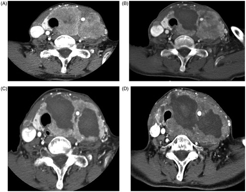
Figure 2. Gradual reduction of papillary thyroid macrocarcinoma after two sessions of RFA (A) Initial ultrasonography (US) of a 77-year-old woman revealed 8.0 cm predominantly cystic mass in the left thyroid gland. (B) After first RFA, US revealed a smaller but persistent, 2.4 cm ill-defined ablation zone in the left thyroid gland. (C) After second RFA, US revealed a smaller, 1.9 cm ill-defined ablation zone in the left thyroid gland. (D) At 12-months follow-up after two sessions of RFA, US revealed a smaller, 1.7 cm ill-defined ablation zone in the left thyroid gland. (E) At 18-months follow-up, US revealed a smaller with decreased calcification, 1.4 cm ill-defined ablation zone in the left thyroid gland.
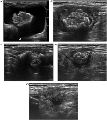
Figure 3. Gradual reduction and complete disappearance of papillary thyroid microcarcinoma after one session of radiofrequency ablation. (A) Initial US of a 49-year-old woman revealed a 0.8 cm mass in the left thyroid gland proven as papillary thyroid carcinoma on CNB. (B) At one-year follow-up after one session of RFA, US revealed a smaller, 0.4-cm ill-defined ablation zone in the left thyroid gland. (C) At 24-months follow-up after RFA, US revealed complete disappearance of ablation zone with subtle scar in the left thyroid.
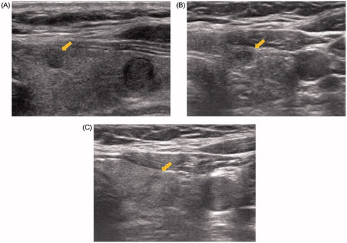
Table 2. Outcome of radiofrequency ablation with primary thyroid cancerTable Footnotea.
Treatment parameters are listed in . In group 1, treatment durations were longer and power was much higher than those in the other groups because we used a longer active tip for large tumour sizes. Compared with groups 2 and 3, treatment duration was longer and power was higher in group 2 because of a large tumour size. Compared with the other groups, energy per millilitre in group 2 was low, probably because of a large haemorrhagic portion of the tumour.
Table 3. Treatment parameters of primary thyroid cancer.
No local tumour recurrences or metastases were detected in the six patients with papillary thyroid carcinoma treated with RFA at a mean follow-up of 19.3 months (range, 17–24). All the patients tolerated RFA. No major complications were experienced, and no patient needed hospitalisation after treatment.
Discussion
Our study demonstrated the efficacy of RFA according to the histologic types of primary thyroid carcinoma with a mean follow-up of 19.3 months. RFA was tolerated in all patients with primary thyroid carcinoma without major complications or procedure-related deaths. In terms of efficacy, RFA effectively reduced tumour size and improved clinical problems in patients with well-differentiated papillary carcinoma regardless of tumour size (i.e. papillary micro- or macro-carcinoma). However, RFA was not effective for anaplastic carcinoma with respect to improvement in cosmetic or symptomatic problems. Therefore, RFA can be applied to patients with well-differentiated papillary thyroid carcinoma, who are at a high-surgical risk.
Several recent studies have suggested the feasibility of RFA for low-risk papillary thyroid carcinoma [Citation22,Citation23]. Zhang et al. [Citation22] reported a mean volume tumour reduction ratio of 96% at one year and complete tumour disappearance of 10.2% during 7.8 months of follow-up. There was no local tumour recurrence or metastasis to lymph nodes in the neck; however, mean follow-up was only 7.8 months. In a four-year follow-up study, Kim et al. [Citation23] reported a mean volume reduction rate of 98.5% and complete disappearance rate of 66.7% during 48.5 months of follow-up. No local tumour recurrence or metastasis to lymph nodes in the neck occurred during a long-term follow-up of 48 months. Also, several studies have been performed on the efficacy of treating primary thyroid carcinoma with LA and MWA. No local tumour recurrence or metastasis to lymph nodes in the neck has been reported during a mean of 11–13 months of follow-up after LA [Citation24] and MWA [Citation10].
Several cases have been reported on the treatment of primary thyroid carcinoma [Citation11,Citation25]. Valcavi et al. [Citation11] reported the results of three patients with low-risk papillary thyroid carcinoma who underwent surgery after LA. LA was also effective for local tumour control; however, at surgery after LA, multiple minute tumours in the thyroid gland and microscopic metastases in the central neck lymph nodes were detected. This result indicated that US-guided ablations, including RFA, MWA and LA are effective for the management of primary thyroid carcinoma, but they may have limited efficacy in the control of regional microscopic metastasis or minute multifocal carcinoma. Therefore, the operator should carefully evaluate lymph node metastasis or minute multifocal carcinoma within the thyroid gland using US or CT images before the ablations. Moreover, RFA in primary thyroid carcinoma should be limited to patients with a high-surgical risk.
Treatment of inoperable anaplastic or medullary thyroid carcinoma with RFA is controversial [Citation12,Citation13,Citation26,Citation27]. Few case reports involve the use of RFA for anaplastic thyroid carcinoma. Cakir et al. [Citation27] demonstrated symptom improvement after LA of anaplastic carcinoma; however, the patient died suddenly. Miyabayashi et al. [Citation12] showed no change in tumour size after RFA of treated advanced cancers, but the tumour became softer after treatment. Pacella et al. [Citation13] also described the limitation of LA for anaplastic carcinoma. In our study, there were no effects of RFA on volume reduction or symptom improvement in patients with anaplastic thyroid carcinoma owing to the rapid doubling time of anaplastic. Therefore, application of RFA in anaplastic thyroid carcinoma or advanced medullary carcinoma is questionable.
Our study revealed no complications; however, previous studies [Citation10,Citation22] reported voice changes after LA and RFA of primary papillary microcarcinoma. Although voice change is the only complication reported after ablation of primary thyroid carcinoma, there have been various complications after ablation of recurrent thyroid carcinoma or benign thyroid nodules [Citation28–30]. Baek et al. [Citation30] reported that the complication rate of RFA for benign thyroid nodules is low. However, various complications may occur, including voice changes, nodule rupture, hypothyroidism, brachial plexus injury, haematoma, vomiting and skin burn, among others. Other large-population, single-centre studies and meta-analyses also showed various complications [Citation28,Citation29]. To prevent these complications, use of a small active tip or the hydrodissection technique has been suggested [Citation18].
Our study has some limitations. First, the number of treated patients was small. Another limitation was the retrospective design of this study and its performance at a single centre. A future prospective, large-population study is necessary to confirm our results.
In conclusion, RFA achieved excellent local tumour control for papillary macro- and micro-carcinoma in primary thyroid carcinoma; however, its clinical effect on anaplastic carcinoma is questionable. RFA may be useful when patients refuse surgery or if they have medical contraindications for undergoing surgery.
Disclosure statement
No potential conflict of interest was reported by the authors.
References
- Haugen BR, Alexander EK, Bible KC, et al. (2016). 2015 American thyroid association management guidelines for adult patients with thyroid nodules and differentiated thyroid cancer: the American thyroid association guidelines task force on thyroid nodules and differentiated thyroid cancer. Thyroid 26:1–133.
- Baek JH, Kim YS, Sung JY, et al. (2011). Locoregional control of metastatic well-differentiated thyroid cancer by ultrasound-guided radiofrequency ablation. AJR Am J Roentgenol 197:W331–6.
- Dupuy DE, Monchik JM, Decrea C, Pisharodi L. (2001). Radiofrequency ablation of regional recurrence from well-differentiated thyroid malignancy. Surgery 130:971–7.
- Kim JH, Yoo WS, Park YJ, et al. (2015). Efficacy and safety of radiofrequency ablation for treatment of locally recurrent thyroid cancers smaller than 2 cm. Radiology 276:909–18.
- Lim HK, Baek JH, Lee JH, et al. (2015). Efficacy and safety of radiofrequency ablation for treating locoregional recurrence from papillary thyroid cancer. Eur Radiol 25:163–70.
- Mauri G, Cova L, Ierace T, et al. (2016). Treatment of metastatic lymph nodes in the neck from papillary thyroid carcinoma with percutaneous laser ablation. Cardiovasc Intervent Radiol 39:1023–30.
- Mauri G, Cova L, Tondolo T, et al. (2013). Percutaneous laser ablation of metastatic lymph nodes in the neck from papillary thyroid carcinoma: preliminary results. J Clin Endocrinol Metab 98:E1203–7.
- Papini E, Bizzarri G, Bianchini A, et al. (2013). Percutaneous ultrasound-guided laser ablation is effective for treating selected nodal metastases in papillary thyroid cancer. J Clin Endocrinol Metab 98:E92–7.
- Yue W, Chen L, Wang S, Yu S. (2015). Locoregional control of recurrent papillary thyroid carcinoma by ultrasound-guided percutaneous microwave ablation: a prospective study. Int J Hyperthermia 31:403–8.
- Yue W, Wang S, Yu S, Wang B. (2014). Ultrasound-guided percutaneous microwave ablation of solitary T1N0M0 papillary thyroid microcarcinoma: initial experience. Int J Hyperthermia 30:150–7.
- Valcavi R, Piana S, Bortolan GS, et al. (2013). Ultrasound-guided percutaneous laser ablation of papillary thyroid microcarcinoma: a feasibility study on three cases with pathological and immunohistochemical evaluation. Thyroid 23:1578–82.
- Miyabayashi C, Ooiwa A, Katakura M, et al. (2005). A successful treatment of percutaneous radio frequency ablation for advanced thyroid cancer. Gan to Kagaku Ryoho 32:1875–7.
- Pacella CM, Bizzarri G, Spiezia S, et al. (2004). Thyroid tissue: US-guided percutaneous laser thermal ablation. Radiology 232:272–80.
- Shin JH, Baek JH, Chung J, et al. (2016). Ultrasonography diagnosis and imaging-based management of thyroid nodules: revised Korean Society of Thyroid Radiology consensus statement and recommendations. Korean J Radiol 17:370–95.
- Na DG, Baek JH, Jung SL, et al. (2017). Core needle biopsy of the thyroid: 2016 consensus statement and recommendations from Korean Society of Thyroid Radiology. Korean J Radiol 18:217–37.
- Na DG, Lee JH, Jung SL, et al. (2012). Radiofrequency ablation of benign thyroid nodules and recurrent thyroid cancers: consensus statement and recommendations. Korean J Radiol 13:117–25.
- Ahn HS, Kim SJ, Park SH, Seo M. (2016). Radiofrequency ablation of benign thyroid nodules: evaluation of the treatment efficacy using ultrasonography. Ultrasonography 35:244–52.
- Park HS, Baek JH, Park AW, et al. (2017). Thyroid radiofrequency ablation: updates on innovative devices and techniques. Korean J Radiol 18:615–23.
- Park HS, Baek JH, Choi YJ, Lee JH. (2017). Innovative techniques for image-guided ablation of benign thyroid nodules: combined ethanol and radiofrequency ablation. Korean J Radiol 18:461–9.
- Yoon HM, Baek JH, Lee JH, et al. (2014). Combination therapy consisting of ethanol and radiofrequency ablation for predominantly cystic thyroid nodules. AJNR Am J Neuroradiol 35:582–6.
- Deandrea M, Sung JY, Limone P, et al. (2015). Efficacy and safety of radiofrequency ablation versus observation for nonfunctioning benign thyroid nodules: a randomized controlled international collaborative trial. Thyroid 25:890–6.
- Zhang M, Luo Y, Zhang Y, Tang J. (2016). Efficacy and safety of ultrasound-guided radiofrequency ablation for treating low-risk papillary thyroid microcarcinoma: a prospective study. Thyroid 26:1581–7.
- Kim JH, Baek JH, Sung JY, et al. (2017). Radiofrequency ablation of low-risk small papillary thyroidcarcinoma: preliminary results for patients ineligible for surgery. Int J Hyperthermia 33:212–19.
- Zhou W, Jiang S, Zhan W, et al. (2017). Ultrasound-guided percutaneous laser ablation of unifocal T1N0M0 papillary thyroid microcarcinoma: preliminary results. Eur Radiol 27:2934–40.
- Sun J, Liu X, Zhang Q, et al. (2016). Papillary thyroid carcinoma treated with radiofrequency ablation in a patient with hypertrophic cardiomyopathy: a case report. Korean J Radiol 17:558–61.
- Owen RP, Silver CE, Ravikumar TS, et al. (2004). Techniques for radiofrequency ablation of head and neck tumors. Arch Otolaryngol Head Neck Surg 130:52–6.
- Cakir B, Topaloglu O, Gul K, et al. (2007). Ultrasound-guided percutaneous laser ablation treatment in inoperable aggressive course anaplastic thyroid carcinoma: the introduction of a novel alternative palliative therapy–second experience in the literature. J Endocrinol Invest 30:624–5.
- Kim C, Lee JH, Choi YJ, et al. (2017). Complications encountered in ultrasonography-guided radiofrequency ablation of benign thyroid nodules and recurrent thyroid cancers. Eur Radiol 27:3128–37.
- Chung SR, Suh CH, Baek JH, et al. (2017). Safety of radiofrequency ablation of benign thyroid nodules and recurrent thyroid cancers: a systematic review and meta-analysis. Int J Hyperthermia 33:920–30.
- Baek JH, Lee JH, Sung JY, et al. (2012). Complications encountered in the treatment of benign thyroid nodules with US-guided radiofrequency ablation: a multicenter study. Radiology 262:335–42.