
Abstract
Background: Papillary thyroid microcarcinoma (PTMC) has high incidence and low disease-specific mortality. However, active surveillance is not accepted by most patients owing to high physical or psychological pressures. The emergence of ablation technologies is supplanting traditional surgery. Our goal was to compare the clinical outcomes of microwave ablation (MWA) and surgery for T1aN0M0 PTMC.
Methods: A total of 92 consecutive patients with T1aN0M0 PTMC were studied retrospectively. Forty-six patients had been treated with MWA, and the other 46 had undergone surgery. MWA was performed using extensive ablation extending from the nodule’s lower pole to the upper pole. Surgery was performed by total thyroidectomy or thyroid lobectomy. We compared the two groups in terms of mean length of stay, cost, mean blood loss, surgical incision, operating room (OR) time, quality of life (QOL) assessment, complications, and therapeutic efficacy over a follow-up period of 42 months.
Results: The mean length of stay, cost, mean blood loss, surgical incisions, OR time, and complications in the MWA group were significantly lower than those of the surgery group. The QOL after MWA was higher than it was after surgery. The nodule volume decreased significantly from 53.61 ± 48.43 mm3 to 4.84 ± 6.55 mm3 (p < .001) at the 42-month follow-up, exhibiting a percentage volume reduction of 81.33 ± 36.87%. No recurrence or metastasis occurred in either group during the follow-up period.
Conclusions: MWA may be considered a minimally invasive alternative to surgery for solitary T1aN0M0 PTMC with low incidence of complications and good therapeutic effect.
Introduction
In recent decades, papillary thyroid microcarcinoma (PTMC) has apparently increased in incidence owing to widespread screening and the technical improvements in thyroid ultrasonography and fine-needle aspiration biopsy (FNAB) [Citation1–3]. Because disease-specific mortality rates of PTMC have been reported to be <1% [Citation4], the American Thyroid Association (ATA) guidelines [Citation4] recommended active surveillance, but the possibility of lymph node and distant metastases increased physical and psychological pressure. If a fraction of patients cannot accept active surveillance, the appropriate treatment becomes the subject of worldwide controversy [Citation5]. The advantage of traditional surgery is the low recurrence rate. However, complications from surgery may exceed the degree of morbidity of the disease itself. With advances in medical technology, various methods of thermal ablation have emerged as new strategies for treatment [Citation6–10].
Compared to percutaneous ethanol injection (PEI), radiofrequency ablation (RFA) or percutaneous laser ablation (PLA) [Citation11–15], microwave ablation (MWA) has many advantages, including higher intratumoral temperatures, ability to ablate larger volumes, and faster ablation speed. For patients who are poor surgical candidates or who unwilling to undergo surgery or surveillance, MWA may be a less-invasive alternative. An initial study confirmed that MWA can also treat solitary T1aN0M0 PTMC [Citation8]. There were earlier studies of thermal ablation versus surgery for benign thyroid nodules and recurrent thyroid cancers [Citation16–18]. However, there has been no data comparing thermal ablation with surgery results for PTMC.
The purpose of the present study was to compare the clinical outcomes of surgery and MWA.
Materials and methods
Patients
This retrospective study was approved by the institutional review board at the Beijing Friendship Hospital of Capital Medical University. Informed consent was obtained from all patients before surgery or MWA. From February 2014 to August 2017, of the 82 patients treated with ultrasound (US)-guided MWA, we excluded 36 patients who were lost to follow-up (20 patients) or who had incomplete information (16 patients). The remaining 46 patients were enrolled in this study as Group B. Of 482 patients with PTMC who underwent surgery, we selected 46 patients in chronological order for Group A beginning in February 2014. No demographic data differed significantly between the two groups prior to treatment ().
Table 1. Demographic characteristics of the subjects.
All of the enrolled patients fulfilled the following criteria: (i) solitary PTMC was confirmed by US-guided core needle biopsy (CNB); (ii) the lesion was solitary and measured ≤10 mm in greatest dimension; (iii) the lesion did not contact or distort the thyroid capsule; (iv) there was no extra-thyroidal spread (trachea or esophagus); (v) there was no lymph node involvement on imaging studies (such as US, computed tomography, magnetic resonance imaging); and (vi) no history of neck irradiation. Surgical patients (Group A) requested surgery because of conscious disturbance or neck extension disorders, and they could not tolerate MWA. MWA patients (Group B) were ineligible for or refused to undergo surgery because of high thyroid surgical risk or other reasons. The exclusion criteria were (i) biopsy showing another type of carcinoma or coexisting thyroid malignancies such as medullary carcinoma; (ii) imaging examinations revealing cervical lymphadenopathy or distant metastasis; (iii) pregnancy; (iv) patients with severe heart, respiratory, liver diseases or renal failure; and (v) coagulation disorders with severe bleeding tendency. All patients were forbidden to take antiplatelet or anticoagulant medications for at least 1 week before the procedure or operation.
Equipment
MW ablation instrument and procedures
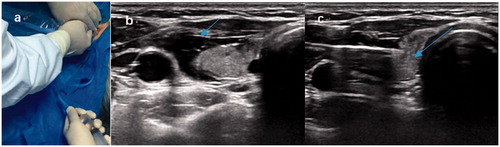
Before each MWA procedure, all Group B patients underwent US examination and contrast-enhanced US (CEUS) to determine the site, vascularity and size (a was lateral diameter, b was anteroposterior diameter, and c was longitudinal diameter) of the nodule by two radiologists with more than 10 years’ experience. Cervical adenopathy was ruled out. Two doctors (L.Q. and Y.L.) who had more than 3 years’ experience in this procedure performed the MWA. The patient was placed in the supine position with the neck fully exposed under continuous electrocardiographic, respiratory, and blood pressure monitoring. Routine sterile skin preparation was performed, and 1% lidocaine was used for topical anaesthesia. A mixture of 1% lidocaine and physiological saline solution was carefully injected into the thyroid capsule to achieve a hydrodissection technique () to protect the major structures (carotid vessels, trachea, the recurrent laryngeal nerve, and esophagus) from thermal injury. The method of continuous injection (5–10 ml/min) was adopted for the nodules close to the vital organs during the whole ablation procedure (). This method was first proposed and applied by US.
Figure 1. Hydrodissection by continuous injection. (a) Surgical assistant injects a mixture of 1% lidocaine and physiological saline solution into the surrounding thyroid capsule by continuous injection (volume: 50 ml, speed: 10–20 ml/min) during the ablation period, (b) achieving ‘hydrodissection’ (arrow) to protect the vital organs from thermal injury. (c) At the beginning of ablation, an MWA antenna was inserted into the nodule’s lower pole.
MWA was performed using the moving-shot technique by multipoint and multidimensional ablation from the nodule’s lower pole to its upper pole. A 17-gauge ablation needle was inserted under ultrasonic guidance into the nodule’s lower pole. The nodule and adjacent thyroid gland were included in the ablated zone (5 mm) to prevent marginal recurrence. A power output of 30 W at 2450 MHz for 20–120 s was routinely used, inducing coagulation necrosis in the tumour. The therapy was sustained until the entire nodule was hyperechoic. When withdrawing the antenna, the needle track was coagulated to prevent tumour cell seeding. During the whole procedure, we intermittently spoke to the patient to monitor the status of phonation. After the procedure, a nurse closely monitored the patients’ post-procedural state for 60 min and compressed the patients’ incision for 10 min to avoid bleeding.
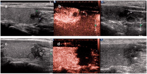
Mean blood loss was measured by withdrawing the syringe (100 ml). The size of the incision and procedure time were also recorded. CEUS was repeated to evaluate the ablation area 1 h after the end of MWA (). We also recorded length of stay (LOS) and cost of ablation.
Figure 2. (a) Longitudinal ultrasound image demonstrating a 5 × 6×8 mm hypoechoic nodule with an irregular margin and microcalcifications located in the right thyroid lobe. (b) Contrast-enhanced ultrasound (CEUS) of the nodule showing irregular hypoenhancement. (c) Ultrasound showing a heterogeneous hypoechoic area with hyperechoic foci 1h after the end of MWA. (d) CEUS was performed to evaluate the 14 × 14 × 17 mm extensive ablation area 1 h after MWA.
Surgical procedure
The patients in Group A underwent surgery by general surgeons with 10 years’ clinical experience under anaesthesia. The surgery methods and decision-making included total thyroidectomy (TT) and thyroid lobectomy (TL) according to the ATA guidelines [Citation4]. Our study included nine patients treated by TT and 37 patients treated by TL. Mean blood loss, size of incision, and operating room (OR) time were recorded. We also recorded mean LOS and cost of surgery.
Post-procedural observation and follow-up
The size, volume, vascularity, and characteristics of each tumour were carefully evaluated 1 h after the procedure and during each follow-up (1, 3, 6, 12, 18, 30 and 42 months). Three diameters of the tumour were measured, and the tumour volume was calculated with the following equation:
(1)
The volume reduction was calculated as:
(2)
Postoperative assessment of every patient was performed by US examination, looking for development of recurrent and metastatic tumours, or lymph node involvement, combined with other imaging examinations as needed. Complications (MWA: dysphagia, haematoma, voice change and hoarseness; surgery: dysphagia, haematoma, voice change, hoarseness and postoperative hyperthyroidism) were carefully monitored after the procedures and during each follow-up period.
Quality-of-life follow-up
Both groups of patients (A and B) underwent postoperative quality-of-life (QOL) assessment [Citation19,Citation20]. The scoring criteria were as follows: (i) physical function: life situation post-procedure (scale ranging from 1 to 5); (ii) limitations in activity (scale ranging from 1 to 3); (iii) bodily pain: (scale ranging from 1 to 6); (iv) if the patient had pain, the extent of its impact on work and life was graded (scale ranging from 1 to 6). Higher scores indicated better QOL.
Statistical methods
SPSS (version 19; IBM, Armonk, New York) statistical software was used to analyse the data. Quantitative data measurements were expressed as the mean ± standard deviation (SD) and range. Comparison of the two groups was done by the Mann–Whitney U-test. Comparison of qualitative data from the two groups was done by the Chi-square test or Fisher’s exact test. Comparison of changes in the mean diameter and volume of each tumour before MWA and at each follow-up was done by the repeated measures test of analysis of variance (ANOVA). The threshold for statistically significant differences was p < .05.
Results
Overall comparison of surgery versus MWA
No recurrences or lymph node metastases were observed in the surgical group or the MWA group. The mean LOS was significantly longer in the surgical group (7.47 ± 2.94 versus 1.3 ± 0.51, p < .001). In China, the cost of surgery and related fees were significantly higher in the surgical group; the cost of surgery was 15342.36 ± 2226.39 (RMB), and that of MWA was 9996.5 ± 586.47 (RMB). The surgical incision in the MWA group was significantly smaller than in the operation group (2.35 ± 0.57 mm versus 66.44 ± 14.01 mm, p < .001). The mean blood loss was higher in the surgery group than in the MWA group (33.10 ± 38.98 ml versus 1.54 ± 0.69 ml, p < .001). Physical function and bodily pain scores after surgery and MWA were 5.00 ± 1.01 and 10.30 ± 2.34 3–4 and 5.84 ± 0.42 and 10.97 ± 1.26, respectively. Physical function but not pain was significantly better in the MWA group ().
Table 2. Overall comparison of surgery versus MWA.
Complications observed following surgery versus MW
All patients tolerated their treatments. During the MWA procedures, 26 patients felt pain or a sensation of heat in the neck. When the procedure was ended, this symptom disappeared. Ten patients reported regional discomfort and neck swelling that resolved 1–2 weeks after the procedure without treatment. Hoarseness lasting 1–3 months occurred in two patients, and one patient’s symptoms disappeared without treatment. One patient felt that the voice had slightly changed during the follow-up. No dysphagia, permanent hoarseness, hypothyroidism, hypoparathyroidism, haematoma, or injury to surrounding organs were observed. During the 3- to 4-year follow-up, all patients in the MWA group maintained normal thyroid hormone levels.
In the surgical cases, hoarseness of varying severity was reported in eight patients. Four patients reported dysphagia and hoarseness. Seven patients with hoarseness recovered without treatment 3–6 months after the surgery, and only one patient with permanent unilateral laryngeal nerve paralysis and transient hypoparathyroidism was reported. One patient with transient hyperthyroidism was reported in Group A. Five patients reported transient hypoparathyroidism in Group A and recovered within one week. One patient had dysphagia and hyperthyroidism. Nine patients had permanent hypothyroidism after TT, and six patients had transient hypothyroidism after TL ().
Table 3. Complications observed following surgery versus MWA.
Efficacy of nodule change at follow-up
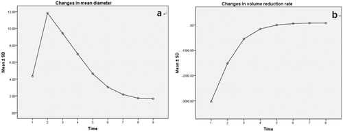
Changes in mean diameter, nodule volume, and percentage volume reduction 1 h, and 1, 3, 6, 12, 18, 30, and 42 months after MWA are shown in and . The US and CEUS appearances after treatment are shown in during the follow-up period. The mean diameter before the MWA procedure was 4.34 ± 1.37 mm (ranging from 2.33 to 7.33 mm). The mean diameter 30 months after the MWA procedure was 1.73 ± 1.19 mm (ranging from 0 to 4.33 mm). The mean diameter at 30 months follow-up significantly changed (p < .001). Change was not clear after the MWA procedure at 30 and 42 months (p = .044). The nodule volume at 42 months follow-up decreased significantly from 53.61 ± 48.43 mm3 to 4.84 ± 6.55 mm3 (p < .001), and the percentage volume reduction was 81.33 ± 36.87%. Seven patients’ nodules completely disappeared in the MWA group. The downward trend of nodule volume change was significant at 1, 3, 6, 12, and 18 months (). The percentage volume reduction was not significantly different between 30 and 42 months (p = .071) (). Moreover, biopsy of unchanged nodules after MWA showed necrosis with inflammatory cells.
Figure 3. Microwave ablation (MWA) treatment and follow-up of one case of papillary thyroid microcarcinoma (PTMC). (a) Longitudinal ultrasound image demonstrating a 3 × 3×4 mm hypoechoic nodule with an irregular margin located in the left thyroid lobe before MWA. (b) One month after MWA, the ablation area was 10 × 10 × 9 mm and showed a well-defined margin. (c) Two years after MWA, the ablation area shrank to 2 × 8×1 mm with a well-defined margin. (d,e) CEUS showing a heterogeneous hypoechoic pattern.
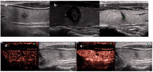
Table 4. Changes in mean diameter (mm) before and 1, 3, 6, 12, 18, 30, and 42 months after MWA.
Table 5. Changes in nodule volume and volume reduction rate (mm3) before and 1 hour, and 1, 3, 6, 12, 18, 30, 42 months after MWA.
Discussion
The 2016 Chinese expert consensus and guidelines for the diagnosis and treatment of PTMC suggested that surgical treatment should be determined with the patient’s appropriate participation [Citation21]. Moreover, the 2015 ATA guidelines recommended active surveillance (AS) for PTMC without metastases or local invasion, especially in patients who have high surgical risk, short lifespan, or who require emergency operations [Citation4]. However, Kwon et al. [Citation22] reported that 14% of 192 patients showed volume increasing 50% during follow-up. One had a new neck node metastasis. Tae Yong et al. [Citation23] also reported that QOL was no better than surgery due to anxiety and fear in patients with AS. Patients have to bear enormous psychological burdens and low life quality with AS.
In recent years, US-guided ablation technology [Citation6–10] has been recommended as an alternative to surgery. Ablative techniques including PEI [Citation14,Citation15], RFA [Citation9,Citation10,Citation24], PLA [Citation6,Citation7] and MWA [Citation8] have been proposed for benign and malignant nodules. PEI was first proposed as a non-surgical treatment of thyroid nodules, primarily including local recurrence or focal distant metastases of well-differentiated thyroid cancer (WTC). No recurrent disease or life-threatening complications were experienced in the group treated by PEI [Citation14,Citation15]. Compared to PEI, LA had the lower risk of side-effect in surrounding cervical structures [Citation25]. LA was subsequently tested in the palliative treatment of local recurrence or lymph node metastases of papillary thyroid carcinoma. Local control was achieved in 16/20 lymph nodes (80%) at the 12-month follow-up [Citation26] and 40/46 (86.9%) lymph nodes at the 30 ± 11 month follow-up [Citation27]. In addition, Zhou et al. [Citation6] reported all 30 patients with solitary PTMC were effectively treated by LA. RFA also showed excellent results for the treatment of benign thyroid nodules and PTMC [Citation9,Citation17,Citation28]. Zhang et al. [Citation9] reported that all 92 patients with PTMC treated by RFA had recurrent and metastatic lymph nodes during follow-up. In addition to excellent treatment safety, the main advantages of thermal ablation are that it is less invasive, more cost-effective, takes less time than surgery, and no longer requires hospitalisation [Citation16].
Compared with PEI, RFA and PLA, MWA had a faster rate of temperature increase and a larger ablation area [Citation13,Citation24,Citation29]. In our study, the incision and mean blood loss of the MWA group were substantially smaller than those of the surgery group. In the MWA group, the mean procedure time was significantly shorter (10.19 ± 8.35 min), the mean LOS was significantly shorter (1.3 ± 0.51 d), and the cost was also lower than surgery, similar to the results reported by Che et al. [Citation16].
Regarding the importance and effectiveness of long-term follow-up, Kim et al. [Citation30] reported a significant reduction of mean volume and complete disappearance ablation zones in the longest follow-up (48.5 ± 12.3 months) ablation study. All patients had at least 42 months follow-up in our study. MWA was confirmed as a feasible and effective treatment for PTMC [Citation8,Citation31]. Yue et al. [Citation8] reported on MWA-treated PTMC with mean follow-up of 11 months. Zhang et al. [Citation9] confirmed successful treatment with PLA, but their follow-up time was <20 months. In the first hour post-ablation, nodule volume increased to 978.23 ± 605.74 mm3 on CEUS. Compared with RFA, the larger change of nodule volume after MWA was consistent with results reported in Yu et al.’s [Citation29] description of liver tumours. The possible reason may be related to different needle paths used in previous reports. In our study, the needle path in the nodule realised one-way multipoint from lower pole toward upper pole may create a larger and more complete ablation area and may improve the effectiveness of ablation [Citation32]. Nodule volume decreased 81.33% over the 42 months of follow-up. The mean ablation area was 978.23 ± 605.74 mm3 immediately after MWA and the volume decreased to 12.43 ± 19.98 mm3 at 18 months. Zhang et al. [Citation9] reported changes in the mean ablation area from 749.8 ± 594.4 mm3 to 9.9 ± 19.0 mm3. Seven nodules (15.2%) disappeared during the late follow-up. The other nodules showed a sclerotic, non-enhancing pattern on CEUS (), similar results in Yue et al.’s [Citation8] report. Our nodule disappearance rate was lower than the rate reported by Zhang et al: 10 (10.2%) tumours completely resolved with RFA [Citation9]. This could be explained by the higher central temperature and greater carbonisation in the MWA than in RFA. The carbonised tissue in the ablated nodules was difficult to dissolve. In previous larger population multicenter prospective study, Cheng et al. [Citation24] also reported RFA showed better effect on nodule volume reduction ratio than did MWA. For the other unchanged nodules on 30-month follow-up after MWA, we confirmed the absence of malignant cells in the fibrotic and necrotic area by FNAB and CNB, similar to findings in other studies [Citation6–8].
The complication rate (4.3%) of MWA was substantially lower than that of surgery (32.6%). MWA had many advantages in terms of low electrical and thermal conductivity and less susceptibility to heat sink effects [Citation6–9,Citation13,Citation32]. In our study, only two patients (4.3%) had transient hoarseness. Kim et al. [Citation33] reported 1.2% (11/875) patients had voice change in an RFA study. In our study, the overall complication rate 4.3% (2/46) was higher than the 3.5% (31/875) of Kim et al.’s study [Citation33], but lower than the 19% (4/21) patients who had hoarseness in Yue et al.’s MWA study [Citation8]. Moreover, Chung et al. [Citation34] reported 2.38% as the overall RFA complication rate. No complications in the vital organs of the neck were observed. MWA can minimise the nerve and vessel injury rate because real-time US imaging can monitor the adjacent vessels and nerves. Hydrodissection effectively prevents thermal damage [Citation24,Citation32]. In addition, we slightly changed the hydrodissection technique by continuous injection to protect vulnerable structures around the thyroid gland from thermal damage (). This technique change may help to explain the lower complication rate in our study compared to the other MWA study.
Figure 4. (a) Changes in mean diameter at each follow-up. (b) Changes in volume reduction ratio at each follow-up.
The complications of surgery included dysphagia, transient or permanent hoarseness, haematoma, and transient hypoparathyroidism. In our study, eight patients had transient hoarseness and one patient had permanent hoarseness. The recurrent laryngeal nerve and parathyroids may be damaged/removed during surgery [Citation35]. Nine patients had permanent hypothyroidism after TT, and six patients had transient hypothyroidism after TL. Nine patients undergoing TT have to take lifelong thyroid hormone supplementation. Most patients undergoing TL suffered unstable thyroid hormone level changes. However, all patients had normal thyroid hormone levels 1 month after MWA. A recent study verified that patients undergoing any kind of thyroid resection had 14–45% complication rates [Citation36,Citation37]. Moreover, in terms of physical function and bodily pain, the QOL scores for MWA were higher than those for surgery.
Our study has several limitations. The first limitation was the retrospective nature. Second, the number of cases was relatively small, and the follow-up time for recurrence and metastasis was short. Third, US and the other examinations could not validly exclude inhibiting lymphatic metastasis [Citation38]. Even if initially we did not find any lymph node involvement, it was possible to detect lymph node involvement during the follow-up period. Finally, MWA treatment was limited to solitary PTMC. However, multifocality cannot be absolutely excluded without histological examinations. In addition, some patients’ nodules were <5 mm. We could use FNAB for the highly suspicious nodules, however, current guidelines do not recommend biopsy in nodules <5 mm.
In conclusion, compared with surgery, MWA was a less invasive treatment for PTMC with a lower incidence of complications, good therapeutic effect, low LOS, and low cost in China. Hence, MWA could be considered to be a treatment alternative to surgery for solitary T1aN0M0 PTMC.
Acknowledgements
We thank Libby Cone, MD, MA from Liwen Bianji, Edanz Group China (www.liwenbianji.cn/ac) and the Taylor and Francis editing service for editing a draft of this article.
Disclosure statement
No potential conflict of interest was reported by the authors.
Additional information
Funding
References
- Jegerlehner S, Bulliard JL, Aujesky D, et al. Overdiagnosis and overtreatment of thyroid cancer: a population-based temporal trend study. PLoS One. 2017;12:e0179387.
- Jung KW, Won YJ, Oh CM, et al. Prediction of cancer incidence and mortality in Korea, 2017. Cancer Res Treat. 2017;49:306–312.
- Deen MH, Burke KM, Janitz A, et al. Cancers of the thyroid: overview and statistics in the United States and Oklahoma. J Okla State Med Assoc. 2016;109:333–338.
- Haugen BR, Alexander EK, Bible KC, et al. 2015 American Thyroid Association management guidelines for adult patients with thyroid nodules and differentiated thyroid cancer: the American Thyroid Association guidelines task force on thyroid nodules and differentiated thyroid cancer. Thyroid. 2016;26:1–133.
- McLeod DSA, Sawka AM, Cooper DS. Controversies in primary treatment of low-risk papillary thyroid cancer. Lancet. 2013;381:1046–1057.
- Zhou W, Jiang S, Zhan W, et al. Ultrasound-guided percutaneous laser ablation of unifocal T1N0M0 papillary thyroid microcarcinoma: preliminary results. Eur Radiol. 2017;27:2934–2940.
- Papini E, Guglielmi R, Gharib H, et al. Ultrasound-guided laser ablation of incidental papillary thyroid microcarcinoma: a potential therapeutic approach in patients at surgical risk. Thyroid. 2011;21:917–920.
- Yue W, Wang S, Yu S, et al. Ultrasound-guided percutaneous microwave ablation of solitary T1N0M0 papillary thyroid microcarcinoma: initial experience. Int J Hyperthermia. 2014;30:150–157.
- Zhang M, Luo Y, Zhang Y, et al. Efficacy and safety of ultrasound-guided radiofrequency ablation for treating low-risk papillary thyroid microcarcinoma: a prospective study. Thyroid. 2016;26:1581–1587.
- Park HS, Baek JH, Park AW, et al. Thyroid radiofrequency ablation: updates on innovative devices and techniques. Korean J Radiol. 2017;18:615–623.
- Simon CJ, Dupuy DE, Mayo-Smith WW. Microwave ablation: principles and applications. Radiographics. 2005;25(Suppl 1):S69–S83.
- Carrafiello G, Lagana D, Mangini M, et al. Microwave tumors ablation: principles, clinical applications and review of preliminary experiences. Int J Surg. 2008;6(Suppl 1):S65–S69.
- Lubner MG, Brace CL, Hinshaw JL, et al. Microwave tumor ablation: mechanism of action, clinical results, and devices. J Vasc Interv Radiol. 2010;21:S192–S203.
- Monchik JM, Donatini G, Iannuccilli J, et al. Radiofrequency ablation and percutaneous ethanol injection treatment for recurrent local and distant well-differentiated thyroid carcinoma. Ann Surg. 2006;244:296–304.
- Shin JE, Baek JH, Lee JH. Radiofrequency and ethanol ablation for the treatment of recurrent thyroid cancers: current status and challenges. Curr Opin Oncol. 2013;25:14–19.
- Che Y, Jin S, Shi C, et al. Treatment of benign thyroid nodules: comparison of surgery with radiofrequency ablation. Am J Neuroradiol. 2015;36:1321–1325.
- Na DG, Lee JH, Jung SL, et al. Radiofrequency ablation of benign thyroid nodules and recurrent thyroid cancers: consensus statement and recommendations. Korean J Radiol. 2012;13:117–125.
- De Bernardi IC, Floridi C, Muollo A, et al. Vascular and interventional radiology radiofrequency ablation of benign thyroid nodules and recurrent thyroid cancers: literature review. Radiol Med. 2014;119:512–520.
- Gou J, Cheng W, Lei J, et al. Health-related quality-of-life assessment in surgical patients with papillary thyroid carcinoma: a single-center analysis from Mainland China. Medicine (Baltimore). 2017;96:e8070.
- Lubitz CC, De Gregorio L, Fingeret AL, et al. Measurement and variation in estimation of quality of life effects of patients undergoing treatment for papillary thyroid carcinoma. Thyroid. 2017;27:197–206.
- Gao M, Ge M, Ji Q, et al. 2016 Chinese expert consensus and guidelines for the diagnosis and treatment of papillary thyroid microcarcinoma. Cancer Biol Med. 2017;14:203–211.
- Kwon H, Oh HS, Kim M, et al. Active surveillance for patients with papillary thyroid microcarcinoma: a single center’s experience in Korea. J Clin Endocrinol Metab. 2017;102:1917–1925.
- Kim TY, Shong YK. Active surveillance of papillary thyroid microcarcinoma: a mini-review from Korea. Endocrinol Metab. 2017;32:399–406.
- Cheng Z, Che Y, Yu S, et al. US-guided percutaneous radiofrequency versus microwave ablation for benign thyroid nodules: a prospective multicenter study. Sci Rep. 2017;7:9554.
- Pacella CM, Papini E. Image-guided percutaneous ablation therapies for local recurrences of thyroid tumors. J Endocrinol Invest. 2013;36:61–70.
- Mauri G, Cova L, Tondolo T, et al. Percutaneous laser ablation of metastatic lymph nodes in the neck from papillary thyroid carcinoma: preliminary results. J Clin Endocrinol Metab. 2013;98:E1203–E1207.
- Mauri G, Cova L, Ierace T, et al. Treatment of metastatic lymph nodes in the neck from papillary thyroid carcinoma with percutaneous laser ablation. Cardiovasc Intervent Radiol. 2016;39:1023–1030.
- Sim JS, Baek JH, Lee J, et al. Radiofrequency ablation of benign thyroid nodules: depicting early sign of regrowth by calculating vital volume. Int J Hyperthermia. 2017;33:905–910.
- Yu J, Liang P, Yu X, et al. A comparison of microwave ablation and bipolar radiofrequency ablation both with an internally cooled probe: results in ex vivo and in vivo porcine livers. Eur J Radiol. 2011;79:124–130.
- Kim JH, Baek JH, Sung JY, et al. Radiofrequency ablation of low-risk small papillary thyroid carcinoma: preliminary results for patients ineligible for surgery. Int J Hyperthermia. 2017;33:212–219.
- Yue W, Chen L, Wang S, et al. Locoregional control of recurrent papillary thyroid carcinoma by ultrasound-guided percutaneous microwave ablation: a prospective study. Int J Hyperthermia. 2015;31:403–408.
- Morelli F, Sacrini A, Pompili G, et al. Microwave ablation for thyroid nodules: a new string to the bow for percutaneous treatments? Gland Surg. 2016;5:553–558.
- Kim C, Lee JH, Choi YJ, et al. Complications encountered in ultrasonography-guided radiofrequency ablation of benign thyroid nodules and recurrent thyroid cancers. Eur Radiol. 2017;27:3128–3137.
- Chung SR, Suh CH, Baek JH, et al. Safety of radiofrequency ablation of benign thyroid nodules and recurrent thyroid cancers: a systematic review and meta-analysis. Int J Hyperthermia. 2017;33:920–930.
- Lynch J, Parameswaran R. Management of unilateral recurrent laryngeal nerve injury after thyroid surgery: a review. Head Neck. 2017;39:1470–1478.
- Ding B, Yu JF, Sun W, et al. Surgical safety analysis of retaining the glands in papillary thyroid microcarcinoma. Eur Rev Med Pharmacol Sci. 2017;21:234–238.
- Price AK, Randle RW, Schneider DF, et al. Papillary thyroid microcarcinoma: decision-making, extent of surgery, and outcomes. J Surg Res. 2017;218:237–245.
- Liu Z, Zeng W, Liu C, et al. Diagnostic accuracy of ultrasonographic features for lymph node metastasis in papillary thyroid microcarcinoma: a single-center retrospective study. World J Surg Onc. 2017;15:32.