
Abstract
Purpose: Magnetic resonance imaging-guided high-intensity-focused ultrasound (MR-HIFU) is a non-invasive treatment modality that precisely focuses ultrasound energy within a tumour and can be customised to result in a wide range of local bioeffects. The purpose of this study was to determine the feasibility of using MR-HIFU to treat soft tissue sarcoma (STS) in dogs.
Materials and methods: Medical records of dogs admitted to the Virginia-Maryland College of Veterinary Medicine from 1 January 2012 to 31 December 2016 were searched for a diagnosis of sarcoma with available cross-sectional imaging of the tumour (MRI or CT). Fifty-three (53) dogs were eligible for inclusion. Tumor tissue (in bone as well as in soft tissue) was considered targetable unless: (1) the ultrasound path was completely obstructed by bone or gas and (2) the MR-HIFU target was within the spinal cord or less than 1 cm from the margin of the spinal cord. Tumors were categorised as <50% targetable, ≥50% targetable or non-targetable.
Results: Eighty-one percent of STS (81.1%, 43/53) were targetable. The head/spine tumour sites had the highest proportion of non-targetable tumours (36%, 9/25). The majority of truncal and axillary tumours were ≥50% targetable (88.9%, 16/18) ,and all extremity tumours were considered ≥50% targetable (100%, 5/5).
Conclusions: The majority of STS were targetable. This is the first study to evaluate MR-HIFU targetability of canine STS. HIFU has potential as a therapeutic modality for treating STS in dogs, and this veterinary application is a possible model for treatment of naturally occurring STS in humans.
Introduction
Soft tissue sarcomas (STS) in dogs account for 15% of all skin tumours and 7% of all subcutaneous tumours [Citation1] with a standardised incidence rate of 122 cases/100 000 dogs/year [Citation2]. STS are mesenchymal cell tumours, which may arise in any anatomical site including muscle, adipose, neurovascular, fascial and fibrous tissue. STS tumour types include fibrosarcoma, peripheral nerve sheath tumour (PNST), undifferentiated sarcoma, liposarcoma, myxosarcoma, leiomyosarcoma, rhabdomyosarcoma, synovial cell sarcoma and lymphangiosarcoma [Citation1]. Sarcomas metastasise hematogenously in up to 20% of cases [Citation1]. STS tumours are locally aggressive and can proliferate through and along fascial planes making excision with clean margins difficult. Wide excision is the foundation of treatment [Citation3]. However, local recurrence is common and radical surgery such as limb amputation is often required to achieve adequate surgical margins [Citation1].
Magnetic resonance imaging-guided high intensity focused ultrasound (MR-HIFU) is a non-invasive treatment that may offer a limb-sparing alternative to conventional therapy. The technology relies on MR-guided heating of tissue. This approach provides the advantage of exquisite spatial delineation of heated tissue and real-time monitoring of thermal dosing [Citation4]. The technology precisely focuses ultrasound energy within the tumour with local bioeffects ranging from a change in vascular permeability to tissue thermal ablation and/or liquefaction, to immunomodulatory effects and enhancement of drug treatments [Citation5]. By varying sonication parameters, it is possible to customise the therapeutic action within this range of bioeffects [Citation6–9]. Precise spatio-temporal control of treatment effects may allow MR-HIFU to offer a non-invasive alternative to both surgery and radiation therapy without the sequelae of either of these conventional therapies.
MR-HIFU technology requires an examination of target anatomy to determine feasibility of treatment. Determining an approach to tumour targeting and treatment is a necessary step towards clinical application. HIFU must have a window for treatment, and within this window, acoustic coupling is required between the transducer and the targeted tissue. HIFU is affected by bone, air and gas in the same manner as B-mode ultrasound used for diagnostic imaging. However, these anatomic structures and tissue interfaces can limit treatment access and must be considered when positioning the patient to attain a treatment window. An appropriate HIFU treatment window is also critical to avoid unintended off-target tissue heating. Healthy bone, bowel or other air-filled structures may cause strong reflection and absorption of ultrasound energy, possibly resulting in off-target thermal injury [Citation10]. Sensitive anatomic structures adjacent to target tissue such as spinal cord, brain, or skin have increased risk of thermal damage [Citation11]. Structures such as the nose, abdominal organs (spleen, liver, kidney and urinary bladder), urethra and mucous membranes may also be in close proximity to the ultrasound beam and warrant additional consideration during MR-HIFU treatment planning.
Clinical trials have sought to balance the benefits of MR-HIFU and its requirements for optimal treatment. While the results in human clinical trials are promising [Citation12–14], few studies have been done in veterinary medicine. Spontaneously arising tumours in dogs, including STS, are recognised as a clinically relevant model for studying similar tumours in man and could help advance research in this area [Citation15,Citation16]. The response of STS to radiation, hyperthermia and chemotherapy in dogs has been previously reported [Citation17–20] and parallels the treatment of STS with MR-HIFU. The hyperthermia and chemotherapeutic methodologies established by Hauck et al. using liposome-encapsulated doxorubicin in canine STS define a basis for a clinical MR-HIFU hyperthermia treatment protocol. While treatment of neoplasia has been performed in canine models [Citation21] using ultrasound-guided HIFU, the treatment of STS using MR-HIFU in dogs has not been evaluated.
Recently, paediatric neoplastic tumours were evaluated for anatomic location and targetability with MR-HIFU [Citation22]. Investigation of limb-sparing STS treatment options in dogs may provide insight to treating similar tumours in people and clinical treatment of veterinary patients. The purpose of this study is to evaluate the feasibility of targeting STS in dogs using MR-HIFU. Our hypothesis is that most canine STS (>50%) are targetable using a commercially available, clinical MR-HIFU system.
Materials and methods
Case selection
The study follows a retrospective design. Institutional animal care and use committee approval was not required. Medical records from 1 January 2012 to 31 December 2016 were searched for a diagnosis of sarcoma in dogs admitted to the Virginia-Maryland College of Veterinary Medicine (VMCVM). Dogs were included in this study if they met the following criteria: 1. Available ante mortem cross-sectional imaging (CT or MR) of the reported neoplasia and 2. A neoplastic mass confirmed as STS by cytology, histology or necropsy or with a clinical diagnosis of PNST. The clinical diagnosis of PNST without histopathology was based on physical exam, neurologic exam and cross-sectional imaging diagnosis. Patient data recorded for each dog included breed, neuter status and age. A total of 118 patients with a diagnosis of sarcoma were identified, 61 of whom were diagnosed with STS. Eight dogs (8/61) were excluded because no mass was identified on cross-sectional imaging. Six (6/8) patients were imaged subsequent to tumour excision, and two (2/8) dogs were identified with evidence of possible tumour recurrence at the incision site. Cytology of the tumour site was not performed at the time of imaging; however, both patients were identified with pulmonary metastatic neoplasia. A total of 53 dogs met inclusion criteria of which two (2/53) dogs had recurrent STS with discrete masses, verified by biopsy and histopathology. Of the included five (5/53) suspected PNST, one (1/5) had histopathology. Twelve (12) patients had one MRI study each, and 49 patients had CT studies (three patients with two CT studies and one patient with three CT studies). The most recently acquired imaging study was used for targeting evaluation in patients with multiple MRI or CT studies.
Image analysis
Cross-sectional studies evaluated included both CT and MRI studies of the affected anatomic region. CT studies were acquired on a 16-slice multidetector helical scanner (Toshiba Aquilion 16; Toshiba Medical Systems, Markham, ON, USA), and postcontrast thin-slice sequences (1.0 and 2.0 mm) were evaluated. Postcontrast sequences were acquired immediately (one patient) or 3 min following the administration of non-ionic iodinated positive contrast (iopromide 370 mgI/mL, Ultravist; Bayer Healthcare Pharmaceuticals, Wayne, NJ, USA) at 0.46 mL/kg. MR image sets were acquired on a 1.5 T scanner (Intera 1.5 T; Philips, Best, the Netherlands) using standard 2D and 3D sequences (T1w, T2w, STIR, T2w* and T1w pre- and postcontrast). Postcontrast sequences were acquired after intravenous administration of gadolinium-based contrast agent (gadopentate dimeglumine, 469.01 mg/mL, Magnevist; Bayer Healthcare Pharmaceuticals, Wayne, NJ, USA) at a dose of 0.02 mL/kg. Data obtained with sequences that subjectively imparted the best lesion detail were used to evaluate targetability in this study.
A workstation with image analysis software (OsiriX v7.5.1; OsiriX Foundation, Geneva, Switzerland) was used to evaluate the tumour dimensions in three perpendicular planes to verify proximity to relevant anatomic structures. Selected 3D MRI and postcontrast CT imaging datasets were analysed using modified clinical MR-HIFU software (Sonalleve Treatment Planning Software R3.2L3; Profound Medical Inc., Mississauga, Canada).
Targetability
All studies were evaluated in three planes to identify a potential acoustic window to accommodate the HIFU beam and to construct a planned treatment volume (PTV) of each tumour. A three-dimensional, ellipsoidal PTV was centred on the mass and sized to encompass all or the majority of the mass while minimising inclusion of surrounding normal tissue. The PTV was selected to exclude soft tissue margins outside of the total tumour volume (TTV), though minimal overlap could not be avoided in some cases due to mismatch of the available ellipsoidal PTV and irregular tumour shape. A HIFU treatment target (i.e. a 4 mm diameter treatment cell) was positioned at the centre of the PTV (automatically identified by the Sonalleve treatment planning software), and the beam was aligned to the target cell to optimise the treatment window based on tumour location. The modified (research) version of the Sonalleve treatment planning software allowed the beam/target cell to be freely positioned relative to the dataset. This feature was used to select a patient position that allowed the greatest unobstructed access to the target.
The TTV was estimated by utilising additional ellipsoid ROIs as necessary. Tumors were categorised as <50% targetable or ≥50% targetable based on the estimated percentage of targetable tumour (% targetable = PTV/TTV). Images were reviewed by a veterinary radiology resident (MCS) and a biomedical engineer with MR-HIFU expertise (PSY) to determine by consensus if the tumour (either in its entirety or a portion) could be targeted with MR-HIFU.
Tumor tissue (in bone as well as in soft tissue) was considered targetable unless:
The ultrasound path was completely obstructed by bone or by gas (nasal or oral cavity, lung, bowel).
The HIFU target was within the spinal cord or ≤1 cm from the margin of the spinal cord.
Lesions in close proximity to the brain or spinal cord constitute a risk of injury to the central nervous system, and a 1 cm safety margin was therefore instituted as in previous studies [Citation22,Citation23]. Tumor proximity to critical structures, such as skin, was used as a selection criterion in other studies [Citation22,Citation24]. Distances to these critical structures were evaluated herein but not used as exclusion criteria. While MR-HIFU is challenged by respiratory and cardiac motion [Citation25,Citation26], these factors were not evaluated or used as exclusion criteria in this study.
Tumor volume and critical structures
Based on the current clinical Sonalleve V2 system configuration, the optimal and maximum target depths were defined as ≤8 and 11 cm from the skin to tumour margin in the far field, respectively (). The percentages of tumours whose PTV and TTV were within optimal and maximum target depths were quantified.
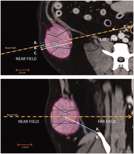
Figure 1. (Top image) Overview of STS evaluation and critical structure measurements in the near field. Each STS was evaluated in three planes. This image illustrates an axial projection (CT image) of an STS tumour present in the right flank. The target cell (4 mm) is positioned centrally within the constructed PTV, and the HIFU beam is aligned to the target cell. Measurements included (A) the closest distance from the skin to any tumour margin, with (B) the distance from the skin to the centre of the tumour in the near-field, and (C) the distance from the skin to the deepest tumour margin in the far field, both acquired along the HIFU beam path. (Bottom image) Overview of STS evaluation and critical structure measurements in the far field. Dorsal projection of the same tumour in the right flank. The target cell is unchanged in location and remains the same point of reference for measurements along the beam axis. Critical structure measurements for bone were acquired from (A) the centre of the tumour to nearest bone structure in the far field and (B) the tumour margin to nearest bone structure in the far field.
An optimal treatment volume of ≤200 cm3 was estimated from the ∼56 cm3/h rate of ablation demonstrated by an experienced operator in uterine fibroids [Citation27]. Tumors were evaluated for proximity of sensitive anatomic structures (i.e. vessels, nerve, bone and bowel) along the ultrasound beam path to the margin and to the centre of the TTV and/or PTV (). Four general assessments were made regarding the proximity of critical structures. These assessments were categorised by tumour type, tumour site and targetability (<50% targetable, ≥50% targetable or non-targetable):
Is TTV impacted by bone in the near field (zero intersection of the HIFU beam with bone in the near field)? Yes or no.
Can the entire tumour be treated without hitting bone (zero intersection of the HIFU beam with bone in near field or 4 cm from bone in the far field)? Yes or no.
Is targeting with 1 cm clean margins possible (no critical structures ≤1 cm of the PTV)? Yes or no.
Is the closest distance from the skin to any tumour margin ≤1 cm? Yes or no.
Statistical analysis
The relative frequency of tumour type, tumour site and targetability (<50% targetable, ≥50% targetable or non-targetable) was determined. Existence of an association between categorical variables was determined using the Chi square analysis test for independence (if n ≥ 5) or Fisher’s exact test (if n < 5). All analyses were completed at a significance level of 0.05 using SAS 9.4 (SAS Institute, Inc., Cary, NC, USA).
Results
A total of 53 dogs with STS were evaluated: 33 castrated males, 2 intact males, 17 spayed females and 1 intact female. The median body weight was 31.5 kg (range: 3.4–65.1 kg), with 9 dogs weighing less than 15 kg, 16 dogs ranging 15–30 kg and 28 dogs weighing more than 30 kg. The most common STS tumour types () were unclassified STS (32.1%, 17/53) and fibrosarcomas (30.2%, 16/53), while the most common STS tumour sites included the head (30.2%, 16/53), truncal tumours (24.5%, 13/53) and spine/paraspinal tumours (17%, 9/53).
Table 1. Total number and relative percentage of canine STS by both tumour type and tumour site.
Targetability of STS
Overall, the majority of STS tumours were targetable (81.1%, 43/53; . Of the targetable tumours, 72.1% (31/43) had ≥50% of the tumour volume available for MR-HIFU. A total of 18.9% (10/53) of evaluated tumours were not targetable (). The majority of non-targetable tumours were located at the spine/paraspinal sites (6/10) and the head (3/10). PNST were the least targetable tumour type, with only one of the five tumours being <50% targetable (). Two recurrent STS (one rhabdomyosarcoma and one anaplastic sarcoma) were considered ≥50% targetable, while one case of disseminated anaplastic sarcoma was considered non-targetable. Images representative of ≥50% targetable, <50% targetable and non-targetable STS categories are demonstrated in .
Figure 2. Anatomic distribution of targetable STS tumours by body regions. Pie charts placed at sites labelled head, truncal/axillary, spine/paraspinal, appendicular and abdomen/disseminated illustrate the distribution of all soft tissue sarcomas (53) as, ≥50% targetable (green), <50% targetable (yellow) and non-targetable (red) tumour categories. The spine/paraspinal and head sites had the largest relative percentage of non-targetable STS. Image adapted with permission [Citation55].
![Figure 2. Anatomic distribution of targetable STS tumours by body regions. Pie charts placed at sites labelled head, truncal/axillary, spine/paraspinal, appendicular and abdomen/disseminated illustrate the distribution of all soft tissue sarcomas (53) as, ≥50% targetable (green), <50% targetable (yellow) and non-targetable (red) tumour categories. The spine/paraspinal and head sites had the largest relative percentage of non-targetable STS. Image adapted with permission [Citation55].](/cms/asset/1ccbcd70-30ca-44de-94f7-19813fbb7294/ihyt_a_1489072_f0002_c.jpg)
Figure 3. Distribution of STS tumour types with the relative percentage of targetability. STS tumour types that are ≥50% targetable are depicted in green and STS tumours <50% targetable are depicted in yellow, while STS deemed non-targetable are illustrated in red. PNST had the largest relative percentage of tumours that were not targetable followed by anaplastic sarcoma, unclassified soft tissue sarcoma and fibrosarcoma.
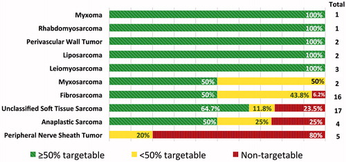
Figure 4. Representative examples of canine STS at seven anatomic sites. The PTV was selected as an ellipsoid (purple). A 4 mm diameter treatment cell (green/blue) and the resulting approximate outline of the HIFU beam (yellow) were aligned with the centre of the PTV to optimise the treatment window based on tumour location, and the patient was positioned to allow the greatest unobstructed access to the target. The orange rectangle around the target represents a safety zone in which high temperatures can occur. (A) Head: ≥50% targetable, left maxillary fibrosarcoma with a portion of the tumour invading the infraorbital foramen and left orbit (green arrow); (B) head: <50% targetable, unclassified STS of the rostral right mandible with geographic bone lysis; (C) head: not targetable, unclassified STS within the right nasal cavity; (D) truncal: ≥50% targetable, subcutaneous unclassified STS dorsal to the pelvis; (E) truncal: <50% targetable, unclassified STS medial to the left scapula; (F) appendicular: ≥50% targetable, subcutaneous anaplastic sarcoma of the left distolateral humerus; (G) spine: ≥50% targetable, unclassified STS in the right cervical region with invasion of the spinal canal at C6 (green arrow); (H) spine: <50% targetable, left paraspinal myxosarcoma involving ribs 10, 11 and 12 and invasion of the spinal canal at T10–T11 resulting in extradural spinal cord compression (green arrow); (I) spine: Not targetable, PNST involving the right spinal nerve segment at C4–C5 with extension to the spinal cord (green arrow); (J) abdominal: ≥50% targetable, extramural small intestinal fibrosarcoma; the ellipsoid ROI approximates the PTV, however, additional targets could be selected during therapy to achieve more conformal treatment; (K) axillary: ≥50% targetable, left axillary myxosarcoma partially incorporating the left first rib (green arrow); and (L) axillary: <50% targetable, left axillary PNST.
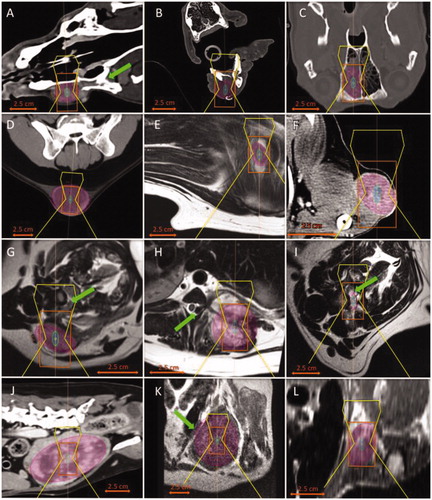
Table 2. Total number and relative percentage of targetable canine STS by both tumour type and tumour site.
Optimal treatment volume and target depth
Based on the volume of the entire tumour described by the TTV variable (), the majority of the 53 sarcomas were within optimal treatment volume of ≤200 cm3 (71.7%, 38/53), optimal target depth of ≤8 cm (73.6%, 39/53) or met both criteria (69.8%, 37/53). Of the 43 targetable sarcomas, based on TTV, the majority were within optimal treatment volume of ≤200 cm3 (67.4%, 29/43), optimal target depth of ≤8 cm or met both criteria (65.1%, 28/43). Seventy-nine percent of targetable tumours (34/43) and 83.0% of all tumours were within a maximum target depth of 11 cm.
Table 3. Number and relative percentage of STS for both total tumour volume (TTV) and planned treatment volume (PTV) that met the criteria for optimal treatment volume (≤200 cm3), optimal target depth (≤8 cm), maximum target depth (≤11 cm), or met both criteria for optimal volume and optimal target depth (≤200 cm3 and ≤8 cm), and optimal treatment volume and maximum target depth (≤200 cm3 and ≤11 cm).
If critical structures are taken into account as delineated by the PTV (), this may reduce the treatable tumour volume in some cases. Therefore, based on PTV, a similar number of tumours were within the optimal treatment volume of ≤200 cm3 (75.5%, 40/53) or met both criteria of optimal target depth and optimal treatment volume (71.1%, 38/53). Of the targetable tumours, most PTVs were within optimal treatment volume (69.8%, 30/43) and met criteria for both optimal treatment volume of ≤200 cm3 and optimal target depth of ≤8 cm (65.1%, 28/43).
Critical structures
Targeting the TTV was impacted by bone in the near field in 49.1% (26/53) of all cases and an association was identified between the presence of bone in the near field and both tumour site (p < .0001) and targetability (p < .0001). The entire tumour (TTV) could be treated without hitting bone in only 1.9% (1/53) of all cases and only 5.7% (3/53) of all tumours could be targeted with 1.0 cm clean margins, avoiding critical structures (skin, major nerve, major vessel or bone). Skin was present ≤1 cm from tumour margins in the majority of STS (69.8%, 37/53), and its presence was associated with tumour type (p ≤ .0007), tumour site (p < .0001) and targetability (p ≤ .0007), as shown in .
Table 4. Contingency coefficients summary for the geometric/anatomical constraints imposed by nearby critical structures when compared by STS tumour type, tumour site and targetability.
Discussion
This study provides an initial evaluation of the targetability of STS with a clinical MR-HIFU system in a population of canine patients. In the study population, the majority of STS were targetable by MR-HIFU, consistent with our hypothesis. The most frequent STS types were unclassified STS, fibrosarcoma and PNST and the most common tumour sites were the head, truncal and spine/paraspinal locations. The least targetable tumour types were PNST, which were frequently located in spine or axillary regions making them less accessible due to overlying bone, adjacent spinal cord or other major critical structures. Multiple factors may affect the targeting and treatment of canine STS. Below, we discuss the impact of variables, which may determine the targetability of STS with MR-HIFU.
Impact on targetability
Tumor depth
The majority of STS were within an optimum target depth of 8 cm and 83% of STS were within a maximum target depth of 11 cm. This suggests that the majority of tumours are within the therapeutic capability of the commercial, clinical MR-HIFU system used for the evaluation of tumour treatment. It is interesting to note that the most common anatomic site for STS in this study was the head, which differs from previous reports of the most common STS sites being the limbs and trunk [Citation1,Citation3,Citation28]. The predominance of this tumour site may be due to a clinical preference for cross-sectional imaging of head tumours compared to tumours located in the trunk or limbs. Though bone is present in the far field, many of these tumours were also partly within bone, and most were determined to be partially targetable. In these cases, diseased bone in the far field may be used advantageously to increase the thermal effects on targeted tissue. However, sensitive structures such as the nose or nasal mucous membranes are at the interface of gas and bone, which may induce thermal damage, and therefore must be taken into consideration. Currently, radiation therapy is reported to provide the best control for canine nasal tumours [Citation29]; thus, it is unknown whether MR-HIFU is a viable treatment option in these cases and further clinical study is required.
Tumor volume
In this study population, the majority of sarcomas based on TTV were ≤200 cm3 (71.7%) and 67.4% of targetable STS met this criterion. In a similar study of paediatric tumours, only 55% of targetable STS were less than 200 cm3 [Citation22]. A limiting factor in treating large tumour volumes is extended treatment time. Traditional point-by-point HIFU utilises small treatment volumes of ∼0.15 cm3, resulting in a relatively slow rate of sonication (0.46 cm3/min, not including cooling periods between sonications) [Citation30]. For large tumours, this technique requires multiple treatment planes. In contrast, volumetric ablation utilised by the Sonalleve system uses electronic beam steering and image-based real-time feedback control and can sonicate cells from 4 to 16 mm in diameter (10–40 mm in length) [Citation31]. This approach allows much faster treatment, especially when using the larger cell sizes. For example, using ablation volume and duration reported by Enholm et al. [Citation31], ablation speed for volumetric sonications is estimated to range 0.09–5.7 cm3/min for a 4–16 mm cell sizes. For the largest cells, volumetric ablation is thus more than 12 times faster than point-by-point ablation (not counting cooling periods between sonications). The possibility of higher ablation speeds using volumetric ablation is also supported by a study reporting the successful treatment of two large uterine fibroids (12.3 and 13.0 cm in diameter). Approximately 72% (431.4 mL) and 88.5% (569.8 mL) of the tumour volumes were ablated. Including cooling time between sonications, these fibroids were ablated with a treatment speed of 158.8–179 cm3/h (or 2.65 cm3 to 2.98 cm3/min) [Citation32]. Further treatments by the same group determined that despite longer cooling times required by larger treatment cells, volumetric sonications can be performed with rates that are several-fold faster than point-by-point ablation [Citation27].
Complications related to volumetric ablation may include increased near-field heating and possible thermal damage to intervening tissues, such as skin burns [Citation33–37], due to more energy deposition per sonication. Therefore, larger treatment cells typically require longer cooling periods between sonications. Cooling times may be reduced by sequential sonications of treatment cells that are positioned further apart, thus reducing cumulative near-field energy densities and near-field heating. Furthermore, direct skin cooling (DISC) concepts have been designed to help prevent thermal skin injury when used with MR-HIFU [Citation38]. However, the impact of DISC technology on the speed of MR-HIFU treatments has not yet been widely explored. Large tumours may also be addressed by serial MR-HIFU treatments or by repositioning the patient during a treatment session to gain better access to tumour tissue.
Critical structures
In this study, only 5.7% (3/53) of all STS could be targeted with 1.0 cm clean margins due to adjacent critical structures (skin, major nerve, major vessel or bone) and only 1.9% (1/53) of all STS could be treated without hitting bone. Thus, if the 1 cm safety margin were adhered to, three STS could be completely treated while the remainder of targetable STS would be candidates for partial treatment. The 1 cm margin is not universally accepted. For example, Lang and Wu relied on a 0.2 cm margin in thyroid ablations [Citation39]. While a 1 cm margin was not used to determine targetability in this study, this criterion may affect the ability to treat the entire tumour. MR-HIFU treatment protocols for specific STS applications are still being developed, which will better define ‘treatable tumours’. Recommendations for the surgical excision of STS in veterinary patients are aggressive – wide surgical excision with minimum margins of 2–3 cm laterally and one fascial plane deep to the tumour [Citation1,Citation3,Citation40].
Appendicular tumours, in particular, are difficult sites in which to achieve wide surgical margins due to the close proximity of neurovascular structures and limited overlying skin available for primary closure [Citation41,Citation42]. While complete tumour treatment is the primary clinical goal in both veterinary and human patients, marginal excision with radiation or chemotherapeutics has had good clinical outcomes with a reduction in patient morbidity and may represent a limb-sparing option for some patients [Citation41–45]. Similar multimodal strategies may be employed using MR-HIFU ablation or hyperthermia with adjunctive treatments. Alternately, in the case of a high-grade tumour that has already metastasised to the lungs or lymph nodes, partial tumour treatment may result in palliative effects of the neoplasia by delaying progression, decreasing pain or reducing the size of the tumour [Citation3,Citation46,Citation47]. Thus, minimally invasive treatment methods such as MR-HIFU may provide both primary and adjunctive treatment options for STS.
Similarly, a large percentage of STS (69.8%) were within 1.0 cm of skin. Among those near the skin, 94.6% were otherwise targetable or partially targetable. The mean distance from the skin to targetable tumours in paediatric sarcomas was 4.1 cm [Citation22], which represents a fundamental difference from canine STS in this study. In Shim’s paediatric study, lesions within 1 cm of skin were considered non-targetable due to the possibility of skin burn. Local effects of HIFU may range from acute minor skin damage to more serious tissue necrosis [Citation48]. However, to our knowledge, a thermal dose threshold for skin damage in dogs has not been determined. Skin burns increase patient morbidity and provide a conduit for infection, while cosmesis is an additional concern in people. Skin burns are also a concern when implementing radiation therapy and an acute radiation morbidity scoring scheme has been suggested to evaluate a multitude of radiation-induced side effects in veterinary patients [Citation49]. The Sonalleve system used to evaluate targetability in this study is now supplied with built-in DISC technology, which may broaden the application of MR-HIFU for treating superficial tumours, large tumour volumes and prevent device related adverse effects [Citation38]. Additional studies are required to evaluate the feasibility, safety and efficacy of treatment utilising hyperthermia or tumour ablation at various anatomic locations and the effect on skin, or adjacent superficial structures in veterinary patients.
Motion
Motion affecting the targeting of STS was not addressed in this study. Tumors that are in sites affected by respiratory or cardiac motion, such as the thorax and abdomen, may require motion compensation to enable accurate targeting and treatment with MR-HIFU. Proton resonance frequency shift (PRFS)-based thermometry is the MRI method typically used for monitoring ablation temperatures and thermal doses in MR-HIFU. Motion is one of the most significant challenges in its clinical application [Citation50]. Intrascan motion occurs when motion occurs during MR image acquisition resulting in ghosting and blurring artefacts and thus a poor-quality image, while interscan motion occurs due to motion of a target between consecutive images. Intrascan motion artefacts can be reduced by increasing the speed of image acquisition, but are not specific to PRFS-based temperature imaging. However, interscan motion results in artefacts in the PRFS-based temperature maps due to misregistration of phase differences from the baseline phase image to the current phase image. Respiration is a major source of interscan motion and can be addressed with respiratory gating or positive pressure ventilation. Additionally, variable MR thermometry techniques may help improve accuracy and reduce the effects of motion [Citation50].
Patient-specific factors
Patient size
The study population of dogs with STS consisted primarily of medium to large or giant-breed dogs, which is consistent with previous reports [Citation1,Citation3,Citation40,Citation51]. The effects of patient breed or size on targetability were not evaluated for statistical correlation in this study; however, some general conclusions may be drawn. Depending on the location of the tumour, larger patients may have a target window that is less affected by adjacent critical structures, particularly in the far field. This would allow MR-HIFU treatment with less risk of off-target thermal injury. Alternately, a larger patient may have STS masses that are deeper targets or may have a larger tumour volume, thereby limiting the ability to treat the tumour.
Treatment planning
Multiplanar (3D) tumour treatment planning is an important component of MR-HIFU tumour treatment. Because veterinary patients are under general anaesthesia for cross-sectional imaging procedures, preplanning optimum treatment windows and patient positioning reduces anaesthesia time and imaging time. Determining the entry and exit point of the HIFU beam and the presence of reflective tissue interfaces (i.e. gas and bone) are important considerations to avoid off-target tissue damage. Near-field structures may block HIFU access to the tumour and far field structures such as lung, bowel or other organs may be at risk for thermal damage. Anticipating these potential challenges helps plan for ways to prevent treatment complications and off-target tissue injury. The use of MR-safe temperature probes for direct temperature monitoring and/or the use of an acoustic conduit such as acoustic gel, cold water bags or gel pads, combined with frequent monitoring of at-risk sites may help reduce off-target tissue heating and prevent injury [Citation52]. Creative strategies such as inducing a hydrothorax to avoid lung injury when treating a body wall mass with MR-HIFU have been utilised in human patients [Citation26].
Positioning/coupling
Ultrasound coupling and penetration require removal of hair and debris from the site, which can be problematic. In the authors’ experience, hair removal gels or closely shaving the area help improve site preparation and acoustic coupling. Acoustic coupling to the appropriate body part can also be difficult. Acoustic gel or gel pads can be utilised to help maintain contact of the body part to the HIFU bath. MRI coil positioning in stand-alone HIFU units can problematic and a table-integrated body coil and HIFU transducer improve the ability to obtain an adequate treatment window. Specially designed interfaces to couple the patient to the HIFU unit and to properly position the body coil may be necessary in veterinary patients.
Study limitations
Limitations of this study include its retrospective nature, which may have introduced selection bias as not all dogs with STS may have been imaged. For example, dogs with smaller STS may have undergone surgical excision rather than cross-sectional imaging. Additionally, the theoretical targeting of STS with modified software helps alleviate variability in patient size and tumour location to achieve an adequate acoustic window; however, this may be more difficult in actual practice. Patient positioning will likely require more trial and error when using standard equipment; thus, variation in patient size and tumour location may require unforeseen additional modifications of clinical MR-HIFU hardware and software for use in veterinary patients. Another limitation was that five of the PNST did not have histopathology. Biopsies of many PNST lesions are often not pursued due to inaccessible locations that my result in increased patient morbidity. However, determining the anatomic location and the ability to physically target the tumour with MR-HIFU was more critical for the purpose of this study than the actual clinical outcome based on tumour type.
Not all metastatic or recurrent lesions were assessed for targetability. Of the four dogs that had metastatic or recurrent disease, only two patients had discrete masses that could be assessed. Two patients were excluded with evidence of tumour recurrence in which both had a diffuse area of contrast enhancement on imaging but neither had an identifiable mass that could be accurately delineated for targeting with MR-HIFU. Similarly, one patient deemed non-targetable had disseminated masses in the abdomen and thorax obscured by critical structures that made MR-HIFU an impractical option based on such widespread disease.
Insights and opportunities for clinical translation
The canine patient population examined in this work consists mostly of primary STS, and therefore, these canine patients could allow the evaluation of treatments with curative intent, providing a much needed parallel to human clinical trials. Treatment of malignant tumours with MR-HIFU in human clinical trials [Citation53,Citation54] usually involves patients with advanced locally recurrent and/or metastatic disease. Our analysis shows that only a small percentage of canine STS could be treated without harming critical structures within 1 cm of tumour margins. Unlike conventional surgical excision, this non-invasive technology allows tissue in the near field to be spared. However, in order to include margins around the tumour that are commonly accepted in surgical practice, use of MR-HIFU would necessitate collateral damage to peritumoral tissue.
While it is important that MR-HIFU can be used in treatments of advanced malignant disease, these treatments are palliative in nature. An opportunity exists to explore the feasibility, safety and efficacy of promising therapies in veterinary patients with less advanced disease that may result in potential for cure or lasting treatment response. Further studies are necessary to determine if incomplete treatment of the tumour with MR-HIFU is beneficial when combined with immunotherapy or liposomal chemotherapy or if immunomodulatory effects of HIFU are further optimised.
Conclusions
This study demonstrated that the majority of spontaneously arising STS in canine veterinary patients are targetable using a commercial, clinical MR-HIFU system. The least targetable tumours were PNST, and most non-targetable tumours were located at the spine/paraspinal site. While our analysis demonstrates that complete tumour treatment with no collateral damage is only possible in 5.7% of patients with STS, a variable degree of collateral damage is likely an inevitable outcome regardless of the treatment modality employed. Side effects stemming from more extensive treatment must be balanced against patient treatment goals. MR-HIFU presents a versatile, non-invasive alternative or adjuvant to radiation therapy. Additional research is warranted to determine MR-HIFU protocols for the clinical treatment of veterinary patients.
Disclosure statement
No potential conflict of interest was reported by the authors.
References
- Liptak JM, Forrest LJ. Soft tissue sarcomas. In: Vail DM, Withrow SJ, editors. Withrow and MacEwen’s small animal clinical oncology. 4th ed. Saint Louis: W.B. Saunders; 2007. p. 425–454.
- Dobson JM, Samuel S, Milstein H, et al. Canine neoplasia in the UK: estimates of incidence rates from a population of insured dogs. J Small Anim Pract. 2002;43:240–246.
- Ehrhart N. Soft-tissue sarcomas in dogs: a review. J Am Anim Hosp Assoc. 2005;41:241–246.
- McDannold HK, Wolf D, et al. MRI evaluation of thermal ablation of tumors with focused ultrasound. J Magn Reson Imaging. 1998;8:91–100.
- Ellens NPK, Hynynen K. High-intensity focused ultrasound for medical therapy. In: Gallego-Juárez JA, Graff KF, editors. Power ultrasonics. Oxford: Woodhead Publishing; 2015. p. 661–693.
- Eranki A, Farr N, Partanen A, et al. Boiling histotripsy lesion characterization on a clinical magnetic resonance imaging-guided high intensity focused ultrasound system. PLoS One. 2017;12:e0173867.
- Julianna CS, Oleg AS, Vera AK, et al. Ultrasonic atomization of tissue and its role in tissue fractionation by high intensity focused ultrasound. Phys Med Biol. 2012;57:8061.
- Khokhlova VA, Fowlkes JB, Roberts WW, et al. Histotripsy methods in mechanical disintegration of tissue: towards clinical applications. Int J Hyperthermia. 2015;31:145–162.
- Wang YN, Khokhlova T, Bailey M, et al. Histological and biochemical analysis of mechanical and thermal bioeffects in boiling histotripsy lesions induced by high intensity focused ultrasound. Ultrasound Med Biol. 2013;39:424–438.
- Hipp E, Partanen A, Karczmar GS, et al. Safety limitations of MR-HIFU treatment near interfaces: a phantom validation. J Appl Clin Med Phys. 2012;13:168–3739.
- Yarmolenko PS, Moon EJ, Landon C, et al. Thresholds for thermal damage to normal tissues: an update. Int J Hyperthermia. 2011;27:320–343.
- Hindley J, Gedroyc WM, Regan L, et al. MRI guidance of focused ultrasound therapy of uterine fibroids: early results. AJR Am J Roentgenol. 2004;183:1713–1719.
- Fennessy FM, Tempany CM, McDannold NJ, et al. Uterine leiomyomas: MR imaging-guided focused ultrasound surgery–results of different treatment protocols. Radiology. 2007;243:885–893.
- Catane R, Beck A, Inbar Y, et al. MR-guided focused ultrasound surgery (MRgFUS) for the palliation of pain in patients with bone metastases – preliminary clinical experience. Ann Oncol. 2006;18:163–167.
- Pinho SS, Carvalho S, Cabral J, et al. Canine tumors: a spontaneous animal model of human carcinogenesis. Transl Res. 2012;159:165–172.
- MacEwen EG. Spontaneous tumors in dogs and cats: models for the study of cancer biology and treatment. Cancer Metastasis Rev. 1990;9:125–136.
- Dewhirst MW, Sim DA, Sapareto S, et al. Importance of minimum tumor temperature in determining early and long-term responses of spontaneous canine and feline tumors to heat and radiation. Cancer Res. 1984;44:43–50.
- Hauck ML, LaRue SM, Petros WP, et al. Phase I trial of doxorubicin-containing low temperature sensitive liposomes in spontaneous canine tumors. Clin Cancer Res. 2006;12:4004–4010.
- Thrall DE, Rosner GL, Azuma C, et al. Using units of CEM 43 °C T90, local hyperthermia thermal dose can be delivered as prescribed. Int J Hyperthermia. 2000;16:415–428.
- Gillette SM, Dewhirst MW, Gillette EL, et al. Response of canine soft tissue sarcomas to radiation or radiation plus hyperthermia: a randomized phase II study. Int J Hyperthermia. 1992;8:309–320.
- Schade GR, Keller J, Ives K, et al. Histotripsy focal ablation of implanted prostate tumor in an ACE-1 canine cancer model. J Urol. 2012;188:1957–1964.
- Shim J, Staruch RM, Koral K, et al. Pediatric sarcomas are targetable by MR-guided high intensity focused ultrasound (MR-HIFU): anatomical distribution and radiological characteristics. Pediatr Blood Cancer. 2016;63:1753–1760.
- Hurwitz MD, Ghanouni P, Kanaev SV, et al. Magnetic resonance-guided focused ultrasound for patients with painful bone metastases: phase III trial results. J Natl Cancer Inst. 2014;106(5). DOI:10.1093/jnci/dju082.
- Furusawa H, Namba K, Thomsen S, et al. Magnetic resonance-guided focused ultrasound surgery of breast cancer: reliability and effectiveness. J Am Coll Surg. 2006;203:54–63.
- Aubry JF, Pauly KB, Moonen C, et al. The road to clinical use of high-intensity focused ultrasound for liver cancer: technical and clinical consensus. J Ther Ultrasound. 2013;1:13.
- Wijlemans JW, de Greef M, Schubert G, et al. Intrapleural fluid infusion for MR-guided high-intensity focused ultrasound ablation in the liver dome. Acad Radiol. 2014;21:1597–1602.
- Kim YS, Keserci B, Partanen A, et al. Volumetric MR-HIFU ablation of uterine fibroids: role of treatment cell size in the improvement of energy efficiency. Eur J Radiol. 2012;81:3652–3659.
- Bray JP, Polton GA, McSporran KD, et al. Canine soft tissue sarcoma managed in first opinion practice: outcome in 350 cases. Vet Surg. 2014;43:774–782.
- LaRue SM, Gillette EL. Radiation therapy. In: Vail DM, Withrow SJ, editors. Withrow & MacEwen's small animal clinical oncology. 4th ed. Saint Louis: W.B. Saunders; 2007. p. 193–210.
- Gorny KR, Hangiandreou NJ, Hesley GK, et al. MR guided focused ultrasound: technical acceptance measures for a clinical system. Phys Med Biol. 2006;51:3155–3173.
- Enholm JK, Kohler MO, Quesson B, et al. Improved volumetric MR-HIFU ablation by robust binary feedback control. IEEE Trans Biomed Eng. 2010;57:103–113.
- Kim YS, Bae DS, Kim BG, et al. A faster nonsurgical solution very large fibroid tumors yielded to a new ablation strategy. Am J Obstet Gynecol. 2011;205:292.e1–295.
- Wu F, Wang ZB, Chen WZ, et al. Extracorporeal high intensity focused ultrasound ablation in the treatment of patients with large hepatocellular carcinoma. Ann Surg Oncol. 2004;11:1061–1069.
- Wu F, Wang ZB, Cao YD, et al. A randomised clinical trial of high-intensity focused ultrasound ablation for the treatment of patients with localised breast cancer. Br J Cancer. 2003;89:2227–2233.
- Li C, Zhang W, Fan W, et al. Noninvasive treatment of malignant bone tumors using high-intensity focused ultrasound. Cancer. 2010;116:3934–3942.
- Li C, Wu P, Zhang L, et al. Osteosarcoma: limb salvaging treatment by ultrasonographically guided high-intensity focused ultrasound. Cancer Biol Ther. 2009;8:1102–1108.
- Chen W, Zhu H, Zhang L, et al. Primary bone malignancy: effective treatment with high-intensity focused ultrasound ablation. Radiology. 2010;255:967–978.
- Ikink ME, van Breugel JM, Schubert G, et al. Volumetric MR-guided high-intensity focused ultrasound with direct skin cooling for the treatment of symptomatic uterine fibroids: proof-of-concept study. Biomed Res Int. 2015;2015:1.
- Lang BH, Wu ALH. High intensity focused ultrasound (HIFU) ablation of benign thyroid nodules – a systematic review. J Ther Ultrasound. 2017;5:11.
- Dernell WS, Withrow SJ, Kuntz CA, et al. Principles of treatment for soft tissue sarcoma. Clin Tech Small Anim Pract. 1998;13:59–64.
- Prpich CY, Santamaria AC, Simcock JO, et al. Second intention healing after wide local excision of soft tissue sarcomas in the distal aspects of the limbs in dogs: 31 cases (2005–2012). J Am Vet Med Assoc. 2014;244:187–194.
- Bray JP. Soft tissue sarcoma in the dog – part 1: a current review. J Small Anim Pract. 2016;57:510–519.
- Forrest LJ, Chun R, Adams WM, et al. Postoperative radiotherapy for canine soft tissue sarcoma. J Vet Intern Med. 2000;14:578–582.
- Demetriou JL, Brearley MJ, Constantino-Casas F, et al. Intentional marginal excision of canine limb soft tissue sarcomas followed by radiotherapy. J Small Anim Pract. 2012;53:174–181.
- Stefanello D, Morello E, Roccabianca P, et al. Marginal excision of low-grade spindle cell sarcoma of canine extremities: 35 dogs (1996–2006). Vet Surg. 2008;37:461–465.
- Gianfelice D, Gupta C, Kucharczyk W, et al. Palliative treatment of painful bone metastases with MR imaging-guided focused ultrasound. Radiology. 2008;249:355–363.
- Dababou S, Marrocchio C, Rosenberg J, et al. A meta-analysis of palliative treatment of pancreatic cancer with high intensity focused ultrasound. J Ther Ultrasound. 2017;5:9.
- Dewhirst MW, Viglianti BL, Lora-Michiels M, et al. Basic principles of thermal dosimetry and thermal thresholds for tissue damage from hyperthermia. Int J Hyperthermia. 2003;19:267–294.
- Ladue T, Klein MK. Veterinary Radiation Therapy Oncology G. Toxicity criteria of the veterinary radiation therapy oncology group. Vet Radiol Ultrasound. 2001;42:475–476.
- Rieke V, Butts Pauly K. MR thermometry. J Magn Reson Imaging. 2008;27:376–390.
- Heller DA, Stebbins ME, Reynolds TL, et al. A retrospective study of 87 cases of canine soft tissue sarcomas. Int J Appl Res Vet Med. 2005;3:81–87.
- Bucknor MD, Rieke V. MRgFUS for desmoid tumors within the thigh: early clinical experiences. J Ther Ultrasound. 2017;5:4.
- Hu X, Cai H, Zhou M, et al. New clinical application of high-intensity focused ultrasound: local control of synovial sarcoma. World J Surg Oncol. 2013;11:265.
- Cheung TT, Poon RT, Yau T, et al. High-intensity focused ultrasound as a treatment for colorectal liver metastasis in difficult position. Int J Colorectal Dis. 2012;27:987–988.
- Dog Image [Clipart]. Website: classroomclipart.com; 2018 [cited 2018 February 20]. Available from: https://classroomclipart.com/clipart-view/Clipart/Animals/Dog_Clipart/dogs_great_dane_jpg.htm