
Abstract
There is substantial research being conducted on the relationships between the gut microbiome, the immune response and health and disease. Environmental temperature and heat stress are known to modify the gut microbiome. Changes in core temperature have been linked, in multiple phyla, to altered microbiome composition and function. This raises the question of whether local/regional or whole body thermal therapies which target tumors in the abdomen, peritoneal cavity, or pelvis influence the gut microbiome. To date, there is little information on whether thermal therapy exerts positive or negative effects on the microbiome. This is an intriguing question since there is growing interest in the immunological impact of various thermal therapies. The goal of this brief review is to highlight research on how environmental conditions, particularly temperature (internal as well as external temperatures) influences the gut microbiome. Given the potential for temperature shifts to modulate gut microbe function and composition, it is likely that various forms of thermal therapy, including hyperthermic intraperitoneal chemotherapy (HIPEC), deep regional, and whole body hyperthermia influence the microbiome in ways that are currently not appreciated. More research is needed to determine whether thermal therapy induced changes in the microbiome occur, and whether they are beneficial or detrimental to the host. Currently, although approaches to microbiome modification such as dietary intervention, fecal transfer, probiotics and prebiotics are being developed, the potential of temperature manipulation has, as yet, not been explored. Therefore, new research could reveal whether perturbations of the microbiome composition that have negative health consequences (dysbiosis) could be an important target for treatment by thermal medicine.
Introduction
Thermal medicine has evolved to include a variety of technologies that are linked by the goal of using temperature manipulation in tissues or fluids to reduce the severity pathological conditions, especially cancer. Substantial preclinical and clinical evidence exists that demonstrate that thermal manipulation of tissues containing tumors can result in long-lasting suppression of tumor growth when used alone or in combination with other therapies [Citation1–5]. Whole body thermal therapy is being tested for the treatment of cancer [Citation6], inflammatory diseases such as rheumatoid arthritis [Citation7], and even psychological conditions such as depression [Citation8]. Importantly, in several of these conditions, there is the potential for the temperature of large regions of the GI tract (including cecum and rectum), located in areas directly heated or in close contact with the major bolus of heat delivered to a tumor, to experience significant hyperthermia. As outlined below, the gut microbial content (the microbiota and its genome, often referred to as just the microbiome) has recently been discovered to have a major impact on host biology, including the immune system. Conversely, the host and its physiological responses to environmental factors are now recognized as major determinants of the microbiome. Indeed, the microbiome is exquisitely sensitive to a variety of external factors. Studies are accumulating showing that an important determinant of microbiome composition and function in plants and animals ranging from kelp, to insects, to amphibians, to poultry are environmental and core body temperatures [Citation9–12]. We suggest here the possibility that various thermal therapies being used world-wide have the potential for altering microbiome composition, an effect which could have either positive or negative consequences for health and response to therapies. Therefore learning more about how thermal therapies being used for cancer and other diseases influence the gut microbiome is an important area for future research.
Substantial recent evidence demonstrates critical interactions between the immune system, the gut microbiome and cancer
That the microbiota and the immune systems, which co-evolved, have regulatory interactions is well established [Citation13], however, we are just beginning to appreciate how this relationship plays out in cancer development and response to therapies. Recent observations have supported the conclusion that the overall response to both radiotherapy [Citation14–21] and chemotherapy [Citation22–28] depend on induction of an antitumor immune response. Additionally, immunotherapies are inherently dependent on the quality of the immune response [Citation29]. Therefore, in light of the mutual interaction of the immune system and the microbiome as well as the recognized importance of the antitumor immune response in tumor control, it is important to consider how changes in the microbiome affect the antitumor immune response and ultimately may regulate response of tumors to therapies, especially immunotherapies.
One of the first groups to look at the role of the microbiome in response to anticancer therapies found that the antitumor effect of cyclophosphamide was dependent on the gut microbiome and was lost in antibiotic treated or germ-free/specific pathogen free mice; furthermore, they identified an accumulation of a specific population of TH17 cells in the spleens of treated mice [Citation30]. Another group found that responses to both platinum-based chemotherapy and CpG-oligonucleotide based immunotherapy were significantly reduced following antibiotic depletion of the microbiome in C57BL/6 mice and this loss was dependent on changes in the inflammatory cells in the tumor microenvironment [Citation31]. In nude mice, engraftment of a rat xenograft tumor altered the microbiome, and treatment (by gavage) with the plant-extract Saponin (GpS) inhibited tumor growth and changed the gut microbiome, such that the microbiome of the treated tumor-bearing mice differed from that of tumor-bearing control mice [Citation32]. Interestingly, GpS did not alter the microbiome of nontumor bearing mice over the 10-day treatment period [Citation32]. These authors concluded that further investigations were warranted to determine whether the effects of GpS on the microbiome (in addition to direct effects on the tumor) are a component of the anticancer effect of this treatment. These data support the conclusion that the microbiome does play a role in the response of tumors to chemotherapy and we can expect that in the future the differential roles of direct effects on tumor cells versus immune effects will become better understood. To date, similar studies in the context of radiotherapy remain to be done [Citation33].
Given that the microbiome plays such a role in modulating the immune response, it is important to consider how the microbiome (and factors which may potentially perturb the normal microbiome) may affect responses to current, exciting immunotherapies such as checkpoint inhibitors, which currently have a response rate of about 20% [Citation29]. Within the last 2–3 years, it has become clear that not only are the microbiomes of responders and nonresponders different, but that fecal transfer can actually convey the associated phenotype to recipient mice. It has been reported that the microbiome influences the response to checkpoint inhibitors in mice and patients: anti-CTLA4 [Citation34], anti-PD-1 in melanoma [Citation35,Citation36], and other epithelial tumors [Citation37]. Zitvogel’s group examined a large patient cohort (with lung, renal, and urethral solid tumors) and found that patients who received antibiotics at the time of anti-PD-1 administration had faster relapses and lower overall survival than other patients who did not receive antibiotics and they identified certain species of bacteria associated with this result that when administered to mice, conveyed sensitivity to anti-PD-1 [Citation37]. Wargo’s group conducted similar studies on melanoma patients, identified a favorable bacterial profile in responders, and showed that this was associated with a more favorable immune contexture in the tumors of patients and recipient mice [Citation35]. At the same time, Gajewski’s group reported similar findings in a different group of melanoma patients, also finding that a favorable microbiome profile in responders was associated with enhanced T-cell responses and better tumor control that could be conveyed to a mouse model of melanoma [Citation36]. Interestingly, each group identified different “favorable” bacteria, indicating that certain, unidentified, mechanisms may be shared by different bacterial species, but that the response to anti-PD-1 is clearly related to the status of the microbiome in both mice and humans. Because of this clear relationship between the status of the microbiome and the antitumor immune response and tumor immune contexture and response to therapy, it is important to consider factors that can perturb or sculpt the microbiome. These data could point to ways that manipulation of the microbiome could be developed as a therapeutic approach to improve responses to cancer therapies [Citation38–40].
The composition of the gut microbiome is influenced by many environmental variables
The composition and relative proportions of the microbiome are easily perturbed and can be altered by a variety of factors. One of the earliest factors to be identified was diet. Given the known contributions of the microbiota to digestion, it is not surprising that differences in the microbiome of lean versus obese mice and human volunteers were identified and that transfer of microbiota from either lean or obese mice to recipient mice phenocopied the donor weight gains, indicating that the microbiota of obese mice were better able to harvest energy from the diet [Citation41,Citation42]. Analysis of the microbiota revealed a difference in the relative proportions of Firmicutes and Bacteroidetes with obese individuals having a higher proportion of Firmicutes [Citation41,Citation42]. The role of the microbiota in obesity continues to be a major topic of investigation.
Recently, there has been a growing awareness of how environmental factors influence the outcomes of studies using preclinical mouse models. The choices for these parameters (including density, lighting, noise, room temperature) are mandated by The Guide for Care and Use of Laboratory Animals [Citation43] and are usually not reported in the literature. However, it is clear that many of these choices have the potential to affect the microbiota in these mice. For instance, a comparison of deer mice captured from the wild versus deer mice raised in captivity showed that their microbiomes differed significantly, with the wild mice having higher diversity in their gut microbiome communities. When captured mice were let go into the wild and then recaptured a few weeks later, their microbiome had rapidly changed to become like that of mice in the wild demonstrating how sensitive the microbiome is to changes in the environment [Citation44]. In another study, even transfer to a different, cross-campus facility altered the microbiome [Citation45]. As concerns have arisen over the reproducibility of experiments, both in the same lab over time and between labs, the potential role of the microbiome in experimental reproducibility has been raised [Citation46]. It was found that the same strain of mice obtained from different sources [Citation47] had different microbiomes and this is an independent source of variability between experiments [Citation48]. One approach to this problem is development of protocols for standardizing the microbiome of control and experimental mice [Citation49]. On the other hand, differences in experimental outcomes which depend on the composition of the microbiome, can tell us a lot about how the microbiome impacts disease processes and accompanying immune responses. For instance, Parker et al. [Citation50] point out that this could affect conclusions drawn about associations between certain disease phenotypes and the microbiome and furthermore, these authors suggest that experiments be repeated at different facilities with records of environmental factors and microbiome analyses. In an example of a compelling study demonstrating this effect, one group looked at severity of malarial infection in C57Bl/6 mice purchased from different vendors and found that not only did the microbiome differ, but mice varied widely in susceptibility to plasmodium infection and subsequent mortality; interestingly, given the known involvement of the microbiome in immunity, resistant mice had an increased humoral immune response [Citation51]. This group further demonstrated that treatment with certain bacterial species could confer some degree of protection, suggesting manipulation of the microbiome as a therapeutic approach.
In addition to diet, several other environmental factors can alter the microbiome, including but not limited to water, bedding, light (type and circadian effects), temperature, caging type and how often they are changed, humidity and even the people that come into contact with the mice. The impact of two different bedding materials and four methods of water purification (eight combinations of commonly used choices) on gut microbiome were recently investigated [Citation52]; this compared cecal and fecal microbiota and found that these two factors could alter the microbiota (primarily in cecal samples) and should be taken into consideration. In a study of the effects of chronic noise, Cui et al. found a significant effect on the microbiome including an increase in the Firmicutes: Bacteroidetes ratio, and, in turn, an acceleration of aging related changes in a mouse model of Alzeheimer’s disease [Citation53]. Another environmental variable that is subject to alterations is light exposure. Routinely, animal facilities maintain mice in 12 h of light/12 h of darkness and this is associated with a circadian oscillation in the microbiome which is lost when animals are kept in D/D, and the D/D animals also show an alteration in microbial composition, with an elevation in Clostridia in the small intestine [Citation54]. This points out that there is likely a time-of-day effect when sampling the microbiome [Citation55]. Different types of light exposure can have an effect; mice exposed to UV irradiation showed a skewing of the microbiome towards increased Firmicutes: Bacteroidetes ratio [Citation56]. Furthermore, Circadian disruption can alter the gut microbiome [Citation57] and sensitivity to antitumor therapies [Citation58].
In addition to these environment factors, the methods of collection and storage can also impact the bacterial taxa that are detected. This is particularly relevant to experiments in which the samples will be stored and processed at a later time, such as when samples are collected during field work or longitudinally over the duration of an experiment. Recently published studies have highlighted the biased profiles that the result from different storage methods when compared to results from analysis of freshly obtained samples; for instance, storage in EDTA reduces α-diversity and storage under hypoxic conditions alters the relative abundance of all detected phyla [Citation59]. These authors found that although each condition causes some change, storage at –80 °C results in the fewest [Citation59]. A second study tested methods for storage of field specimens (including freeze/thaw, –20 °C, 4 °C, ambient temperature, and several preservatives and identified three with alteration no higher than those found in technical replicates (95% ethanol, Whatman FTA cards, and OMNIgene Gut) [Citation60]. From these studies it is clear that the methods of collection and storage should be thoughtfully selected and reported.
Evidence that temperature can influence the composition and function of microbiome
In terms of housing choices, there have been a handful of studies which demonstrate that the room temperature in which laboratory mice are housed has the potential to alter gut microbiome. One of these studies examined changes resulting from imposed cold stress (such as housing mice at 4 °C for several days) and preliminary results of this study showed that this exposure can altered the microbiome of research mice [Citation61]. Our lab has previously reported that the mildly cool temperatures of subthermoneutral housing are sufficient to cause suppression of the antitumor immune response and a study by Giles et al. [Citation62] is, to date, the only study which has reported that housing mice at 22 °C increased the Firmicutes: Bacteroidetes ratio compared to when mice are housed at 30 °C. Worthman et al. recently reported that cool housing leads to increased production of bile acids which in turn alter the microbiome to promote thermogenesis in lab mice [Citation63]. Neither of these articles looked at tumor bearing mice. This is an important area that needs more research to clarify the relationships between housing temperature choice, microbiome composition, immune system development, immune responses, and related effects of other stressors in preclinical mouse models, all of which have potential to significantly effect the outcomes of preclinical disease research.
There have been numbers of studies demonstrating that environmental temperature can affect the composition of the bacterial microbiome in a large variety of organisms. This includes land dwelling animals (including arachnids such as deer ticks [Citation64]), insects and vertebrates (including amphibians) as well as rodents and humans. Additionally, there have been studies on the impact of ocean temperatures on the microbiome functionality of members of diverse phyla including fish and sea urchins [Citation65], and even plants such as kelp [Citation12]. These studies have revealed that the gut microbiome is very sensitive to ambient and internal temperature. These studies are generating increasing recent interest because of the importance of being able to predict the impact of both short-term weather changes and more long-term climate shifts on the health of these species, and their relationship to humans. One very recent study reveals that tropical fish are able to adapt better to changes in their thermal environment by a coordinated adaptation process involving not only the host fish, but also the microbiome, and this interaction is thought to help fish and other organisms better survive temperature extremes [Citation66] (see also commentary in ScienceDaily [Citation67]).
In addition to microbiome composition itself, multiple aspects of gut function, including digestive status, in vertebrates are temperature dependent and it is likely that temperature induced alterations to the gut may directly influence the microbiota which may, in turn, be influenced by the relationship between digestion and environmental temperature. For example, a recent study [Citation9] shows that gut microbiota mediate the relationship between temperature and digestive efficiency and energy assimilation, gut passage time and metabolic response to feeding in ectotherms. Even a biobehavioral response such as “huddling” to conserve body heat is sufficient to alter microbial composition in small mammals and studies have revealed a “co-evolutionary mechanism between gut microbiota and host behavior during the cold to save energy during the winter in endotherms. Huddling is a long conserved interactive behavioral strategy that many mammals use to maximize their survival in harsh environments. Zhang et al. hypothesized that the ability of huddling to alter energy usage and thermoregulation could shape cecal microbiota in small mammals, such as voles [Citation68]. In their study, voles were maintained either in a group where they could huddle or as separate individuals and exposed to warm (23 ± 1 °C) and cold (4 ± 1 °C) air temperature. Their research revealed that remodeling of gut microbiota was associated not only with host core temperature but also huddling activity which ultimately serves to orchestrate overall host metabolic and thermal homeostasis; most intriguingly, huddling remodels gut microbiota to reduce energy requirements in a small mammal species during cold exposure.
What are mechanisms by which temperature affects intestinal microbes? A major focus is on the sensitivity of the gut intestinal barrier to temperature, but the mechanisms by which heat stress alters intestinal permeability are not fully understood. Just a single layer of epithelial cells (enterocytes) connected by tight junctions forms the intestinal barrier that controls transport of molecules from the luminal compartment (containing microbial populations) to the lamina propria and the blood vessels which supply the epithelial barrier. Important factors seem to be inflammation and hypoxia, each of which can regulate intestinal “tight junction” (TJ) proteins (such as occludin and claudins) along with proteins such as heat shock proteins, hypoxia-inducible factor (HIF) [Citation69,Citation70].
It is important to note that in the face of increased body temperature in response to environmental heat mammals direct blood to the surface of the body to maximize radiant heat loss, and this change in directed blood flow is accompanied by vasoconstriction the GI tract [Citation71]. This may exert tensional stress on tight junctions which enhances leakiness but also may change the conformation of transport proteins. In one study using growing pigs, heat stress reduced the intestinal barrier integrity and favored intestinal glucose transport [Citation72]. Heat stress (generated by exposure to an environmental temperature of 35 °C for 24 h) was associated with an increased rectal temperature of ∼1.6 °C and increased respiration rates of twofold [Citation72]. Numerous other studies have shown that heat stress is associated with an increase in intestinal permeability, not only in whole animal studies, but also from using isolated intestinal segments, which show that heat stress results in increased permeability to endotoxin or dextran (reviewed in Dokladny et al. [Citation73]). In mice using running wheels in a warm environment, body temperature was elevated to ∼39.5 °C and exhibited elevated intestinal permeability compared with control animals at a 4 h time point [Citation74]. Thus, exercise in the heat is sufficient to alter intestinal epithelial barrier function and tight junction proteins. However, while Dokladny et al. [Citation73] state that there is evidence that prolonged exercise or heat stress can produce an increase in gut permeability, more research is needed on this topic, and using different animal models.
In humans, for ethical reasons, there is little direct experimental evidence for the onset of intestinal permeability under classic (i.e., nonexertional) hyperthermic conditions. Instead, most data are available from studies of patients being treated for heat stroke. Among these individuals, frequently a core temperature of 42 °C is reached. Plasma endotoxin, which is used as an indirect measure of intestinal permeability, has been observed to be elevated [Citation75]. Plasma endotoxin levels were observed to decrease after cooling for approximately an hour but remained higher than in individuals who have been at thermal neutral conditions.
The potential for thermal therapies currently in place for the treatment of cancer and other diseases to influence the gut microbiome may be high
Together these data related to the impact of elevated temperature on gut function and microbial compositions raise intriguing questions regarding the impact of thermal therapies for cancer. For example, 42 °C is often within the target temperature for local or regional hyperthermia treatments to the abdomen. Moreover, in hyperthermic intraperitoneal chemotherapy (HIPEC) applications, the intestines may be directly bathed in heated chemotherapeutic fluids for an hour or more. Whole body hyperthermia protocols routinely keep mice at an elevated core temperature for several hours [Citation7,Citation76,Citation77]. To date, there have been no analyses of gut microbiome even in experimental tumor bearing mice treated with hyperthermia protocols.
A variety of thermal therapies are being tested not only for cancer treatment, but also for the treatment of inflammatory diseases, and even psychological conditions such as depression [Citation8]. Thermal therapies include external and internal heating devices that have been used for decades for the treatment of several different types of cancers, both superficial in location (i.e., melanoma, chest wall breast cancer recurrences) and tumors of deeper occurrence, as bladder, cervix, abdominal tumors, sarcomas and other cancer and now adding to a growing base of correlative information about the properties of heating. Many of these treatments have been delivered to nonextremity tumors, which are located in the abdomen, or retroabdominal positions and as such, come very close to regions of the small and large intestine. Careful thermal dosimetry has been conducted in these trials which provide a wealth of information on the characteristics of deep heating. For example, Juang et al. [Citation78] measured thermal dose not only to the region of the tumor, which had a mean thermal dose of 21.3 ± 16.5 CEM43, but they also measured the thermal dose to the rectum which was 1.6 ± 1.2 CEM43 (CEM43 is a unit of standardization of thermal dose and indicates the “cumulative equivalent minutes at 43 °C”, it is a measure of thermal exposure plus damage [Citation79]). These data indicate that while most of the thermal dose can be steered toward the tumor, there is also a temperature shift in the regions surrounding the tumor, including in the GI tract. In addition to regional direct heating, temperature of the surrounding tissues also increases due to the passage of normothermic blood through the tumors as its being heated, with the heat being deposited in adjacent cooler tissues. As a result, it is very likely that protocols that are aiming to treat cancer have already been affecting the microbiome.
Another major series of deep hyperthermia for cancer involves cervical cancer as a target.
In another example, Lee et al. have measured the effect of modulated electrohyperthermia on the temperature and blood flow in human cervical cancer [Citation80]. Their data that regional heating of the pelvic volume not only increased the tumor and peritumoral temperature (measured directly with a temperature probe inserted into the cervical os, but it also increased blood flow into and out of the heating region as measured using three-dimensional color Doppler ultrasound by determining peak systolic velocity/end-diastolic velocity ration (S/D ration) and the resistance index (RI) within blood vessels. Using this heating protocol, all patients exhibited an increased peritumoral temperature of at least 1–2 °C, while three patients had an increase of approximately 3.5 °C, which was maintained for at least 30 min.
Conclusions and future studies
The gut microbiome exerts multiple levels of control on the immune system, inflammation, metabolism and energy usage and more, all of which influences the homeostatic balance. Disruption in the interactions between the microbiome and the host can lead to increased risk of several diseases, including cancer, autoimmunity, diabetes and other diseases. Environmental factors have a major impact on the homeostatic control of the microbiome and temperature shifts in core body emerge as a major factor which can impact microbiome function, in part, because of the sensitivity of the gut epithelium to temperature.
Thermal medicine applications, particularly those treatments which deliver local/regional heating to pelvic, abdominal, or cervical cavities, could be impacting the gut microbiome and thus present important opportunities to assess the impact of temperature on the human gut microbiome composition and function (. Thermal dosimetry or blood flow measurements amply demonstrate the potential for heated blood to warm regions of the GI tract. Similarly, in mouse studies, whole body hyperthermia as well as local heating procedures is likely creating heat stress or mild heat stroke-like conditions which could be affecting the gut microbiome.
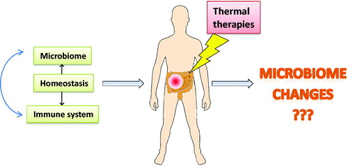
Figure 1. Under normal conditions, the microbiome and the immune system interact to regulate each other and maintain a homeostatic balance. The composition of the microbiome is sensitive to environmental factors such as diet and a variety of other stressors (including temperature) and dysbiosis is associated with several disease states. It has been reported that the composition of the microbiome directly regulates response to anticancer chemotherapy and immunotherapy. However, the effect of thermal therapy of the microbiome and antitumor immune response is unknown.
At the current time, we do not know whether thermal medicine applications can alter the microbiome and this is an important question for future research, not only in patients, but also in animal models, particularly mice, that are used to study thermal applications for diseases such as cancer, arthritis, diabetes and other diseases. Conversely, many humans suffer from conditions related to a dysfunctional microbiome. It could be very important to test whether heat treatments are able to modulate the microbiome and help reestablish a more “healthy” condition. Without a doubt, new research focusing on cellular and molecular mechanisms by which hyperthermia influences the microbiome of the gut is needed. At the present time, thermal therapy applications offer one of the only opportunities to obtain bodily fluids or fecal samples from patients undergoing hyperthermia to areas containing segments of the gut. This type of study may reveal significant clinical and therapeutic implications.
Disclosure statement
No potential conflict of interest was reported by the authors.
Additional information
Funding
References
- van der Zee J, van Rhoon GC. Cervical cancer: radiotherapy and hyperthermia. Int J Hyperthermia. 2006;22:229–234.
- Issels RD, Lindner LH, Verweij J, et al. Effect of neoadjuvant chemotherapy plus regional hyperthermia on long-term outcomes among patients with localized high-risk soft tissue sarcoma: the EORTC 62961-ESHO 95 randomized clinical trial. JAMA Oncol. 2018;4:483–492.
- Mauri G, Nicosia L, Xu Z, et al. Focused ultrasound: tumour ablation and its potential to enhance immunological therapy to cancer. Br Inst Radiol. 2018;91:20170641.
- Jones EL, Oleson JR, Prosnitz LR, et al. Randomized trial of hyperthermia and radiation for superficial tumors. J Clin Oncol. 2005;23:3079–3085.
- Pavlov MJ, Ceranic MS, Latincic SM, et al. Cytoreductive surgery and hyperthermic intraperitoneal chemotherapy for the treatment of advanced epithelial and recurrent ovarian carcinoma: a single center experience. Int J Hyperthermia. 2018;34:564–569.
- Bull JM, Scott GL, Strebel FR, et al. Fever-range whole-body thermal therapy combined with cisplatin, gemcitabine, and daily interferon-alpha: a description of a phase I-II protocol. Int J Hyperthermia. 2008;24:649–662.
- Lee CT, Kokolus KM, Leigh ND, et al. Defining immunological impact and therapeutic benefit of mild heating in a murine model of arthritis. PLoS One. 2015;10:e0120327.
- Janssen CW, Lowry CA, Mehl MR, et al. Whole-body hyperthermia for the treatment of major depressive disorder: a randomized clinical trial. JAMA Psychiatry. 2016;73:789–795.
- Fontaine SS, Novarro AJ, Kohl KD. Environmental temperature alters the digestive performance and gut microbiota of a terrestrial amphibian. J Exp Biol. 2018;221:187559.
- Sohail MU, Hume ME, Byrd JA, et al. Molecular analysis of the caecal and tracheal microbiome of heat-stressed broilers supplemented with prebiotic and probiotic. Avian Pathol. 2015;44:67–74.
- Thapa S, Zhang Y, Allen MS. Effects of temperature on bacterial microbiome composition in Ixodes scapularis ticks. Microbiologyopen. 2019;8:e00719.
- Minich JJ, Morris MM, Brown M, et al. Elevated temperature drives kelp microbiome dysbiosis, while elevated carbon dioxide induces water microbiome disruption. PLoS One. 2018;13:e0192772.
- Hooper LV, Littman DR, Macpherson AJ. Interactions between the microbiota and the immune system. Science. 2012;336:1268–1273.
- Demaria S, Ng B, Devitt ML, et al. Ionizing radiation inhibition of distant untreated tumors (abscopal effect) is immune mediated. Int J Radiat Oncol Biol Phys. 2004;58:862–870.
- Sharp HJ, Wansley EK, Garnett CT, et al. Synergistic antitumor activity of immune strategies combined with radiation. Front Biosci. 2007;12:4900–4910.
- Shiao SL, Coussens LM. The tumor-immune microenvironment and response to radiation therapy. J Mammary Gland Biol Neoplasia. 2010;15:411–421.
- Demaria S, Formenti SC. Role of T lymphocytes in tumor response to radiotherapy. Front Oncol. 2012;2:95.
- Ludgate CM. Optimizing cancer treatments to induce an acute immune response: radiation abscopal effects, PAMPs, and DAMPs. Clin Cancer Res. 2012;18:4522–4525.
- Zeng J, Harris TJ, Lim M, et al. Immune modulation and stereotactic radiation: improving local and abscopal responses. Biomed Res Int. 2013;2013:1.
- Zitvogel L, Galluzzi L, Smyth MJ, et al. Mechanism of action of conventional and targeted anticancer therapies: reinstating immunosurveillance. Immunity. 2013;39:74–88.
- Gerber SA, Lim JY, Connolly KA, et al. Radio-responsive tumors exhibit greater intratumoral immune activity than nonresponsive tumors. Int J Cancer. 2014;134:2383–2392.
- Apetoh L, Ghiringhelli F, Tesniere A, et al. Toll-like receptor 4-dependent contribution of the immune system to anticancer chemotherapy and radiotherapy. Nat Med. 2007;13:1050–1059.
- Mattarollo SR, Loi S, Duret H, et al. Pivotal role of innate and adaptive immunity in anthracycline chemotherapy of established tumors. Cancer Res. 2011;71:4809–4820.
- Bracci L, Schiavoni G, Sistigu A, et al. Immune-based mechanisms of cytotoxic chemotherapy: implications for the design of novel and rationale-based combined treatments against cancer. Cell Death Differ. 2014;21:15–25.
- de Biasi AR, Villena-Vargas J, Adusumilli PS. Cisplatin-induced antitumor immunomodulation: a review of preclinical and clinical evidence. Clin Cancer Res. 2014;20:5384–5391.
- Emens LA, Middleton G. The interplay of immunotherapy and chemotherapy: harnessing potential synergies. Cancer Immunol Res. 2015;3:436–443.
- Galluzzi L, Buque A, Kepp O, et al. Immunological effects of conventional chemotherapy and targeted anticancer agents. Cancer Cell. 2015;28:690–714.
- Zitvogel L, Kepp O, Kroemer G. Immune parameters affecting the efficacy of chemotherapeutic regimens. Nat Rev Clin Oncol. 2011;8:151–160.
- Galluzzi L, Chan TA, Kroemer G, et al. The hallmarks of successful anticancer immunotherapy. Sci Transl Med. 2018;10:eaat7807.
- Viaud S, Saccheri F, Mignot G, et al. The intestinal microbiota modulates the anticancer immune effects of cyclophosphamide. Science. 2013;342:971–976.
- Iida N, Dzutsev A, Stewart CA, et al. Commensal bacteria control cancer response to therapy by modulating the tumor microenvironment. Science. 2013;342:967–970.
- Chen L, Tai WC, Brar MS, et al. Tumor grafting induces changes of gut microbiota in athymic nude mice in the presence and absence of medicinal Gynostemma saponins. PLoS One. 2015;10:e0126807.
- Al-Qadami G, Van Sebille Y, Le H, et al. Gut microbiota: implications for radiotherapy response and radiotherapy-induced mucositis. Expert Rev Gastroenterol Hepatol. 2019;13:485–496.
- Vetizou M, Pitt JM, Daillere R, et al. Anticancer immunotherapy by CTLA-4 blockade relies on the gut microbiota. Science. 2015;350:1079–1084.
- Gopalakrishnan V, Spencer CN, Nezi L, et al. Gut microbiome modulates response to anti-PD-1 immunotherapy in melanoma patients. Science. 2018;359:97–103.
- Matson V, Fessler J, Bao R, et al. The commensal microbiome is associated with anti-PD-1 efficacy in metastatic melanoma patients. Science. 2018;359:104–108.
- Routy B, Le Chatelier E, Derosa L, et al. Gut microbiome influences efficacy of PD-1-based immunotherapy against epithelial tumors. Science. 2018;359:91–97.
- McQuade JL, Daniel CR, Helmink BA, et al. Modulating the microbiome to improve therapeutic response in cancer. Lancet Oncol. 2019;20:e77–e91.
- Fessler JL, Gajewski TF. The microbiota: a new variable impacting cancer treatment outcomes. Clin Cancer Res. 2017;23:3229–3231.
- Fessler J, Matson V, Gajewski TF. Exploring the emerging role of the microbiome in cancer immunotherapy. J Immunother Cancer. 2019;7:108.
- Ley RE, Turnbaugh PJ, Klein S, et al. Microbial ecology: human gut microbes associated with obesity. Nature. 2006;444:1022–1023.
- Turnbaugh PJ, Ley RE, Mahowald MA, et al. An obesity-associated gut microbiome with increased capacity for energy harvest. Nature. 2006;444:1027–1031.
- National Research Council (US) Committee for the Update of the Guide for the Care and Use of Laboratory Animals. Guide for the cared and use of laboratory animals. 8th ed. Washington, DC: The National Academies Press; 2012.
- Schmidt E, Mykytczuk N, Schulte-Hostedde AI. Effects of the captive and wild environment on diversity of the gut microbiome of deer mice (Peromyscus maniculatus). ISME J. 2019;13:1293–1305.
- Ma BW, Bokulich NA, Castillo PA, et al. Routine habitat change: a source of unrecognized transient alteration of intestinal microbiota in laboratory mice. PLoS One. 2012;7:e47416.
- Franklin CL, Ericsson AC. Microbiota and reproducibility of rodent models. Lab Anim (NY). 2017;46:114–122.
- Hufeldt MR, Nielsen DS, Vogensen FK, et al. Variation in the gut microbiota of laboratory mice is related to both genetic and environmental factors. Comp Med. 2010;60:336–347.
- Servick K. Of mice and microbes. Science. 2016;353:741–743.
- McCoy KD, Geuking MB, Ronchi F. Gut microbiome standardization in control and experimental mice. Curr Protoc Immunol. 2017;117:23.21.21–23.21.13.
- Parker KD, Albeke SE, Gigley JP, et al. Microbiome composition in both wild-type and disease model mice is heavily influenced by mouse facility. Front Microbiol. 2018;9:1598.
- Villarino NF, LeCleir GR, Denny JE, et al. Composition of the gut microbiota modulates the severity of malaria. Proc Natl Acad Sci USA. 2016;113:2235–2240.
- Bidot WA, Ericsson AC, Franklin CL. Effects of water decontamination methods and bedding material on the gut microbiota. PLoS One. 2018;13:e0198305.
- Cui B, Su D, Li W, et al. Effects of chronic noise exposure on the microbiome-gut-brain axis in senescence-accelerated prone mice: implications for Alzheimer's disease. J Neuroinflammation. 2018;15:190.
- Wu G, Tang W, He Y, et al. Light exposure influences the diurnal oscillation of gut microbiota in mice. Biochem Biophys Res Commun. 2018;501:16–23.
- Turner PV. The role of the gut microbiota on animal model reproducibility. Anim Model Exp Med. 2018;1:109–115.
- Ghaly S, Kaakoush NO, Lloyd F, et al. Ultraviolet irradiation of skin alters the faecal microbiome independently of vitamin D in mice. Nutrients 2018;10:1069.
- Deaver JA, Eum SY, Toborek M. Circadian disruption changes gut microbiome taxa and functional gene composition. Front Microbiol. 2018;9:737.
- Antoch MP, Kondratov RV, Takahashi JS. Circadian clock genes as modulators of sensitivity to genotoxic stress. Cell Cycle 2005;4:901–907.
- Jenkins SV, Vang KB, Gies A, et al. Sample storage conditions induce post-collection biases in microbiome profiles. BMC Microbiol. 2018;18:227.
- Song SJ, Amir A, Metcalf JL, et al. Preservation methods differ in fecal microbiome stability, affecting suitability for field studies. mSystems. 2016;1:e00021–16.
- Nicholls HT, Krisko TI, LeClair KB, et al. Regulation of adaptive thermogenesis by the gut microbiome. FASEB J. 2016;30:854–852.
- Giles DA, Moreno-Fernandez ME, Stankiewicz TE, et al. Thermoneutral housing exacerbates nonalcoholic fatty liver disease in mice and allows for sex-independent disease modeling. Nat Med. 2017;23:829–838.
- Worthmann A, John C, Ruhlemann MC, et al. Cold-induced conversion of cholesterol to bile acids in mice shapes the gut microbiome and promotes adaptive thermogenesis. Nat Med. 2017;23:839–849.
- Thapa S, Zhang Y, Allen MS. Effects of temperature on bacterial microbiome composition in Ixodes scapularis ticks. Microbiologyopen. 2018;8:e00719.
- Brothers CJ, Van Der Pol WJ, Morrow CD, et al. Ocean warming alters predicted microbiome functionality in a common sea urchin. Proc Biol Sci. 2018;285:20180340.
- Kokou F, Sasson G, Nitzan T, et al. Host genetic selection for cold tolerance shapes microbiome composition and modulates its response to temperature. Elife. 2018;7:e36398.
- eLife. Tropical fish adapt to cold temperatures in coordination with their microbiome. ScienceDaily [Internet]. [cited 2018 Nov 20]. Available from: https://www.sciencedaily.com/releases/2018/11/181120125843.htm
- Zhang XY, Sukhchuluun G, Bo TB, et al. Huddling remodels gut microbiota to reduce energy requirements in a small mammal species during cold exposure. Microbiome. 2018;6:103.
- Qi H, Wang P, Liu C, et al. Involvement of HIF-1alpha in MLCK-dependent endothelial barrier dysfunction in hypoxia. Cell Physiol Biochem. 2011;27:251–262.
- Yamagata K, Tagami M, Takenaga F, et al. Hypoxia-induced changes in tight junction permeability of brain capillary endothelial cells are associated with IL-1beta and nitric oxide. Neurobiol Dis. 2004;17:491–499.
- Lambert GP. Stress-induced gastrointestinal barrier dysfunction and its inflammatory effects. J Anim Sci. 2009;87:E101–E108.
- Pearce SC, Mani V, Boddicker RL, et al. Heat stress reduces intestinal barrier integrity and favors intestinal glucose transport in growing pigs. PLoS One. 2013;8:e70215.
- Dokladny K, Zuhl MN, Moseley PL. Intestinal epithelial barrier function and tight junction proteins with heat and exercise. J Appl Physiol (1985). 2016;120:692–701.
- Costa KA, Soares AD, Wanner SP, et al. L-arginine supplementation prevents increases in intestinal permeability and bacterial translocation in male Swiss mice subjected to physical exercise under environmental heat stress. J Nutr. 2014;144:218–223.
- Bouchama A, Parhar RS, el-Yazigi A, et al. Endotoxemia and release of tumor necrosis factor and interleukin 1 alpha in acute heatstroke. J Appl Physiol. 1991;70:2640–2644.
- Appenheimer MM, Girard RA, Chen Q, et al. Conservation of IL-6 trans-signaling mechanisms controlling L-selectin adhesion by fever-range thermal stress. Eur J Immunol. 2007;37:2856–2867.
- Sakaguchi Y, Makino M, Kaneko T, et al. Therapeutic efficacy of long duration-low temperature whole body hyperthermia when combined with tumor necrosis factor and carboplatin in rats. Cancer Res. 1994;54:2223–2227.
- Juang T, Stauffer PR, Craciunescu OA, et al. Thermal dosimetry characteristics of deep regional heating of non-muscle invasive bladder cancer. Int J Hyperthermia. 2014;30:176–183.
- Sapareto SA, Dewey WC. Thermal dose determination in cancer therapy. Int J Radiat Oncol Biol Phys. 1984;10:787–800.
- Lee SY, Kim JH, Han YH, et al. The effect of modulated electro-hyperthermia on temperature and blood flow in human cervical carcinoma. Int J Hyperthermia. 2018;34:953–960.